 Movie
Movie Controller

[English] 日本語
 Yorodumi
- EMDB-33738: Cryo-EM structure of Tetrahymena ribozyme conformation 2 undergoi... -
+ Open data

- Basic information
| Entry |  | |||||||||
|---|---|---|---|---|---|---|---|---|---|---|
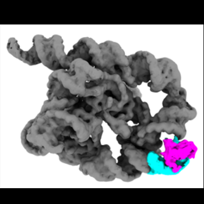
| Title | Cryo-EM structure of Tetrahymena ribozyme conformation 2 undergoing the first-step self-splicing | |||||||||
 Map data Map data | Cryo-EM structure of Tetrahymena ribozyme conformation 2 undergoing the the first-step self-splicing--- unmask | |||||||||
 Sample Sample |
| |||||||||
 Keywords Keywords | Tetrahymena ribozyme / first step of self-splicing / conformation 2 / RNA | |||||||||
| Biological species |  Tetrahymena (eukaryote) Tetrahymena (eukaryote) | |||||||||
| Method | single particle reconstruction / cryo EM / Resolution: 3.18 Å | |||||||||
 Authors Authors | Zhang X / Li S / Pintilie G / Palo MZ / Zhang K / Liu L | |||||||||
| Funding support |  China, 1 items China, 1 items
| |||||||||
 Citation Citation | Journal: Nucleic Acids Res / Year: 2023 Title: Snapshots of the first-step self-splicing of Tetrahymena ribozyme revealed by cryo-EM. Authors: Xiaojing Zhang / Shanshan Li / Grigore Pintilie / Michael Z Palo / Kaiming Zhang /  Abstract: Tetrahymena ribozyme is a group I intron, whose self-splicing is the result of two sequential ester-transfer reactions. To understand how it facilitates catalysis in the first self-splicing reaction, ...Tetrahymena ribozyme is a group I intron, whose self-splicing is the result of two sequential ester-transfer reactions. To understand how it facilitates catalysis in the first self-splicing reaction, we used cryogenic electron microscopy (cryo-EM) to resolve the structures of L-16 Tetrahymena ribozyme complexed with a 11-nucleotide 5'-splice site analog substrate. Four conformations were achieved to 4.14, 3.18, 3.09 and 2.98 Å resolutions, respectively, corresponding to different splicing intermediates during the first enzymatic reaction. Comparison of these structures reveals structural alterations, including large conformational changes in IGS/IGSext (P1-P1ext duplex) and J5/4, as well as subtle local rearrangements in the G-binding site. These structural changes are required for the enzymatic activity of the Tetrahymena ribozyme. Our study demonstrates the ability of cryo-EM to capture dynamic RNA structural changes, ushering in a new era in the analysis of RNA structure-function by cryo-EM. | |||||||||
| History |
|
- Structure visualization
| Supplemental images |
|---|
- Downloads & links
-EMDB archive
| Map data | emd_33738.map.gz | 31.9 MB |  EMDB map data format EMDB map data format | |
|---|---|---|---|---|
| Header (meta data) | emd-33738-v30.xmlemd-33738.xml | 17.9 KB 17.9 KB | Display Display | EMDB header |
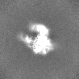
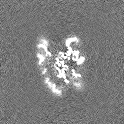
| Images |  emd_33738.png emd_33738.png | 73.6 KB | ||
| Others | emd_33738_additional_1.map.gzemd_33738_half_map_1.map.gzemd_33738_half_map_2.map.gz | 32.8 MB 59.4 MB 59.4 MB | ||
| Archive directory |  http://ftp.pdbj.org/pub/emdb/structures/EMD-33738ftp://ftp.pdbj.org/pub/emdb/structures/EMD-33738 http://ftp.pdbj.org/pub/emdb/structures/EMD-33738ftp://ftp.pdbj.org/pub/emdb/structures/EMD-33738 | HTTPS FTP |
-Related structure data
| Related structure data |  7ycgMC  7yc8C  7ychC  7yciC M: atomic model generated by this map C: citing same article ( |
|---|


-Links
| EMDB pages | EMDB (EBI/PDBe) / EMDataResource |
|---|
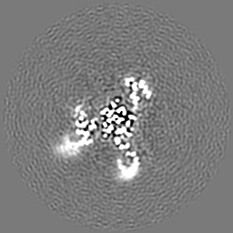
-Map
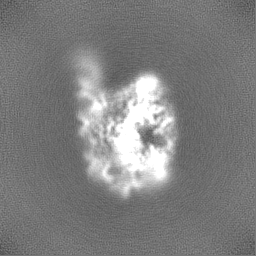
| File | Download / File: emd_33738.map.gz / Format: CCP4 / Size: 64 MB / Type: IMAGE STORED AS FLOATING POINT NUMBER (4 BYTES) | ||||||||||||||||||||||||||||||||||||
|---|---|---|---|---|---|---|---|---|---|---|---|---|---|---|---|---|---|---|---|---|---|---|---|---|---|---|---|---|---|---|---|---|---|---|---|---|---|
| Annotation | Cryo-EM structure of Tetrahymena ribozyme conformation 2 undergoing the the first-step self-splicing--- unmask | ||||||||||||||||||||||||||||||||||||
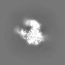
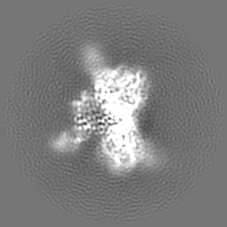
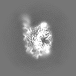
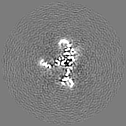
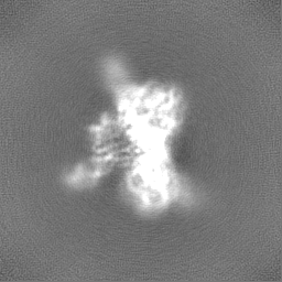
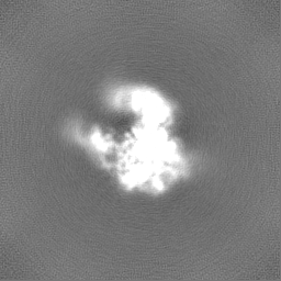
| Projections & slices | Image control
Images are generated by Spider. | ||||||||||||||||||||||||||||||||||||
| Voxel size | X=Y=Z: 0.82 Å | ||||||||||||||||||||||||||||||||||||
| Density |
| ||||||||||||||||||||||||||||||||||||
| Symmetry | Space group: 1 | ||||||||||||||||||||||||||||||||||||
| Details | EMDB XML:
|
 Z (Sec.)
 Y (Row.)
 X (Col.)


-Supplemental data
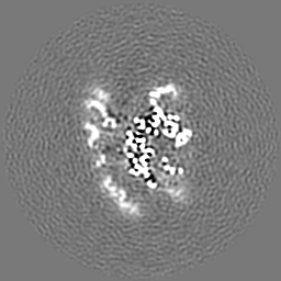
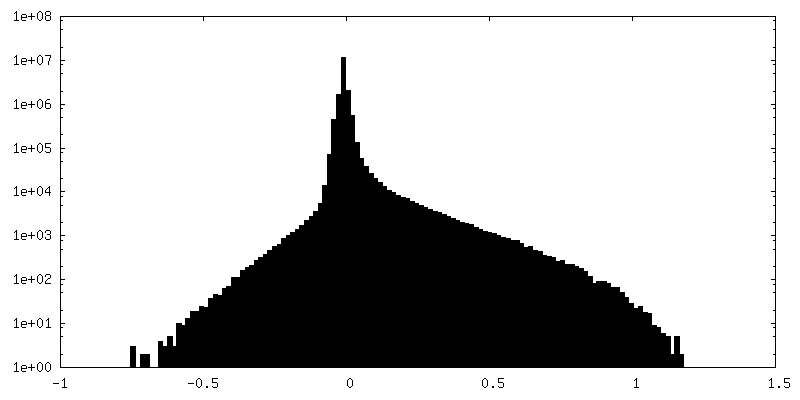
-Additional map: Cryo-EM structure of Tetrahymena ribozyme conformation 2 undergoing...
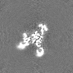
| File | emd_33738_additional_1.map | ||||||||||||
|---|---|---|---|---|---|---|---|---|---|---|---|---|---|
| Annotation | Cryo-EM structure of Tetrahymena ribozyme conformation 2 undergoing the the first-step self-splicing--- sharp | ||||||||||||
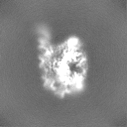
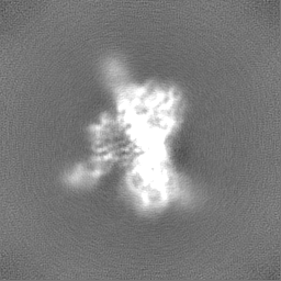
| Projections & Slices |
| ||||||||||||
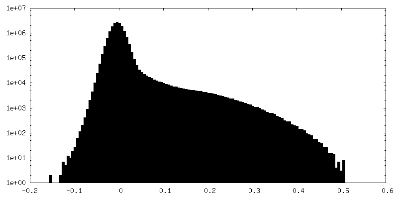
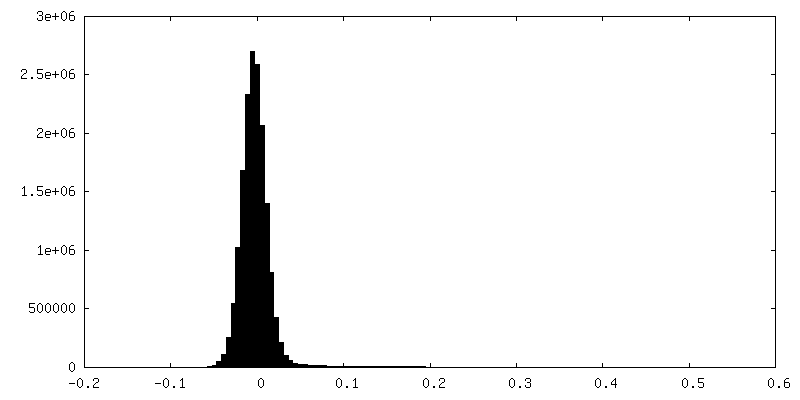
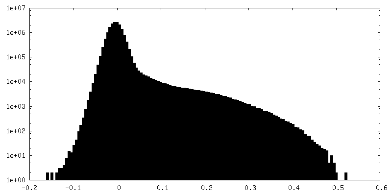
| Density Histograms |
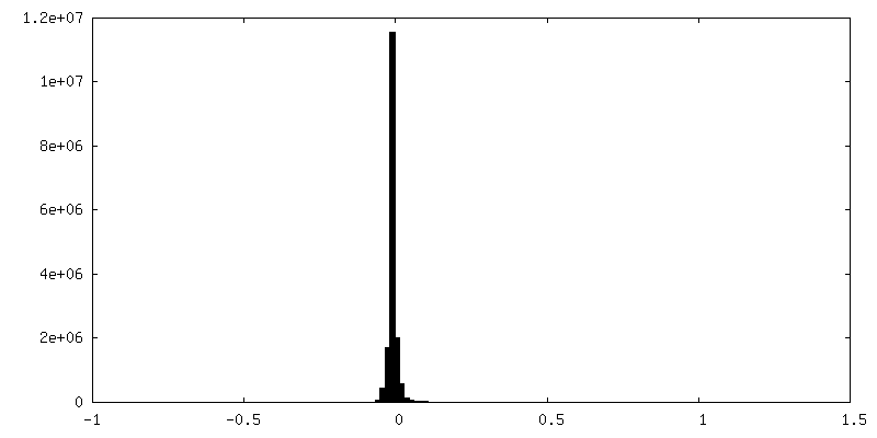
-Half map: Cryo-EM structure of Tetrahymena ribozyme conformation 2 undergoing...
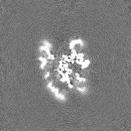
| File | emd_33738_half_map_1.map | ||||||||||||
|---|---|---|---|---|---|---|---|---|---|---|---|---|---|
| Annotation | Cryo-EM structure of Tetrahymena ribozyme conformation 2 undergoing the the first-step self-splicing--- half | ||||||||||||
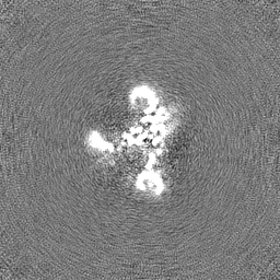
| Projections & Slices |
| ||||||||||||
| Density Histograms |


-Half map: Cryo-EM structure of Tetrahymena ribozyme conformation 2 undergoing...
| File | emd_33738_half_map_2.map | ||||||||||||
|---|---|---|---|---|---|---|---|---|---|---|---|---|---|
| Annotation | Cryo-EM structure of Tetrahymena ribozyme conformation 2 undergoing the the first-step self-splicing--- half | ||||||||||||
| Projections & Slices |
| ||||||||||||
| Density Histograms |

- Sample components
-Entire : Cryo-EM structure of Tetrahymena ribozyme conformation 2 undergoi...
| Entire | Name: Cryo-EM structure of Tetrahymena ribozyme conformation 2 undergoing the first-step self-splicing |
|---|---|
| Components |
|
-Supramolecule #1: Cryo-EM structure of Tetrahymena ribozyme conformation 2 undergoi...
| Supramolecule | Name: Cryo-EM structure of Tetrahymena ribozyme conformation 2 undergoing the first-step self-splicing type: complex / ID: 1 / Parent: 0 / Macromolecule list: #1-#2 |
|---|---|
| Source (natural) | Organism: Tetrahymena (eukaryote) |
| Molecular weight | Theoretical: 130 KDa |
-Macromolecule #1: RNA (5'-R(*CP*CP*CP*UP*CP*UP*AP*AP*AP*CP*C)-3')
| Macromolecule | Name: RNA (5'-R(*CP*CP*CP*UP*CP*UP*AP*AP*AP*CP*C)-3') / type: rna / ID: 1 / Number of copies: 1 |
|---|---|
| Source (natural) | Organism: Tetrahymena (eukaryote) |
| Molecular weight | Theoretical: 3.386082 KDa |
| Sequence | String: CCCUCUAAAC C |
-Macromolecule #2: RNA (393-MER)
| Macromolecule | Name: RNA (393-MER) / type: rna / ID: 2 / Number of copies: 1 |
|---|---|
| Source (natural) | Organism: Tetrahymena (eukaryote) |
| Molecular weight | Theoretical: 127.011883 KDa |
| Sequence | String: GGUUUGGAGG GAAAAGUUAU CAGGCAUGCA CCUGGUAGCU AGUCUUUAAA CCAAUAGAUU GCAUCGGUUU AAAAGGCAAG ACCGUCAAA UUGCGGGAAA GGGGUCAACA GCCGUUCAGU ACCAAGUCUC AGGGGAAACU UUGAGAUGGC CUUGCAAAGG G UAUGGUAA ...String: GGUUUGGAGG GAAAAGUUAU CAGGCAUGCA CCUGGUAGCU AGUCUUUAAA CCAAUAGAUU GCAUCGGUUU AAAAGGCAAG ACCGUCAAA UUGCGGGAAA GGGGUCAACA GCCGUUCAGU ACCAAGUCUC AGGGGAAACU UUGAGAUGGC CUUGCAAAGG G UAUGGUAA UAAGCUGACG GACAUGGUCC UAACCACGCA GCCAAGUCCU AAGUCAACAG AUCUUCUGUU GAUAUGGAUG CA GUUCACA GACUAAAUGU CGGUCGGGGA AGAUGUAUUC UUCUCAUAAG AUAUAGUCGG ACCUCUCCUU AAUGGGAGCU AGC GGAUGA AGUGAUGCAA CACUGGAGCC GCUGGGAACU AAUUUGUAUG CGAAAGUAUA UUGAUUAGUU UUGGAGU GENBANK: GENBANK: X54512.1 |
-Macromolecule #3: GUANOSINE-5'-TRIPHOSPHATE
| Macromolecule | Name: GUANOSINE-5'-TRIPHOSPHATE / type: ligand / ID: 3 / Number of copies: 1 / Formula: GTP |
|---|---|
| Molecular weight | Theoretical: 523.18 Da |
| Chemical component information |  ChemComp-GTP: |
-Macromolecule #4: MAGNESIUM ION
| Macromolecule | Name: MAGNESIUM ION / type: ligand / ID: 4 / Number of copies: 3 / Formula: MG |
|---|---|
| Molecular weight | Theoretical: 24.305 Da |
-Experimental details
-Structure determination
| Method | cryo EM |
|---|---|
 Processing Processing | single particle reconstruction |
| Aggregation state | particle |
-Sample preparation
| Concentration | 1 mg/mL |
|---|---|
| Buffer | pH: 7.5 |
| Vitrification | Cryogen name: ETHANE |
- Electron microscopy
| Microscope | FEI TITAN KRIOS |
|---|---|
| Image recording | Film or detector model: GATAN K3 (6k x 4k) / Average electron dose: 51.0 e/Å2 |
| Electron beam | Acceleration voltage: 300 kV / Electron source:  FIELD EMISSION GUN FIELD EMISSION GUN |
| Electron optics | Illumination mode: FLOOD BEAM / Imaging mode: BRIGHT FIELD / Nominal defocus max: 2.8000000000000003 µm / Nominal defocus min: 0.8 µm |
| Experimental equipment |  Model: Titan Krios / Image courtesy: FEI Company |
