[English] 日本語
 Yorodumi
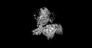
- EMDB-33585: Cryo-EM structure of the octreotide-bound SSTR2-miniGo-scFv16 complex -
+ Open data
- Basic information
| Entry |  | |||||||||
|---|---|---|---|---|---|---|---|---|---|---|
| Title | Cryo-EM structure of the octreotide-bound SSTR2-miniGo-scFv16 complex | |||||||||
 Map data Map data | ||||||||||
 Sample Sample |
| |||||||||
| Function / homology |  Function and homology information Function and homology informationsomatostatin receptor activity / peristalsis / mu-type opioid receptor binding / corticotropin-releasing hormone receptor 1 binding / vesicle docking involved in exocytosis / neuropeptide binding / cellular response to glucocorticoid stimulus / G protein-coupled dopamine receptor signaling pathway / regulation of heart contraction / response to starvation ...somatostatin receptor activity / peristalsis / mu-type opioid receptor binding / corticotropin-releasing hormone receptor 1 binding / vesicle docking involved in exocytosis / neuropeptide binding / cellular response to glucocorticoid stimulus / G protein-coupled dopamine receptor signaling pathway / regulation of heart contraction / response to starvation / G protein-coupled receptor signaling pathway, coupled to cyclic nucleotide second messenger / neuropeptide signaling pathway / negative regulation of insulin secretion / G protein-coupled serotonin receptor binding / forebrain development / muscle contraction / adenylate cyclase-inhibiting G protein-coupled receptor signaling pathway / cerebellum development / Peptide ligand-binding receptors / cellular response to estradiol stimulus / locomotory behavior / PDZ domain binding / G-protein beta/gamma-subunit complex binding / Olfactory Signaling Pathway / adenylate cyclase-modulating G protein-coupled receptor signaling pathway / Activation of the phototransduction cascade / G beta:gamma signalling through PLC beta / Presynaptic function of Kainate receptors / Thromboxane signalling through TP receptor / G protein-coupled acetylcholine receptor signaling pathway / G-protein activation / Activation of G protein gated Potassium channels / Inhibition of voltage gated Ca2+ channels via Gbeta/gamma subunits / Prostacyclin signalling through prostacyclin receptor / Glucagon signaling in metabolic regulation / G beta:gamma signalling through CDC42 / G beta:gamma signalling through BTK / ADP signalling through P2Y purinoceptor 12 / Sensory perception of sweet, bitter, and umami (glutamate) taste / Synthesis, secretion, and inactivation of Glucagon-like Peptide-1 (GLP-1) / photoreceptor disc membrane / Glucagon-type ligand receptors / Adrenaline,noradrenaline inhibits insulin secretion / Vasopressin regulates renal water homeostasis via Aquaporins / G alpha (z) signalling events / Glucagon-like Peptide-1 (GLP1) regulates insulin secretion / cellular response to catecholamine stimulus / ADORA2B mediated anti-inflammatory cytokines production / sensory perception of taste / ADP signalling through P2Y purinoceptor 1 / G beta:gamma signalling through PI3Kgamma / adenylate cyclase-activating dopamine receptor signaling pathway / Cooperation of PDCL (PhLP1) and TRiC/CCT in G-protein beta folding / GPER1 signaling / cellular response to prostaglandin E stimulus / Inactivation, recovery and regulation of the phototransduction cascade / G-protein beta-subunit binding / heterotrimeric G-protein complex / G alpha (12/13) signalling events / extracellular vesicle / signaling receptor complex adaptor activity / Thrombin signalling through proteinase activated receptors (PARs) / GTPase binding / retina development in camera-type eye / Ca2+ pathway / phospholipase C-activating G protein-coupled receptor signaling pathway / cell body / G alpha (i) signalling events / fibroblast proliferation / spermatogenesis / G alpha (s) signalling events / Hydrolases; Acting on acid anhydrides; Acting on GTP to facilitate cellular and subcellular movement / G alpha (q) signalling events / Ras protein signal transduction / cell population proliferation / Extra-nuclear estrogen signaling / neuron projection / G protein-coupled receptor signaling pathway / negative regulation of cell population proliferation / lysosomal membrane / GTPase activity / dendrite / synapse / protein-containing complex binding / GTP binding / signal transduction / extracellular exosome / membrane / metal ion binding / plasma membrane / cytosol / cytoplasm Similarity search - Function | |||||||||
| Biological species |  Homo sapiens (human) Homo sapiens (human) | |||||||||
| Method | single particle reconstruction / cryo EM / Resolution: 3.25 Å | |||||||||
 Authors Authors | Chen S / Zheng S | |||||||||
| Funding support |  China, 1 items China, 1 items
| |||||||||
 Citation Citation |  Journal: Nat Chem Biol / Year: 2023 Journal: Nat Chem Biol / Year: 2023Title: Molecular basis for the selective G protein signaling of somatostatin receptors. Authors: Sijia Chen / Xiao Teng / Sanduo Zheng /  Abstract: G protein-coupled receptors (GPCRs) modulate every aspect of physiological functions mainly through activating heterotrimeric G proteins. A majority of GPCRs promiscuously couple to multiple G ...G protein-coupled receptors (GPCRs) modulate every aspect of physiological functions mainly through activating heterotrimeric G proteins. A majority of GPCRs promiscuously couple to multiple G protein subtypes. Here we validate that in addition to the well-known G pathway, somatostatin receptor 2 and 5 (SSTR2 and SSTR5) couple to the G pathway and show that smaller ligands preferentially activate the G pathway. We further determined cryo-electron microscopy structures of the SSTR2‒G and SSTR2‒G complexes bound to octreotide and SST-14. Structural and functional analysis revealed that G protein selectivity of SSTRs is not only determined by structural elements in the receptor-G protein interface, but also by the conformation of the agonist-binding pocket. Accordingly, smaller ligands fail to stabilize a broader agonist-binding pocket of SSTRs that is required for efficient G coupling but not G coupling. Our studies facilitate the design of drugs with selective G protein signaling to improve therapeutic efficacy. | |||||||||
| History |
|
- Structure visualization
| Supplemental images |
|---|
- Downloads & links
-EMDB archive
| Map data |  emd_33585.map.gz emd_33585.map.gz | 20.9 MB |  EMDB map data format EMDB map data format | |
|---|---|---|---|---|
| Header (meta data) |  emd-33585-v30.xml emd-33585-v30.xml emd-33585.xml emd-33585.xml | 20.5 KB 20.5 KB | Display Display |  EMDB header EMDB header |
| Images |  emd_33585.png emd_33585.png | 26.5 KB | ||
| Others |  emd_33585_half_map_1.map.gz emd_33585_half_map_1.map.gz emd_33585_half_map_2.map.gz emd_33585_half_map_2.map.gz | 20.6 MB 20.6 MB | ||
| Archive directory |  http://ftp.pdbj.org/pub/emdb/structures/EMD-33585 http://ftp.pdbj.org/pub/emdb/structures/EMD-33585 ftp://ftp.pdbj.org/pub/emdb/structures/EMD-33585 ftp://ftp.pdbj.org/pub/emdb/structures/EMD-33585 | HTTPS FTP |
-Validation report
| Summary document |  emd_33585_validation.pdf.gz emd_33585_validation.pdf.gz | 650.5 KB | Display |  EMDB validaton report EMDB validaton report |
|---|---|---|---|---|
| Full document |  emd_33585_full_validation.pdf.gz emd_33585_full_validation.pdf.gz | 650.1 KB | Display | |
| Data in XML |  emd_33585_validation.xml.gz emd_33585_validation.xml.gz | 10.1 KB | Display | |
| Data in CIF |  emd_33585_validation.cif.gz emd_33585_validation.cif.gz | 11.8 KB | Display | |
| Arichive directory |  https://ftp.pdbj.org/pub/emdb/validation_reports/EMD-33585 https://ftp.pdbj.org/pub/emdb/validation_reports/EMD-33585 ftp://ftp.pdbj.org/pub/emdb/validation_reports/EMD-33585 ftp://ftp.pdbj.org/pub/emdb/validation_reports/EMD-33585 | HTTPS FTP |
-Related structure data
| Related structure data |  7y24MC  7y26C  7y27C M: atomic model generated by this map C: citing same article ( |
|---|---|
| Similar structure data | Similarity search - Function & homology  F&H Search F&H Search |
- Links
Links
| EMDB pages |  EMDB (EBI/PDBe) / EMDB (EBI/PDBe) /  EMDataResource EMDataResource |
|---|---|
| Related items in Molecule of the Month |
- Map
Map
| File |  Download / File: emd_33585.map.gz / Format: CCP4 / Size: 22.2 MB / Type: IMAGE STORED AS FLOATING POINT NUMBER (4 BYTES) Download / File: emd_33585.map.gz / Format: CCP4 / Size: 22.2 MB / Type: IMAGE STORED AS FLOATING POINT NUMBER (4 BYTES) | ||||||||||||||||||||||||||||||||||||
|---|---|---|---|---|---|---|---|---|---|---|---|---|---|---|---|---|---|---|---|---|---|---|---|---|---|---|---|---|---|---|---|---|---|---|---|---|---|
| Projections & slices | Image control
Images are generated by Spider. | ||||||||||||||||||||||||||||||||||||
| Voxel size | X=Y=Z: 1.087 Å | ||||||||||||||||||||||||||||||||||||
| Density |
| ||||||||||||||||||||||||||||||||||||
| Symmetry | Space group: 1 | ||||||||||||||||||||||||||||||||||||
| Details | EMDB XML:
|
-Supplemental data
-Half map: #2
| File | emd_33585_half_map_1.map | ||||||||||||
|---|---|---|---|---|---|---|---|---|---|---|---|---|---|
| Projections & Slices |
| ||||||||||||
| Density Histograms |
-Half map: #1
| File | emd_33585_half_map_2.map | ||||||||||||
|---|---|---|---|---|---|---|---|---|---|---|---|---|---|
| Projections & Slices |
| ||||||||||||
| Density Histograms |
- Sample components
+Entire : complex of SSTR2-miniGo-scFv16 bound with octreotide
+Supramolecule #1: complex of SSTR2-miniGo-scFv16 bound with octreotide
+Supramolecule #2: G(o) subunit alpha
+Supramolecule #3: G(I)/G(S)/G(T) subunit beta-1, G(I)/G(S)/G(O) subunit gamma-2
+Supramolecule #4: svFv16, octreotide
+Supramolecule #5: Somatostatin receptor type 2
+Macromolecule #1: Guanine nucleotide-binding protein G(o) subunit alpha
+Macromolecule #2: Guanine nucleotide-binding protein G(I)/G(S)/G(T) subunit beta-1
+Macromolecule #3: Guanine nucleotide-binding protein G(I)/G(S)/G(O) subunit gamma-2
+Macromolecule #4: single Fab chain (svFv16)
+Macromolecule #5: Octreotide
+Macromolecule #6: Somatostatin receptor type 2
-Experimental details
-Structure determination
| Method | cryo EM |
|---|---|
 Processing Processing | single particle reconstruction |
| Aggregation state | particle |
- Sample preparation
| Buffer | pH: 7.5 |
|---|---|
| Grid | Model: Quantifoil R1.2/1.3 / Material: GOLD |
| Vitrification | Cryogen name: ETHANE |
- Electron microscopy
| Microscope | FEI TITAN KRIOS |
|---|---|
| Image recording | Film or detector model: GATAN K3 (6k x 4k) / Average electron dose: 50.0 e/Å2 |
| Electron beam | Acceleration voltage: 300 kV / Electron source:  FIELD EMISSION GUN FIELD EMISSION GUN |
| Electron optics | Illumination mode: FLOOD BEAM / Imaging mode: BRIGHT FIELD / Nominal defocus max: 5.0 µm / Nominal defocus min: 1.0 µm |
| Experimental equipment |  Model: Titan Krios / Image courtesy: FEI Company |
- Image processing
| Final reconstruction | Resolution.type: BY AUTHOR / Resolution: 3.25 Å / Resolution method: FSC 0.143 CUT-OFF / Number images used: 687989 |
|---|---|
| Initial angle assignment | Type: MAXIMUM LIKELIHOOD |
| Final angle assignment | Type: MAXIMUM LIKELIHOOD |
 Movie
Movie Controller


 Z (Sec.)
 Y (Row.)
 X (Col.)


