登録情報 データベース : EMDB / ID : EMD-32842タイトル CryoEM structure of loose sNS1 tetramer 細胞器官・細胞要素 : Structure of Dengue NS1 in Loose tetramer form / 機能・相同性 分子機能 ドメイン・相同性 構成要素
/ / / / / / / / / / / / / / / / / / / / / / / / / / / / / / / / / / / / / / / / / / / / / / / / / / / / / / / / / / / / / / / / / / / / / / / / / / / / / / / / / / / / / / / / / / / / / / / / / / / / / / / / / / / / / / 生物種 手法 / / 解像度 : 8.3 Å Shu B / Lok SM 資金援助 Organization Grant number 国 National Research Foundation (NRF, Singapore) NRF-NRFI2016-01
ジャーナル : Nat Commun / 年 : 2022タイトル : CryoEM structures of the multimeric secreted NS1, a major factor for dengue hemorrhagic fever.著者 : Bo Shu / Justin S G Ooi / Aaron W K Tan / Thiam-Seng Ng / Wanwisa Dejnirattisai / Juthathip Mongkolsapaya / Guntur Fibriansah / Jian Shi / Victor A Kostyuchenko / Gavin R Screaton / Shee-Mei Lok / 要旨 : Dengue virus infection can cause dengue hemorrhagic fever (DHF). Dengue NS1 is multifunctional. The intracellular dimeric NS1 (iNS1) forms part of the viral replication complex. Previous studies ... Dengue virus infection can cause dengue hemorrhagic fever (DHF). Dengue NS1 is multifunctional. The intracellular dimeric NS1 (iNS1) forms part of the viral replication complex. Previous studies suggest the extracellular secreted NS1 (sNS1), which is a major factor contributing to DHF, exists as hexamers. The structure of the iNS1 is well-characterised but not that of sNS1. Here we show by cryoEM that the recombinant sNS1 exists in multiple oligomeric states: the tetrameric (stable and loose conformation) and hexameric structures. Stability of the stable and loose tetramers is determined by the conformation of their N-terminal domain - elongated β-sheet or β-roll. Binding of an anti-NS1 Fab breaks the loose tetrameric and hexameric sNS1 into dimers, whereas the stable tetramer remains largely unbound. Our results show detailed quaternary organization of different oligomeric states of sNS1 and will contribute towards the design of dengue therapeutics. 履歴 登録 2022年2月9日 - ヘッダ(付随情報) 公開 2022年12月21日 - マップ公開 2022年12月21日 - 更新 2024年6月26日 - 現状 2024年6月26日 処理サイト : PDBj / 状態 : 公開
すべて表示 表示を減らす
 ムービー
ムービー コントローラー


 データを開く
 基本情報
基本情報
 マップデータ
 試料
試料 キーワード
キーワード 機能・相同性情報
 Dengue virus 2 (デング熱ウイルス)
 データ登録者
 シンガポール, 1件
  引用
引用
 構造の表示
構造の表示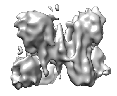
 ダウンロードとリンク
 emd_32842.png
 http://ftp.pdbj.org/pub/emdb/structures/EMD-32842


 Z (Sec.)
 Y (Row.)
 X (Col.)


 試料の構成要素
 Homo sapiens (ヒト)
 解析
解析 電子顕微鏡法
 FIELD EMISSION GUN

