 Movie
Movie Controller

[English] 日本語
 Yorodumi
- EMDB-32838: Tethered peptide activation mechanism of adhesion GPCRs ADGRG2 an... -
+ Open data

- Basic information
| Entry |  | |||||||||
|---|---|---|---|---|---|---|---|---|---|---|
| Title | Tethered peptide activation mechanism of adhesion GPCRs ADGRG2 and ADGRG4 | |||||||||
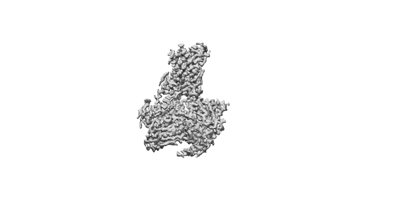
 Map data Map data | Cryo-EM structure of the ADGRG2-beta-Gs complex | |||||||||
 Sample Sample |
| |||||||||
| Function / homology |  Function and homology information Function and homology informationG protein-coupled receptor activity / adenylate cyclase-activating G protein-coupled receptor signaling pathway / Olfactory Signaling Pathway / Activation of the phototransduction cascade / Prostacyclin signalling through prostacyclin receptor / G beta:gamma signalling through PLC beta / Presynaptic function of Kainate receptors / Thromboxane signalling through TP receptor / Glucagon signaling in metabolic regulation / G protein-coupled acetylcholine receptor signaling pathway ...G protein-coupled receptor activity / adenylate cyclase-activating G protein-coupled receptor signaling pathway / Olfactory Signaling Pathway / Activation of the phototransduction cascade / Prostacyclin signalling through prostacyclin receptor / G beta:gamma signalling through PLC beta / Presynaptic function of Kainate receptors / Thromboxane signalling through TP receptor / Glucagon signaling in metabolic regulation / G protein-coupled acetylcholine receptor signaling pathway / G-protein activation / Activation of G protein gated Potassium channels / Inhibition of voltage gated Ca2+ channels via Gbeta/gamma subunits / G beta:gamma signalling through CDC42 / Vasopressin regulates renal water homeostasis via Aquaporins / G beta:gamma signalling through BTK / ADP signalling through P2Y purinoceptor 12 / Synthesis, secretion, and inactivation of Glucagon-like Peptide-1 (GLP-1) / Glucagon-type ligand receptors / Sensory perception of sweet, bitter, and umami (glutamate) taste / photoreceptor disc membrane / G alpha (z) signalling events / Adrenaline,noradrenaline inhibits insulin secretion / Glucagon-like Peptide-1 (GLP1) regulates insulin secretion / ADORA2B mediated anti-inflammatory cytokines production / cellular response to catecholamine stimulus / sensory perception of taste / ADP signalling through P2Y purinoceptor 1 / adenylate cyclase-activating dopamine receptor signaling pathway / G beta:gamma signalling through PI3Kgamma / GPER1 signaling / Cooperation of PDCL (PhLP1) and TRiC/CCT in G-protein beta folding / cellular response to prostaglandin E stimulus / Inactivation, recovery and regulation of the phototransduction cascade / G-protein beta-subunit binding / heterotrimeric G-protein complex / G alpha (12/13) signalling events / extracellular vesicle / signaling receptor complex adaptor activity / Thrombin signalling through proteinase activated receptors (PARs) / GTPase binding / retina development in camera-type eye / Ca2+ pathway / phospholipase C-activating G protein-coupled receptor signaling pathway / G alpha (i) signalling events / fibroblast proliferation / G alpha (s) signalling events / G alpha (q) signalling events / Ras protein signal transduction / cell population proliferation / Extra-nuclear estrogen signaling / cell surface receptor signaling pathway / G protein-coupled receptor signaling pathway / apical plasma membrane / lysosomal membrane / GTPase activity / synapse / protein-containing complex binding / signal transduction / extracellular exosome / membrane / plasma membrane / cytosol / cytoplasm Similarity search - Function | |||||||||
| Biological species |  Homo sapiens (human) / Homo sapiens (human) /  | |||||||||
| Method | single particle reconstruction / cryo EM / Resolution: 2.9 Å | |||||||||
 Authors Authors | He QT / Guo SC / Xiao P / Sun JP / Yu X / Gou L / Kong LL / Zhang L | |||||||||
| Funding support |  China, 2 items China, 2 items
| |||||||||
 Citation Citation | Journal: Nature / Year: 2022 Title: Tethered peptide activation mechanism of the adhesion GPCRs ADGRG2 and ADGRG4. Authors: Peng Xiao / Shengchao Guo / Xin Wen / Qing-Tao He / Hui Lin / Shen-Ming Huang / Lu Gou / Chao Zhang / Zhao Yang / Ya-Ni Zhong / Chuan-Cheng Yang / Yu Li / Zheng Gong / Xiao-Na Tao / Zhi- ...Authors: Peng Xiao / Shengchao Guo / Xin Wen / Qing-Tao He / Hui Lin / Shen-Ming Huang / Lu Gou / Chao Zhang / Zhao Yang / Ya-Ni Zhong / Chuan-Cheng Yang / Yu Li / Zheng Gong / Xiao-Na Tao / Zhi-Shuai Yang / Yan Lu / Shao-Long Li / Jun-Yan He / Chuanxin Wang / Lei Zhang / Liangliang Kong / Jin-Peng Sun / Xiao Yu / Abstract: Adhesion G protein-coupled receptors (aGPCRs) constitute an evolutionarily ancient family of receptors that often undergo autoproteolysis to produce α and β subunits. A tethered agonism mediated by ...Adhesion G protein-coupled receptors (aGPCRs) constitute an evolutionarily ancient family of receptors that often undergo autoproteolysis to produce α and β subunits. A tethered agonism mediated by the 'Stachel sequence' of the β subunit has been proposed to have central roles in aGPCR activation. Here we present three cryo-electron microscopy structures of aGPCRs coupled to the G heterotrimer. Two of these aGPCRs are activated by tethered Stachel sequences-the ADGRG2-β-G complex and the ADGRG4-β-G complex (in which β indicates the β subunit of the aGPCR)-and the other is the full-length ADGRG2 in complex with the exogenous ADGRG2 Stachel-sequence-derived peptide agonist IP15 (ADGRG2(FL)-IP15-G). The Stachel sequences of both ADGRG2-β and ADGRG4-β assume a U shape and insert deeply into the seven-transmembrane bundles. Constituting the FXφφφXφ motif (in which φ represents a hydrophobic residue), five residues of ADGRG2-β or ADGRG4-β extend like fingers to mediate binding to the seven-transmembrane domain and activation of the receptor. The structure of the ADGRG2(FL)-IP15-G complex reveals the structural basis for the improved binding affinity of IP15 compared with VPM-p15 and indicates that rational design of peptidic agonists could be achieved by exploiting aGPCR-β structures. By converting the 'finger residues' to acidic residues, we develop a method to generate peptidic antagonists towards several aGPCRs. Collectively, our study provides structural and biochemical insights into the tethered activation mechanism of aGPCRs. | |||||||||
| History |
|
- Structure visualization
| Supplemental images |
|---|
- Downloads & links
-EMDB archive
| Map data | emd_32838.map.gz | 25.3 MB | EMDB map data format | |
|---|---|---|---|---|
| Header (meta data) | emd-32838-v30.xmlemd-32838.xml | 18.8 KB 18.8 KB | Display Display | EMDB header |
| Images |  emd_32838.png emd_32838.png | 17.9 KB | ||
| Archive directory |  http://ftp.pdbj.org/pub/emdb/structures/EMD-32838ftp://ftp.pdbj.org/pub/emdb/structures/EMD-32838 http://ftp.pdbj.org/pub/emdb/structures/EMD-32838ftp://ftp.pdbj.org/pub/emdb/structures/EMD-32838 | HTTPS FTP |
-Validation report
| Summary document | emd_32838_validation.pdf.gz | 464 KB | Display | EMDB validaton report |
|---|---|---|---|---|
| Full document | emd_32838_full_validation.pdf.gz | 463.6 KB | Display | |
| Data in XML | emd_32838_validation.xml.gz | 4.9 KB | Display | |
| Data in CIF | emd_32838_validation.cif.gz | 5.7 KB | Display | |
| Arichive directory | https://ftp.pdbj.org/pub/emdb/validation_reports/EMD-32838ftp://ftp.pdbj.org/pub/emdb/validation_reports/EMD-32838 | HTTPS FTP |
-Related structure data
| Related structure data | 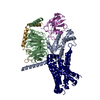 7wuqMC 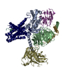 7wuiC 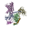 7wujC M: atomic model generated by this map C: citing same article ( |
|---|---|
| Similar structure data |
-Links
| EMDB pages | EMDB (EBI/PDBe) / EMDataResource |
|---|---|
| Related items in Molecule of the Month |


-Map
| File | Download / File: emd_32838.map.gz / Format: CCP4 / Size: 27 MB / Type: IMAGE STORED AS FLOATING POINT NUMBER (4 BYTES) | ||||||||||||||||||||||||||||||||||||
|---|---|---|---|---|---|---|---|---|---|---|---|---|---|---|---|---|---|---|---|---|---|---|---|---|---|---|---|---|---|---|---|---|---|---|---|---|---|
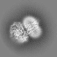
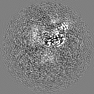
| Annotation | Cryo-EM structure of the ADGRG2-beta-Gs complex | ||||||||||||||||||||||||||||||||||||
| Projections & slices | Image control
Images are generated by Spider. | ||||||||||||||||||||||||||||||||||||
| Voxel size | X=Y=Z: 1 Å | ||||||||||||||||||||||||||||||||||||
| Density |
| ||||||||||||||||||||||||||||||||||||
| Symmetry | Space group: 1 | ||||||||||||||||||||||||||||||||||||
| Details | EMDB XML:
|
 Z (Sec.)
 Y (Row.)
 X (Col.)


-Supplemental data
- Sample components
-Entire : ADGRG2-beta-Gs complex
| Entire | Name: ADGRG2-beta-Gs complex |
|---|---|
| Components |
|
-Supramolecule #1: ADGRG2-beta-Gs complex
| Supramolecule | Name: ADGRG2-beta-Gs complex / type: complex / Chimera: Yes / ID: 1 / Parent: 0 / Macromolecule list: all |
|---|---|
| Molecular weight | Experimental: 134 KDa |
-Supramolecule #2: Gs
| Supramolecule | Name: Gs / type: complex / Chimera: Yes / ID: 2 / Parent: 1 / Macromolecule list: #1-#3 |
|---|---|
| Source (natural) | Organism: Homo sapiens (human) |
| Recombinant expression | Organism:  Baculovirus expression vector pFastBac1-HM Baculovirus expression vector pFastBac1-HM |
-Supramolecule #3: Nanobody-35
| Supramolecule | Name: Nanobody-35 / type: complex / Chimera: Yes / ID: 3 / Parent: 1 / Macromolecule list: #4 |
|---|---|
| Source (natural) | Organism: |
| Recombinant expression | Organism: Escherichia phage EcSzw-2 (virus) |
-Supramolecule #4: Adhesion G-protein coupled receptor G2
| Supramolecule | Name: Adhesion G-protein coupled receptor G2 / type: complex / Chimera: Yes / ID: 4 / Parent: 1 / Macromolecule list: #5 |
|---|---|
| Source (natural) | Organism: |
| Recombinant expression | Organism: Baculovirus expression vector pFastBac1-HM |
-Macromolecule #1: Guanine nucleotide-binding protein G(s) subunit alpha isoforms short
| Macromolecule | Name: Guanine nucleotide-binding protein G(s) subunit alpha isoforms short type: protein_or_peptide / ID: 1 / Number of copies: 1 / Enantiomer: LEVO |
|---|---|
| Source (natural) | Organism: Homo sapiens (human) |
| Molecular weight | Theoretical: 45.683434 KDa |
| Recombinant expression | Organism: Baculovirus expression vector pFastBac1-HM |
| Sequence | String: MGCLGNSKTE DQRNEEKAQR EANKKIEKQL QKDKQVYRAT HRLLLLGAGE SGKNTIVKQM RILHVNGFNG EGGEEDPQAA RSNSDGEKA TKVQDIKNNL KEAIETIVAA MSNLVPPVEL ANPENQFRVD YILSVMNVPD FDFPPEFYEH AKALWEDEGV R ACYERSNE ...String: MGCLGNSKTE DQRNEEKAQR EANKKIEKQL QKDKQVYRAT HRLLLLGAGE SGKNTIVKQM RILHVNGFNG EGGEEDPQAA RSNSDGEKA TKVQDIKNNL KEAIETIVAA MSNLVPPVEL ANPENQFRVD YILSVMNVPD FDFPPEFYEH AKALWEDEGV R ACYERSNE YQLIDCAQYF LDKIDVIKQA DYVPSDQDLL RCRVLTSGIF ETKFQVDKVN FHMFDVGAQR DERRKWIQCF ND VTAIIFV VASSSYNMVI REDNQTNRLQ AALKLFDSIW NNKWLRDTSV ILFLNKQDLL AEKVLAGKSK IEDYFPEFAR YTT PEDATP EPGEDPRVTR AKYFIRDEFL RISTASGDGR HYCYPHFTCA VDTENIRRVF NDCRDIIQRM HLRQYELL |
-Macromolecule #2: Guanine nucleotide-binding protein G(I)/G(S)/G(T) subunit beta-1
| Macromolecule | Name: Guanine nucleotide-binding protein G(I)/G(S)/G(T) subunit beta-1 type: protein_or_peptide / ID: 2 / Number of copies: 1 / Enantiomer: LEVO |
|---|---|
| Source (natural) | Organism: Homo sapiens (human) |
| Molecular weight | Theoretical: 39.48916 KDa |
| Recombinant expression | Organism: Baculovirus expression vector pFastBac1-HM |
| Sequence | String: MHHHHHHLEV LFQGPGSSQS ELDQLRQEAE QLKNQIRDAR KACADATLSQ ITNNIDPVGR IQMRTRRTLR GHLAKIYAMH WGTDSRLLV SASQDGKLII WDSYTTNKVH AIPLRSSWVM TCAYAPSGNY VACGGLDNIC SIYNLKTREG NVRVSRELAG H TGYLSCCR ...String: MHHHHHHLEV LFQGPGSSQS ELDQLRQEAE QLKNQIRDAR KACADATLSQ ITNNIDPVGR IQMRTRRTLR GHLAKIYAMH WGTDSRLLV SASQDGKLII WDSYTTNKVH AIPLRSSWVM TCAYAPSGNY VACGGLDNIC SIYNLKTREG NVRVSRELAG H TGYLSCCR FLDDNQIVTS SGDTTCALWD IETGQQTTTF TGHTGDVMSL SLAPDTRLFV SGACDASAKL WDVREGMCRQ TF TGHESDI NAICFFPNGN AFATGSDDAT CRLFDLRADQ ELMTYSHDNI ICGITSVSFS KSGRLLLAGY DDFNCNVWDA LKA DRAGVL AGHDNRVSCL GVTDDGMAVA TGSWDSFLKI WN |
-Macromolecule #3: Guanine nucleotide-binding protein G(I)/G(S)/G(O) subunit gamma-2
| Macromolecule | Name: Guanine nucleotide-binding protein G(I)/G(S)/G(O) subunit gamma-2 type: protein_or_peptide / ID: 3 / Number of copies: 1 / Enantiomer: LEVO |
|---|---|
| Source (natural) | Organism: Homo sapiens (human) |
| Molecular weight | Theoretical: 7.861143 KDa |
| Recombinant expression | Organism: Baculovirus expression vector pFastBac1-HM |
| Sequence | String: MASNNTASIA QARKLVEQLK MEANIDRIKV SKAAADLMAY CEAHAKEDPL LTPVPASENP FREKKFFCAI L |
-Macromolecule #4: Nanobody-35
| Macromolecule | Name: Nanobody-35 / type: protein_or_peptide / ID: 4 / Number of copies: 1 / Enantiomer: LEVO |
|---|---|
| Source (natural) | Organism: |
| Molecular weight | Theoretical: 13.885439 KDa |
| Recombinant expression | Organism: Escherichia phage EcSzw-2 (virus) |
| Sequence | String: QVQLQESGGG LVQPGGSLRL SCAASGFTFS NYKMNWVRQA PGKGLEWVSD ISQSGASISY TGSVKGRFTI SRDNAKNTLY LQMNSLKPE DTAVYYCARC PAPFTRDCFD VTSTTYAYRG QGTQVTVSS |
-Macromolecule #5: Adhesion G-protein coupled receptor G2,mCherry
| Macromolecule | Name: Adhesion G-protein coupled receptor G2,mCherry / type: protein_or_peptide / ID: 5 Details: residues 1010-1017 = His tag, residues 1018-1020 = linker, residues 1021-1027 = TEV site Number of copies: 1 / Enantiomer: LEVO |
|---|---|
| Source (natural) | Organism: |
| Molecular weight | Theoretical: 77.147891 KDa |
| Recombinant expression | Organism: Baculovirus expression vector pFastBac1-HM |
| Sequence | String: MKTIIALSYI FCLVFAHLTS FGILLDLSRT SLPPSQMMAL TFITYIGCGL SSIFLSVTLV TYIAFEKIRR DYPSKILIQL CAALLLLNL IFLLDSWIAL YNTRGFCIAV AVFLHYFLLV SFTWMGLEAF HMYLALVKVF NTYIRKYILK FCIVGWGIPA V VVSIVLTI ...String: MKTIIALSYI FCLVFAHLTS FGILLDLSRT SLPPSQMMAL TFITYIGCGL SSIFLSVTLV TYIAFEKIRR DYPSKILIQL CAALLLLNL IFLLDSWIAL YNTRGFCIAV AVFLHYFLLV SFTWMGLEAF HMYLALVKVF NTYIRKYILK FCIVGWGIPA V VVSIVLTI SPDNYGIGSY GKFPNGTPDD FCWINSNVVF YITVVGYFCV IFLLNVSMFI VVLVQLCRIK KKKQLGAQRK TS IQDLRSI AGLTFLLGIT WGFAFFAWGP VNVTFMYLFA IFNTLQGFFI FIFYCAAKEN VRKQWRRYLC CGKLRLAENS DWS KTATNG LKKQTVNQGV SSSSNSLQSS CNSTNSTTLL VNSDCSVHAS GNGNASTERN GVSFSVQNGD VCLHDLTGKQ HMFS DKEDS CNGKSRIALR RTSKRGSLHF IEQMHHHHHH HHGSAENLYF QGMVSKGEED NMAIIKEFMR FKVHMEGSVN GHEFE IEGE GEGRPYEGTQ TAKLKVTKGG PLPFAWDILS PQFMYGSKAY VKHPADIPDY LKLSFPEGFK WERVMNFEDG GVVTVT QDS SLQDGEFIYK VKLRGTNFPS DGPVMQKKTM GWEASSERMY PEDGALKGEI KQRLKLKDGG HYDAEVKTTY KAKKPVQ LP GAYNVNIKLD ITSHNEDYTI VEQYERAEGR HSTGGMDELY K |
-Experimental details
-Structure determination
| Method | cryo EM |
|---|---|
 Processing Processing | single particle reconstruction |
| Aggregation state | particle |
-Sample preparation
| Buffer | pH: 7.5 |
|---|---|
| Vitrification | Cryogen name: ETHANE |
- Electron microscopy
| Microscope | FEI TITAN KRIOS |
|---|---|
| Image recording | Film or detector model: GATAN K2 SUMMIT (4k x 4k) / Average electron dose: 64.0 e/Å2 |
| Electron beam | Acceleration voltage: 300 kV / Electron source:  FIELD EMISSION GUN FIELD EMISSION GUN |
| Electron optics | Illumination mode: SPOT SCAN / Imaging mode: BRIGHT FIELD / Nominal defocus max: 2.5 µm / Nominal defocus min: 1.0 µm |
| Experimental equipment |  Model: Titan Krios / Image courtesy: FEI Company |
-Image processing
| Startup model | Type of model: PDB ENTRY PDB model - PDB ID: |
|---|---|
| Final reconstruction | Resolution.type: BY AUTHOR / Resolution: 2.9 Å / Resolution method: FSC 0.143 CUT-OFF / Number images used: 3743997 |
| Initial angle assignment | Type: ANGULAR RECONSTITUTION |
| Final angle assignment | Type: ANGULAR RECONSTITUTION |

