[English] 日本語
 Yorodumi
- EMDB-32764: Cryo-EM Structure of Membrane-bound Fructose Dehydrogenase from G... -
+ Open data
- Basic information
| Entry |  | |||||||||
|---|---|---|---|---|---|---|---|---|---|---|
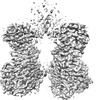
| Title | Cryo-EM Structure of Membrane-bound Fructose Dehydrogenase from Gluconobacter japonicus | |||||||||
 Map data Map data | ||||||||||
 Sample Sample |
| |||||||||
 Keywords Keywords | Complex / OXIDOREDUCTASE Membrane-bound protein / OXIDOREDUCTASE | |||||||||
| Function / homology |  Function and homology information Function and homology informationfructose 5-dehydrogenase / fructose 5-dehydrogenase activity / oxidoreductase activity, acting on CH-OH group of donors / fructose metabolic process / flavin adenine dinucleotide binding / electron transfer activity / iron ion binding / heme binding / plasma membrane Similarity search - Function | |||||||||
| Biological species |  Gluconobacter japonicus (bacteria) Gluconobacter japonicus (bacteria) | |||||||||
| Method | single particle reconstruction / cryo EM / Resolution: 3.8 Å | |||||||||
 Authors Authors | Suzuki Y / Makino F | |||||||||
| Funding support |  Japan, 2 items Japan, 2 items
| |||||||||
 Citation Citation |  Journal: Acs Catalysis / Year: 2023 Journal: Acs Catalysis / Year: 2023Title: Essential Insight of Direct Electron Transfer-Type Bioelectrocatalysis by Membrane-Bound d-Fructose Dehydrogenase with Structural Bioelectrochemistry Authors: Suzuki Y / Makino F / Miyata T / Tanaka H / Namba K / Kano K / Sowa K / Kitazumi Y / Shirai O | |||||||||
| History |
|
- Structure visualization
| Supplemental images |
|---|
- Downloads & links
-EMDB archive
| Map data |  emd_32764.map.gz emd_32764.map.gz | 78.4 MB |  EMDB map data format EMDB map data format | |
|---|---|---|---|---|
| Header (meta data) |  emd-32764-v30.xml emd-32764-v30.xml emd-32764.xml emd-32764.xml | 19.2 KB 19.2 KB | Display Display |  EMDB header EMDB header |
| Images |  emd_32764.png emd_32764.png | 146.7 KB | ||
| Filedesc metadata |  emd-32764.cif.gz emd-32764.cif.gz | 6.9 KB | ||
| Archive directory |  http://ftp.pdbj.org/pub/emdb/structures/EMD-32764 http://ftp.pdbj.org/pub/emdb/structures/EMD-32764 ftp://ftp.pdbj.org/pub/emdb/structures/EMD-32764 ftp://ftp.pdbj.org/pub/emdb/structures/EMD-32764 | HTTPS FTP |
-Validation report
| Summary document |  emd_32764_validation.pdf.gz emd_32764_validation.pdf.gz | 570.5 KB | Display |  EMDB validaton report EMDB validaton report |
|---|---|---|---|---|
| Full document |  emd_32764_full_validation.pdf.gz emd_32764_full_validation.pdf.gz | 570.1 KB | Display | |
| Data in XML |  emd_32764_validation.xml.gz emd_32764_validation.xml.gz | 6 KB | Display | |
| Data in CIF |  emd_32764_validation.cif.gz emd_32764_validation.cif.gz | 6.9 KB | Display | |
| Arichive directory |  https://ftp.pdbj.org/pub/emdb/validation_reports/EMD-32764 https://ftp.pdbj.org/pub/emdb/validation_reports/EMD-32764 ftp://ftp.pdbj.org/pub/emdb/validation_reports/EMD-32764 ftp://ftp.pdbj.org/pub/emdb/validation_reports/EMD-32764 | HTTPS FTP |
-Related structure data
| Related structure data |  7wsqMC  7w2jC C: citing same article ( M: atomic model generated by this map |
|---|---|
| Similar structure data | Similarity search - Function & homology  F&H Search F&H Search |
- Links
Links
| EMDB pages |  EMDB (EBI/PDBe) / EMDB (EBI/PDBe) /  EMDataResource EMDataResource |
|---|---|
| Related items in Molecule of the Month |
- Map
Map
| File |  Download / File: emd_32764.map.gz / Format: CCP4 / Size: 83.7 MB / Type: IMAGE STORED AS FLOATING POINT NUMBER (4 BYTES) Download / File: emd_32764.map.gz / Format: CCP4 / Size: 83.7 MB / Type: IMAGE STORED AS FLOATING POINT NUMBER (4 BYTES) | ||||||||||||||||||||||||||||||||||||
|---|---|---|---|---|---|---|---|---|---|---|---|---|---|---|---|---|---|---|---|---|---|---|---|---|---|---|---|---|---|---|---|---|---|---|---|---|---|
| Projections & slices | Image control
Images are generated by Spider. | ||||||||||||||||||||||||||||||||||||
| Voxel size | X=Y=Z: 0.87 Å | ||||||||||||||||||||||||||||||||||||
| Density |
| ||||||||||||||||||||||||||||||||||||
| Symmetry | Space group: 1 | ||||||||||||||||||||||||||||||||||||
| Details | EMDB XML:
|
-Supplemental data
- Sample components
-Entire : D-fructose dehydrogenase from Gluconobacter japonicus
| Entire | Name: D-fructose dehydrogenase from Gluconobacter japonicus |
|---|---|
| Components |
|
-Supramolecule #1: D-fructose dehydrogenase from Gluconobacter japonicus
| Supramolecule | Name: D-fructose dehydrogenase from Gluconobacter japonicus / type: complex / ID: 1 / Parent: 0 / Macromolecule list: #1-#3 |
|---|---|
| Source (natural) | Organism:  Gluconobacter japonicus (bacteria) Gluconobacter japonicus (bacteria) |
| Molecular weight | Theoretical: 140 KDa |
-Macromolecule #1: Fructose dehydrogenase large subunit
| Macromolecule | Name: Fructose dehydrogenase large subunit / type: protein_or_peptide / ID: 1 / Number of copies: 2 / Enantiomer: LEVO / EC number: ec: 1.1.99.11 |
|---|---|
| Source (natural) | Organism:  Gluconobacter japonicus (bacteria) Gluconobacter japonicus (bacteria) |
| Molecular weight | Theoretical: 59.798309 KDa |
| Recombinant expression | Organism:  Gluconobacter oxydans (bacteria) Gluconobacter oxydans (bacteria) |
| Sequence | String: MSNETLSADV VIIGAGICGS LLAHKLVRNG LSVLLLDAGP RRDRSQIVEN WRNMPPDNKS QYDYATPYPS VPWAPHTNYF PDNNYLIVK GPDRTAYKQG IIKGVGGTTW HWAASSWRYL PNDFKLHSTY GVGRDYAMSY DELEPYYYEA ECEMGVMGPN G EEITPSAP ...String: MSNETLSADV VIIGAGICGS LLAHKLVRNG LSVLLLDAGP RRDRSQIVEN WRNMPPDNKS QYDYATPYPS VPWAPHTNYF PDNNYLIVK GPDRTAYKQG IIKGVGGTTW HWAASSWRYL PNDFKLHSTY GVGRDYAMSY DELEPYYYEA ECEMGVMGPN G EEITPSAP RQNPWPMTSM PYGYGDRTFT EIVSKLGFSN TPVPQARNSR PYDGRPQCCG NNNCMPICPI GAMYNGVYAA IK AEKLGAK IIPNAVVYAM ETDAKNRITA ISFYDPDKQS HRVVAKTFVI AANGIETPKL LLLAANDRNP HGIANSSDLV GRN MMDHPG IGMSFQSAEP IWAGGGSVQM SSITNFRDGD FRSEYAATQI GYNNTAQNSR AGMKALSMGL VGKKLDEEIR RRTA HGVDI YANHEVLPDP NNRLVLSKDY KDALGIPHPE VTYDVGEYVR KSAAISRQRL MDIAKAMGGT EIEMTPYFTP NNHIT GGTI MGHDPRDSVV DKWLRTHDHS NLFLATGATM AASGTVNSTL TMAALSLRAA DAILNDLKQG UniProtKB: Fructose dehydrogenase large subunit |
-Macromolecule #2: Fructose dehydrogenase small subunit
| Macromolecule | Name: Fructose dehydrogenase small subunit / type: protein_or_peptide / ID: 2 / Number of copies: 2 / Enantiomer: LEVO |
|---|---|
| Source (natural) | Organism:  Gluconobacter japonicus (bacteria) Gluconobacter japonicus (bacteria) |
| Molecular weight | Theoretical: 20.106732 KDa |
| Recombinant expression | Organism:  Gluconobacter oxydans (bacteria) Gluconobacter oxydans (bacteria) |
| Sequence | String: MEKIADSGPV QIFLSRRKLL AFSGASLTVA AIGAPSKGST QDVVASNRDS ISDFMQLSAF ATGHKNLDLN IGSALLLAFE AQKHDFSTQ IKALREHITK NNYQDVEALD AAMKDDPLHP TLIQIIRAWY SGVIEDETNA KVYAFEKALM YQPSRDVVVI P TYAHNGPN YWVSEPASVD VMPAF UniProtKB: Fructose dehydrogenase small subunit |
-Macromolecule #3: Fructose dehydrogenase cytochrome subunit
| Macromolecule | Name: Fructose dehydrogenase cytochrome subunit / type: protein_or_peptide / ID: 3 / Number of copies: 2 / Enantiomer: LEVO |
|---|---|
| Source (natural) | Organism:  Gluconobacter japonicus (bacteria) Gluconobacter japonicus (bacteria) |
| Molecular weight | Theoretical: 52.252652 KDa |
| Recombinant expression | Organism:  Gluconobacter oxydans (bacteria) Gluconobacter oxydans (bacteria) |
| Sequence | String: MRYFRPLSAT AMTTVLLLAG TNVRAQPTEP TPASAHRPSI SRGHYLAIAA DCAACHTNGR DGQFLAGGYA ISSPMGNIYS TNITPSKTH GIGNYTLEQF SKALRHGIRA DGAQLYPAMP YDAYNRLTDE DVKSLYAYIM TEVKPVDAPS PKTQLPFPFS I RASLGIWK ...String: MRYFRPLSAT AMTTVLLLAG TNVRAQPTEP TPASAHRPSI SRGHYLAIAA DCAACHTNGR DGQFLAGGYA ISSPMGNIYS TNITPSKTH GIGNYTLEQF SKALRHGIRA DGAQLYPAMP YDAYNRLTDE DVKSLYAYIM TEVKPVDAPS PKTQLPFPFS I RASLGIWK IAARIEGKPY VFDHTHNDDW NRGRYLVDEL AHCGECHTPR NFLLAPNQSA YLAGADIGSW RAPNITNAPQ SG IGSWSDQ DLFQYLKTGK TAHARAAGPM AEAIEHSLQY LPDADISAIV TYLRSVPAKA ESGQTVANFE HAGRPSSYSV ANA NSRRSN STLTKTTDGA ALYEAVCASC HQSDGKGSKD GYYPSLVGNT TTGQLNPNDL IASILYGVDR TTDNHEILMP AFGP DSLVQ PLTDEQIATI ADYVLSHFGN AQATVSADAV KQVRAGGKQV PLAKLASPGV MLLLGTGGIL GAILVVAGLW WLISR RKKR SA UniProtKB: Fructose dehydrogenase cytochrome subunit |
-Macromolecule #4: FLAVIN-ADENINE DINUCLEOTIDE
| Macromolecule | Name: FLAVIN-ADENINE DINUCLEOTIDE / type: ligand / ID: 4 / Number of copies: 2 / Formula: FAD |
|---|---|
| Molecular weight | Theoretical: 785.55 Da |
| Chemical component information |  ChemComp-FAD: |
-Macromolecule #5: FE3-S4 CLUSTER
| Macromolecule | Name: FE3-S4 CLUSTER / type: ligand / ID: 5 / Number of copies: 2 / Formula: F3S |
|---|---|
| Molecular weight | Theoretical: 295.795 Da |
| Chemical component information |  ChemComp-F3S: |
-Macromolecule #6: HEME C
| Macromolecule | Name: HEME C / type: ligand / ID: 6 / Number of copies: 6 / Formula: HEC |
|---|---|
| Molecular weight | Theoretical: 618.503 Da |
| Chemical component information |  ChemComp-HEC: |
-Experimental details
-Structure determination
| Method | cryo EM |
|---|---|
 Processing Processing | single particle reconstruction |
| Aggregation state | particle |
- Sample preparation
| Concentration | 5 mg/mL | |||||||||||||||
|---|---|---|---|---|---|---|---|---|---|---|---|---|---|---|---|---|
| Buffer | pH: 6 Component:
| |||||||||||||||
| Grid | Model: Quantifoil R1.2/1.3 / Material: COPPER / Mesh: 200 / Support film - Material: CARBON / Support film - topology: HOLEY / Support film - Film thickness: 500 / Pretreatment - Type: GLOW DISCHARGE / Pretreatment - Time: 20 sec. / Pretreatment - Atmosphere: AIR | |||||||||||||||
| Vitrification | Cryogen name: ETHANE |
- Electron microscopy
| Microscope | JEOL CRYO ARM 300 |
|---|---|
| Temperature | Min: 80.0 K / Max: 80.0 K |
| Alignment procedure | Coma free - Residual tilt: 0.01 mrad |
| Specialist optics | Energy filter - Name: In-column Omega Filter / Energy filter - Slit width: 20 eV |
| Image recording | Film or detector model: GATAN K3 (6k x 4k) / Number grids imaged: 1 / Number real images: 5575 / Average exposure time: 3.0 sec. / Average electron dose: 2.0 e/Å2 |
| Electron beam | Acceleration voltage: 300 kV / Electron source:  FIELD EMISSION GUN FIELD EMISSION GUN |
| Electron optics | C2 aperture diameter: 40.0 µm / Calibrated defocus max: 2.5 µm / Calibrated defocus min: 0.5 µm / Calibrated magnification: 56754 / Illumination mode: FLOOD BEAM / Imaging mode: BRIGHT FIELD / Cs: 2.7 mm / Nominal defocus max: 2.5 µm / Nominal defocus min: 0.5 µm / Nominal magnification: 60000 |
| Sample stage | Specimen holder model: JEOL CRYOSPECPORTER / Cooling holder cryogen: NITROGEN |
+ Image processing
-Atomic model buiding 1
| Refinement | Space: REAL / Protocol: FLEXIBLE FIT |
|---|---|
| Output model |  PDB-7wsq: |
 Movie
Movie Controller


 Z (Sec.)
 Y (Row.)
 X (Col.)


