 Movie
Movie Controller

[English] 日本語
 Yorodumi
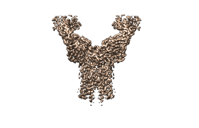
- EMDB-31977: SARS-CoV-2 M protein dimer (long form) in complex with YN7756_1 Fab -
+ Open data

- Basic information
| Entry |  | |||||||||
|---|---|---|---|---|---|---|---|---|---|---|
| Title | SARS-CoV-2 M protein dimer (long form) in complex with YN7756_1 Fab | |||||||||
 Map data Map data | ||||||||||
 Sample Sample |
| |||||||||
 Keywords Keywords | SARS-CoV-2 / M protein / viral structural protein / virus assembly / VIRAL PROTEIN / VIRAL PROTEIN-IMMUNE SYSTEM complex | |||||||||
| Function / homology |  Function and homology information Function and homology informationMaturation of protein M / SARS-CoV-2 modulates autophagy / host cell Golgi membrane / CD28 dependent PI3K/Akt signaling / SARS-CoV-2 targets host intracellular signalling and regulatory pathways / symbiont-mediated suppression of host cytoplasmic pattern recognition receptor signaling pathway via inhibition of MAVS activity / VEGFR2 mediated vascular permeability / protein sequestering activity / PIP3 activates AKT signaling / TRAF3-dependent IRF activation pathway ...Maturation of protein M / SARS-CoV-2 modulates autophagy / host cell Golgi membrane / CD28 dependent PI3K/Akt signaling / SARS-CoV-2 targets host intracellular signalling and regulatory pathways / symbiont-mediated suppression of host cytoplasmic pattern recognition receptor signaling pathway via inhibition of MAVS activity / VEGFR2 mediated vascular permeability / protein sequestering activity / PIP3 activates AKT signaling / TRAF3-dependent IRF activation pathway / Translation of Structural Proteins / Virion Assembly and Release / Induction of Cell-Cell Fusion / structural constituent of virion / Attachment and Entry / viral envelope / SARS-CoV-2 activates/modulates innate and adaptive immune responses / virion membrane / identical protein binding / plasma membrane Similarity search - Function | |||||||||
| Biological species |    Severe acute respiratory syndrome coronavirus 2 Severe acute respiratory syndrome coronavirus 2 | |||||||||
| Method | single particle reconstruction / cryo EM / Resolution: 2.7 Å | |||||||||
 Authors Authors | Zhang Z / Ohto U / Shimizu T | |||||||||
| Funding support | 1 items
| |||||||||
 Citation Citation | Journal: Nat Commun / Year: 2022 Title: Structure of SARS-CoV-2 membrane protein essential for virus assembly. Authors: Zhikuan Zhang / Norimichi Nomura / Yukiko Muramoto / Toru Ekimoto / Tomoko Uemura / Kehong Liu / Moeko Yui / Nozomu Kono / Junken Aoki / Mitsunori Ikeguchi / Takeshi Noda / So Iwata / ...Authors: Zhikuan Zhang / Norimichi Nomura / Yukiko Muramoto / Toru Ekimoto / Tomoko Uemura / Kehong Liu / Moeko Yui / Nozomu Kono / Junken Aoki / Mitsunori Ikeguchi / Takeshi Noda / So Iwata / Umeharu Ohto / Toshiyuki Shimizu /  Abstract: The coronavirus membrane protein (M) is the most abundant viral structural protein and plays a central role in virus assembly and morphogenesis. However, the process of M protein-driven virus ...The coronavirus membrane protein (M) is the most abundant viral structural protein and plays a central role in virus assembly and morphogenesis. However, the process of M protein-driven virus assembly are largely unknown. Here, we report the cryo-electron microscopy structure of the SARS-CoV-2 M protein in two different conformations. M protein forms a mushroom-shaped dimer, composed of two transmembrane domain-swapped three-helix bundles and two intravirion domains. M protein further assembles into higher-order oligomers. A highly conserved hinge region is key for conformational changes. The M protein dimer is unexpectedly similar to SARS-CoV-2 ORF3a, a viral ion channel. Moreover, the interaction analyses of M protein with nucleocapsid protein (N) and RNA suggest that the M protein mediates the concerted recruitment of these components through the positively charged intravirion domain. Our data shed light on the M protein-driven virus assembly mechanism and provide a structural basis for therapeutic intervention targeting M protein. | |||||||||
| History |
|
- Structure visualization
| Supplemental images |
|---|
- Downloads & links
-EMDB archive
| Map data | emd_31977.map.gz | 27.4 MB | EMDB map data format | |
|---|---|---|---|---|
| Header (meta data) | emd-31977-v30.xmlemd-31977.xml | 15.4 KB 15.4 KB | Display Display | EMDB header |
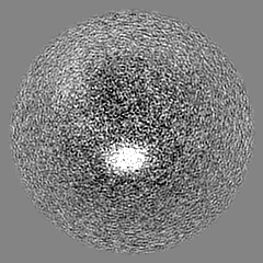
| Images |  emd_31977.png emd_31977.png | 45.6 KB | ||
| Filedesc metadata | emd-31977.cif.gz | 6.3 KB | ||
| Archive directory |  http://ftp.pdbj.org/pub/emdb/structures/EMD-31977ftp://ftp.pdbj.org/pub/emdb/structures/EMD-31977 http://ftp.pdbj.org/pub/emdb/structures/EMD-31977ftp://ftp.pdbj.org/pub/emdb/structures/EMD-31977 | HTTPS FTP |
-Related structure data
| Related structure data |  7vgrMC  7vgsC M: atomic model generated by this map C: citing same article ( |
|---|---|
| Similar structure data | |
| EM raw data | EMPIAR-11168 (Title: Structure of SARS-CoV-2 membrane protein / Data size: 4.4 TB / Data #1: M protein (LMNG/CHS) [micrographs - multiframe] Data #2: M protein (LMNG/CHS) + Fab-E [micrographs - multiframe] Data #3: M protein (LMNG/CHS) + Fab-B [micrographs - multiframe]) |

-Links
| EMDB pages | EMDB (EBI/PDBe) / EMDataResource |
|---|---|
| Related items in Molecule of the Month |


-Map
| File | Download / File: emd_31977.map.gz / Format: CCP4 / Size: 52.7 MB / Type: IMAGE STORED AS FLOATING POINT NUMBER (4 BYTES) | ||||||||||||||||||||||||||||||||||||
|---|---|---|---|---|---|---|---|---|---|---|---|---|---|---|---|---|---|---|---|---|---|---|---|---|---|---|---|---|---|---|---|---|---|---|---|---|---|
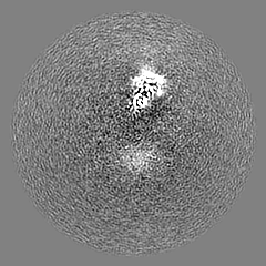
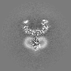
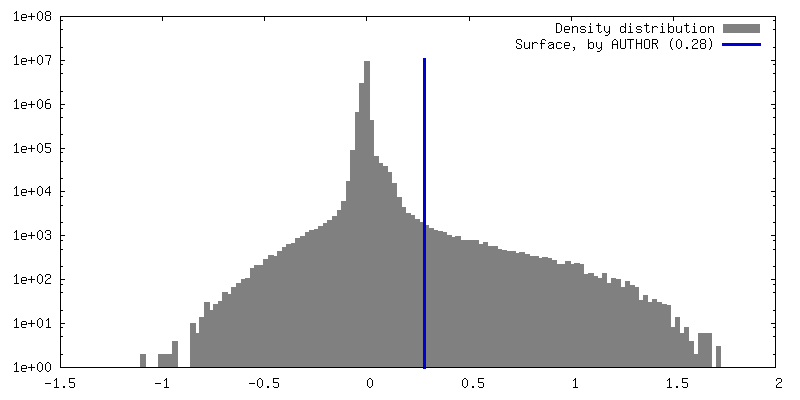
| Projections & slices | Image control
Images are generated by Spider. | ||||||||||||||||||||||||||||||||||||
| Voxel size | X=Y=Z: 1.245 Å | ||||||||||||||||||||||||||||||||||||
| Density |
| ||||||||||||||||||||||||||||||||||||
| Symmetry | Space group: 1 | ||||||||||||||||||||||||||||||||||||
| Details | EMDB XML:
|
 Z (Sec.)
 Y (Row.)
 X (Col.)


-Supplemental data
- Sample components
-Entire : SARS-CoV-2 M protein dimer (long form) in complex with YN7756_1 Fab
| Entire | Name: SARS-CoV-2 M protein dimer (long form) in complex with YN7756_1 Fab |
|---|---|
| Components |
|
-Supramolecule #1: SARS-CoV-2 M protein dimer (long form) in complex with YN7756_1 Fab
| Supramolecule | Name: SARS-CoV-2 M protein dimer (long form) in complex with YN7756_1 Fab type: complex / ID: 1 / Parent: 0 / Macromolecule list: all |
|---|
-Supramolecule #2: YN7756_1 Fab
| Supramolecule | Name: YN7756_1 Fab / type: complex / ID: 2 / Parent: 1 / Macromolecule list: #1-#2 |
|---|---|
| Source (natural) | Organism: |
-Supramolecule #3: Membrane protein
| Supramolecule | Name: Membrane protein / type: complex / ID: 3 / Parent: 1 / Macromolecule list: #3 |
|---|---|
| Source (natural) | Organism: Severe acute respiratory syndrome coronavirus 2 |
-Macromolecule #1: YN7756_1 Fab light chain
| Macromolecule | Name: YN7756_1 Fab light chain / type: protein_or_peptide / ID: 1 / Number of copies: 2 / Enantiomer: LEVO |
|---|---|
| Source (natural) | Organism: |
| Molecular weight | Theoretical: 24.025412 KDa |
| Sequence | String: DIVLTQSPAS LTVSLGQRAT ISCRASESVD SFGNSFMHWY QQKPGQPPKL LIYRASNLES GIPARFSGSG SRTDFTLTIN PVEADDVAT YYCQQSSEDP YTFGGGTKLE IKRADAAPTV SIFPPSSEQL TSGGASVVCF LNNFYPKDIN VKWKIDGSER Q NGVLNSWT ...String: DIVLTQSPAS LTVSLGQRAT ISCRASESVD SFGNSFMHWY QQKPGQPPKL LIYRASNLES GIPARFSGSG SRTDFTLTIN PVEADDVAT YYCQQSSEDP YTFGGGTKLE IKRADAAPTV SIFPPSSEQL TSGGASVVCF LNNFYPKDIN VKWKIDGSER Q NGVLNSWT DQDSKDSTYS MSSTLTLTKD EYERHNSYTC EATHKTSTSP IVKSFNRNEC |
-Macromolecule #2: YN7756_1 Fab heavy chain
| Macromolecule | Name: YN7756_1 Fab heavy chain / type: protein_or_peptide / ID: 2 / Number of copies: 2 / Enantiomer: LEVO |
|---|---|
| Source (natural) | Organism: |
| Molecular weight | Theoretical: 25.114109 KDa |
| Sequence | String: EVQLQQSGAE LVRPGSSVKI SCKGSGYVFS NYWMNWVKQR PGQGLEWIGQ IYPGDGDTNY NGKFKGKATL TADKSSSTAY MQLSSLTSE DSAVYFCASG YLGENYVMDF WGQGTSVTVS SAKTTPPSVY PLAPGSAAQT NSMVTLGCLV KGYFPEPVTV T WNSGSLSS ...String: EVQLQQSGAE LVRPGSSVKI SCKGSGYVFS NYWMNWVKQR PGQGLEWIGQ IYPGDGDTNY NGKFKGKATL TADKSSSTAY MQLSSLTSE DSAVYFCASG YLGENYVMDF WGQGTSVTVS SAKTTPPSVY PLAPGSAAQT NSMVTLGCLV KGYFPEPVTV T WNSGSLSS GVHTFPAVLQ SDLYTLSSSV TVPSSTWPSE TVTCNVAHPA SSTKVDKKIV PRDCGCKPCI CTVPEVSS |
-Macromolecule #3: Membrane protein
| Macromolecule | Name: Membrane protein / type: protein_or_peptide / ID: 3 / Number of copies: 2 / Enantiomer: LEVO |
|---|---|
| Source (natural) | Organism: Severe acute respiratory syndrome coronavirus 2 |
| Molecular weight | Theoretical: 28.257822 KDa |
| Recombinant expression | Organism:  Homo sapiens (human) Homo sapiens (human) |
| Sequence | String: MHHHHHHHHD YKDDDDKENL YFQGMADSNG TITVEELKKL LEQWNLVIGF LFLTWICLLQ FAYANRNRFL YIIKLIFLWL LWPVTLACF VLAAVYRINW ITGGIAIAMA CLVGLMWLSY FIASFRLFAR TRSMWSFNPE TNILLNVPLH GTILTRPLLE S ELVIGAVI ...String: MHHHHHHHHD YKDDDDKENL YFQGMADSNG TITVEELKKL LEQWNLVIGF LFLTWICLLQ FAYANRNRFL YIIKLIFLWL LWPVTLACF VLAAVYRINW ITGGIAIAMA CLVGLMWLSY FIASFRLFAR TRSMWSFNPE TNILLNVPLH GTILTRPLLE S ELVIGAVI LRGHLRIAGH HLGRCDIKDL PKEITVATSR TLSYYKLGAS QRVAGDSGFA AYSRYRIGNY KLNTDHSSSS DN IALLVQ UniProtKB: Membrane protein |
-Experimental details
-Structure determination
| Method | cryo EM |
|---|---|
 Processing Processing | single particle reconstruction |
| Aggregation state | particle |
-Sample preparation
| Concentration | 2.0 mg/mL |
|---|---|
| Buffer | pH: 7.6 |
| Grid | Model: Quantifoil R1.2/1.3 / Material: COPPER |
| Vitrification | Cryogen name: ETHANE |
- Electron microscopy
| Microscope | TFS KRIOS |
|---|---|
| Image recording | Film or detector model: GATAN K3 (6k x 4k) / Average electron dose: 61.9 e/Å2 |
| Electron beam | Acceleration voltage: 300 kV / Electron source:  FIELD EMISSION GUN FIELD EMISSION GUN |
| Electron optics | Illumination mode: OTHER / Imaging mode: BRIGHT FIELD |
| Experimental equipment |  Model: Titan Krios / Image courtesy: FEI Company |
+Image processing
-Atomic model buiding 1
| Refinement | Space: REAL / Protocol: AB INITIO MODEL |
|---|---|
| Output model | PDB-7vgr: |
