90S preribosome / endonucleolytic cleavage to generate mature 3'-end of SSU-rRNA from (SSU-rRNA, 5.8S rRNA, LSU-rRNA) / endonucleolytic cleavage in ITS1 to separate SSU-rRNA from 5.8S rRNA and LSU-rRNA from tricistronic rRNA transcript (SSU-rRNA, 5.8S rRNA, LSU-rRNA) / maturation of SSU-rRNA from tricistronic rRNA transcript (SSU-rRNA, 5.8S rRNA, LSU-rRNA) / maturation of SSU-rRNA / small-subunit processome / maintenance of translational fidelity / rRNA processing / ribosomal small subunit assembly / ribosomal small subunit biogenesis ...90S preribosome / endonucleolytic cleavage to generate mature 3'-end of SSU-rRNA from (SSU-rRNA, 5.8S rRNA, LSU-rRNA) / endonucleolytic cleavage in ITS1 to separate SSU-rRNA from 5.8S rRNA and LSU-rRNA from tricistronic rRNA transcript (SSU-rRNA, 5.8S rRNA, LSU-rRNA) / maturation of SSU-rRNA from tricistronic rRNA transcript (SSU-rRNA, 5.8S rRNA, LSU-rRNA) / maturation of SSU-rRNA / small-subunit processome / maintenance of translational fidelity / rRNA processing / ribosomal small subunit assembly / ribosomal small subunit biogenesis / small ribosomal subunit / small ribosomal subunit rRNA binding / cytosolic small ribosomal subunit / rRNA binding / structural constituent of ribosome / ribosome / translation / ribonucleoprotein complex / mRNA binding / nucleolus / RNA binding / zinc ion binding / nucleus / cytosol 類似検索 - 分子機能
Beta-grasp domain superfamily / Ribosomal protein S26e / Ribosomal protein S26e superfamily / Ribosomal protein S26e / Ribosomal protein S5, eukaryotic/archaeal / Ribosomal protein S21e / Ribosomal protein S21e superfamily / Ribosomal protein S21e / Ribosomal protein S2, eukaryotic / 40S Ribosomal protein S10 ...Beta-grasp domain superfamily / Ribosomal protein S26e / Ribosomal protein S26e superfamily / Ribosomal protein S26e / Ribosomal protein S5, eukaryotic/archaeal / Ribosomal protein S21e / Ribosomal protein S21e superfamily / Ribosomal protein S21e / Ribosomal protein S2, eukaryotic / 40S Ribosomal protein S10 / S27a-like superfamily / Plectin/S10, N-terminal / Plectin/S10 domain / Ribosomal protein S30 / Ribosomal protein S30 / Ribosomal protein S25 / S25 ribosomal protein / Ribosomal protein S2, eukaryotic/archaeal / Ribosomal protein S27a / Ribosomal protein S27a / Ribosomal protein S27a / 40S ribosomal protein S29/30S ribosomal protein S14 type Z / Ribosomal protein S3, eukaryotic/archaeal / 40S ribosomal protein S4, C-terminal domain / 40S ribosomal protein S4 C-terminus / Ribosomal protein S4e, N-terminal, conserved site / Ribosomal protein S4e signature. / Ribosomal protein S19A/S15e / Ribosomal protein S6, eukaryotic / Ribosomal protein S19e / Ribosomal protein S19e / Ribosomal_S19e / Ribosomal protein S17e / Ribosomal protein S17e-like superfamily / Ribosomal S17 / 40S ribosomal protein S1/3, eukaryotes / 40S ribosomal protein S11, N-terminal / Ribosomal_S17 N-terminal / Ribosomal protein S7e / Ribosomal protein S7e / : / Ribosomal protein S4e, N-terminal / RS4NT (NUC023) domain / Ribosomal protein S4, KOW domain / Ribosomal protein S4e / Ribosomal protein S4e, central region / Ribosomal protein S4e, central domain superfamily / Ribosomal family S4e / Ribosomal protein S28e conserved site / Ribosomal protein S28e signature. / Ribosomal protein S6/S6e/A/B/2, conserved site / Ribosomal protein S17, archaeal/eukaryotic / Ribosomal protein S6e signature. / Ribosomal protein S23, eukaryotic/archaeal / Ribosomal protein S24e / Ribosomal protein S24e / Ribosomal protein S8e / Ribosomal protein S27 / Ribosomal protein S27, zinc-binding domain superfamily / Ribosomal protein S27 / Ribosomal protein S3Ae / Ribosomal S3Ae family / Ribosomal S3Ae family / Ribosomal protein S28e / Ribosomal protein S28e / Ribosomal protein S6e / Ribosomal protein S6e / Ribosomal protein S5/S7, eukaryotic/archaeal / Ribosomal protein S6e / Ribosomal protein S13/S15, N-terminal / Ribosomal protein S15P / Ribosomal S13/S15 N-terminal domain / Ribosomal S13/S15 N-terminal domain / Ribosomal protein S4/S9, eukaryotic/archaeal / Ribosomal protein S8e/ribosomal biogenesis NSA2 / Ribosomal protein S8e / Ribosomal protein S14/S29 / Ribosomal protein L7Ae/L30e/S12e/Gadd45 / Ribosomal protein L7Ae/L30e/S12e/Gadd45 family / 50S ribosomal protein L30e-like / Ribosomal protein S2 signature 2. / : / Ribosomal protein S2 signature 1. / Ribosomal protein S3, C-terminal / Ribosomal protein S3, C-terminal domain / Ribosomal protein S3, C-terminal domain superfamily / Ribosomal protein S10 / Ribosomal protein S19/S15 / Ribosomal protein S19/S15, superfamily / Ribosomal protein S19 / Ribosomal protein S5, N-terminal, conserved site / Ribosomal protein S5 signature. / Ribosomal protein S2, conserved site / K homology domain superfamily, prokaryotic type / Ribosomal protein S2 / Ribosomal protein S2, flavodoxin-like domain superfamily / Ribosomal protein S2 / Ribosomal protein S17, conserved site / Ribosomal protein S17 signature. / Ribosomal protein S5 類似検索 - ドメイン・相同性
40S ribosomal protein S30 / 40S ribosomal protein S8 / 40S ribosomal protein S21 / Ribosomal protein S28 / Ribosomal protein S20 / Ribosomal protein S24 / 40S ribosomal protein S6 / Ribosomal protein S29A / 40S ribosomal protein S25 / 40S ribosomal protein S3 ...40S ribosomal protein S30 / 40S ribosomal protein S8 / 40S ribosomal protein S21 / Ribosomal protein S28 / Ribosomal protein S20 / Ribosomal protein S24 / 40S ribosomal protein S6 / Ribosomal protein S29A / 40S ribosomal protein S25 / 40S ribosomal protein S3 / Ribosomal protein S15 / Ribosomal protein S13 / Small ribosomal subunit protein uS2 / 40S ribosomal protein S4 / Ribosomal protein S27 / Ribosomal protein S16 / Ribosomal protein S9 / Small ribosomal subunit protein uS5 / Ribosomal protein S27a / 40S ribosomal protein S26 / Ribosomal protein S17 / Ribosomal protein S10B / Small ribosomal subunit protein eS1 / Ribosomal protein S12 / Ribosomal protein S18 / Ribosomal protein S14 / SSU ribosomal protein S7P / SSU ribosomal protein S12P / 40S ribosomal protein S7 / SSU ribosomal protein S19E / SSU ribosomal protein S17P / SSU ribosomal protein S8P 類似検索 - 構成要素
National Institutes of Health/National Institute of General Medical Sciences (NIH/NIGMS)
GM118070
米国
National Institutes of Health/National Institute Of Allergy and Infectious Diseases (NIH/NIAID)
AI149210
米国
National Institutes of Health/National Institute of General Medical Sciences (NIH/NIGMS)
GM128680
米国
引用
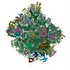
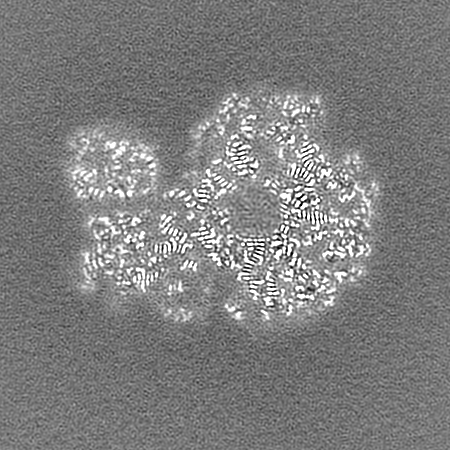
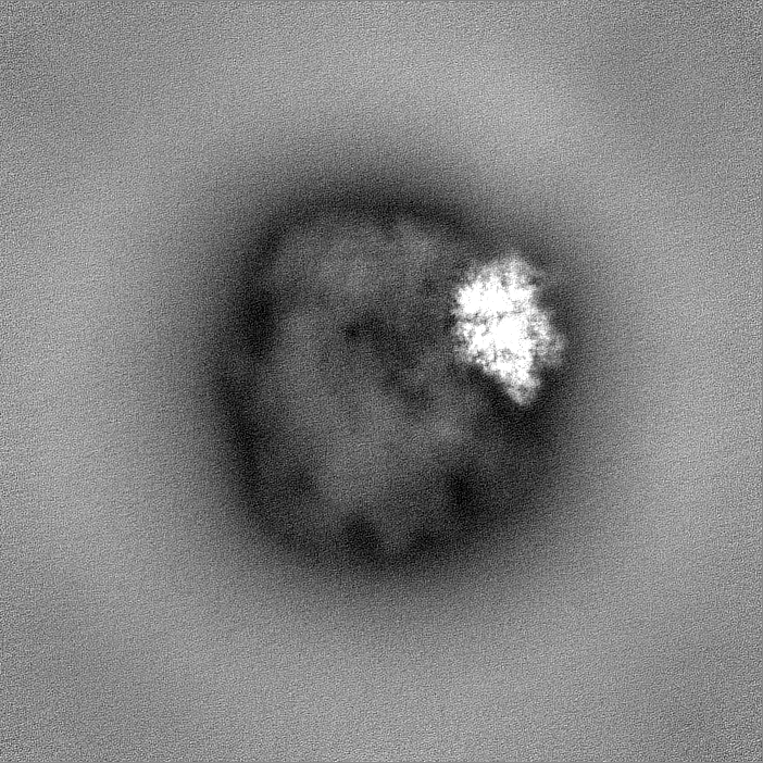
ジャーナル: Structure / 年: 2024 タイトル: The Giardia lamblia ribosome structure reveals divergence in several biological pathways and the mode of emetine function. 著者: Daniel R Eiler / Brian T Wimberly / Danielle Y Bilodeau / J Matthew Taliaferro / Philip Reigan / Olivia S Rissland / Jeffrey S Kieft / 要旨: Giardia lamblia is a deeply branching protist and a human pathogen. Its unusual biology presents the opportunity to explore conserved and fundamental molecular mechanisms. We determined the structure ...Giardia lamblia is a deeply branching protist and a human pathogen. Its unusual biology presents the opportunity to explore conserved and fundamental molecular mechanisms. We determined the structure of the G. lamblia 80S ribosome bound to tRNA, mRNA, and the antibiotic emetine by cryo-electron microscopy, to an overall resolution of 2.49 Å. The structure reveals rapidly evolving protein and nucleotide regions, differences in the peptide exit tunnel, and likely altered ribosome quality control pathways. Examination of translation initiation factor binding sites suggests these interactions are conserved despite a divergent initiation mechanism. Highlighting the potential of G. lamblia to resolve conserved biological principles; our structure reveals the interactions of the translation inhibitor emetine with the ribosome and mRNA, thus providing insight into the mechanism of action for this widely used antibiotic. Our work defines key questions in G. lamblia and motivates future experiments to explore the diversity of eukaryotic gene regulation.
 ムービー
ムービー コントローラー


 データを開く
 基本情報
基本情報
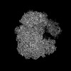
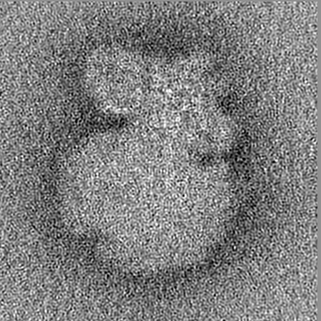
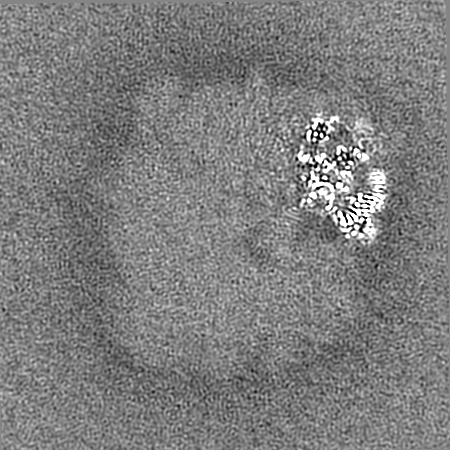
 マップデータ
 試料
試料 キーワード
キーワード 機能・相同性情報
 Giardia intestinalis assemblage A (真核生物)
 データ登録者
 米国, 3件
  引用
引用 構造の表示
構造の表示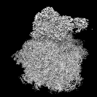
 ダウンロードとリンク
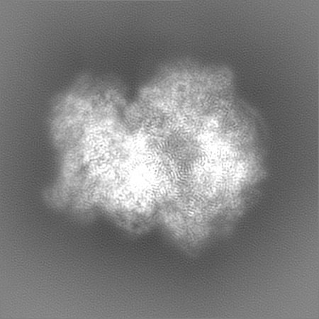
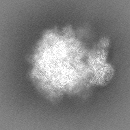
 emd_29495.png
 http://ftp.pdbj.org/pub/emdb/structures/EMD-29495


 Z (Sec.)
 Y (Row.)
 X (Col.)


 試料の構成要素

 解析
解析 電子顕微鏡法
 FIELD EMISSION GUN