[English] 日本語
Yorodumi
- EMDB-29286: Human PTH1R in complex with M-PTH(1-14) and Gs -

+
Open data


ID or keywords:

Loading...

-
Basic information

Entry
Database: EMDB / ID: EMD-29286
TitleHuman PTH1R in complex with M-PTH(1-14) and GsParathyroid hormone 1 receptor
Map dataConsensus map sharpened; rescaled to 0.85 angstrom/px
Sample
  • Complex: Human PTH1R in complex with M-PTH(1-14) and GsParathyroid hormone 1 receptor
    • Protein or peptide: Guanine nucleotide-binding protein G(s) subunit alpha isoforms short
    • Protein or peptide: Guanine nucleotide-binding protein G(I)/G(S)/G(T) subunit beta-1
    • Protein or peptide: Guanine nucleotide-binding protein G(I)/G(S)/G(O) subunit gamma-2
    • Protein or peptide: Nanobody35
    • Protein or peptide: M-PTH(1-14)
    • Protein or peptide: Parathyroid hormone/parathyroid hormone-related peptide receptor
KeywordsGPCR / agonist / hormone / MEMBRANE PROTEIN
Function / homology
Function and homology information


parathyroid hormone receptor activity / G protein-coupled peptide receptor activity / Class B/2 (Secretin family receptors) / osteoblast development / positive regulation of inositol phosphate biosynthetic process / bone mineralization / G protein-coupled receptor signaling pathway, coupled to cyclic nucleotide second messenger / peptide hormone binding / PKA activation in glucagon signalling / hair follicle placode formation ...parathyroid hormone receptor activity / G protein-coupled peptide receptor activity / Class B/2 (Secretin family receptors) / osteoblast development / positive regulation of inositol phosphate biosynthetic process / bone mineralization / G protein-coupled receptor signaling pathway, coupled to cyclic nucleotide second messenger / peptide hormone binding / PKA activation in glucagon signalling / hair follicle placode formation / developmental growth / D1 dopamine receptor binding / intracellular transport / chondrocyte differentiation / Hedgehog 'off' state / positive regulation of cAMP-mediated signaling / adenylate cyclase-activating adrenergic receptor signaling pathway / cell maturation / bone resorption / activation of adenylate cyclase activity / adenylate cyclase activator activity / trans-Golgi network membrane / skeletal system development / Olfactory Signaling Pathway / G-protein beta/gamma-subunit complex binding / Activation of the phototransduction cascade / bone development / G beta:gamma signalling through PLC beta / Presynaptic function of Kainate receptors / Thromboxane signalling through TP receptor / adenylate cyclase-modulating G protein-coupled receptor signaling pathway / G-protein activation / G protein-coupled acetylcholine receptor signaling pathway / Activation of G protein gated Potassium channels / Inhibition of voltage gated Ca2+ channels via Gbeta/gamma subunits / adenylate cyclase-activating G protein-coupled receptor signaling pathway / Prostacyclin signalling through prostacyclin receptor / Glucagon signaling in metabolic regulation / G beta:gamma signalling through CDC42 / ADP signalling through P2Y purinoceptor 12 / G beta:gamma signalling through BTK / Synthesis, secretion, and inactivation of Glucagon-like Peptide-1 (GLP-1) / Sensory perception of sweet, bitter, and umami (glutamate) taste / platelet aggregation / photoreceptor disc membrane / intracellular calcium ion homeostasis / cognition / Adrenaline,noradrenaline inhibits insulin secretion / Glucagon-type ligand receptors / positive regulation of GTPase activity / Vasopressin regulates renal water homeostasis via Aquaporins / G alpha (z) signalling events / cellular response to catecholamine stimulus / Glucagon-like Peptide-1 (GLP1) regulates insulin secretion / ADORA2B mediated anti-inflammatory cytokines production / ADP signalling through P2Y purinoceptor 1 / adenylate cyclase-activating dopamine receptor signaling pathway / G beta:gamma signalling through PI3Kgamma / cellular response to prostaglandin E stimulus / Cooperation of PDCL (PhLP1) and TRiC/CCT in G-protein beta folding / sensory perception of taste / GPER1 signaling / G-protein beta-subunit binding / Inactivation, recovery and regulation of the phototransduction cascade / heterotrimeric G-protein complex / : / G alpha (12/13) signalling events / extracellular vesicle / signaling receptor complex adaptor activity / sensory perception of smell / Thrombin signalling through proteinase activated receptors (PARs) / retina development in camera-type eye / GTPase binding / Ca2+ pathway / phospholipase C-activating G protein-coupled receptor signaling pathway / positive regulation of cold-induced thermogenesis / G alpha (i) signalling events / fibroblast proliferation / G alpha (s) signalling events / G alpha (q) signalling events / basolateral plasma membrane / Ras protein signal transduction / in utero embryonic development / cell population proliferation / Extra-nuclear estrogen signaling / cell surface receptor signaling pathway / receptor complex / apical plasma membrane / G protein-coupled receptor signaling pathway / lysosomal membrane / negative regulation of cell population proliferation / GTPase activity / synapse / positive regulation of cell population proliferation / protein-containing complex binding / GTP binding / signal transduction / protein homodimerization activity / extracellular exosome / membrane
Similarity search - Function
GPCR, family 2, parathyroid hormone receptor / G-protein coupled receptors family 2 signature 1. / Hormone receptor domain / GPCR, family 2, extracellular hormone receptor domain / G-protein coupled receptors family 2 profile 1. / Domain present in hormone receptors / GPCR family 2, extracellular hormone receptor domain superfamily / G-protein coupled receptors family 2 signature 2. / GPCR, family 2, secretin-like, conserved site / GPCR, family 2, secretin-like ...GPCR, family 2, parathyroid hormone receptor / G-protein coupled receptors family 2 signature 1. / Hormone receptor domain / GPCR, family 2, extracellular hormone receptor domain / G-protein coupled receptors family 2 profile 1. / Domain present in hormone receptors / GPCR family 2, extracellular hormone receptor domain superfamily / G-protein coupled receptors family 2 signature 2. / GPCR, family 2, secretin-like, conserved site / GPCR, family 2, secretin-like / 7 transmembrane receptor (Secretin family) / GPCR, family 2-like / G-protein coupled receptors family 2 profile 2. / G-protein alpha subunit, group S / G-alpha domain profile. / Guanine nucleotide binding protein (G-protein), alpha subunit / G protein alpha subunit, helical insertion / G-protein alpha subunit / G protein alpha subunit / G-protein, gamma subunit / G-protein gamma subunit domain profile. / GGL domain / G-protein gamma-like domain superfamily / G-protein gamma-like domain / GGL domain / G protein gamma subunit-like motifs / Guanine nucleotide-binding protein, beta subunit / G-protein, beta subunit / G-protein beta WD-40 repeat / WD40 repeat, conserved site / Trp-Asp (WD) repeats signature. / WD domain, G-beta repeat / WD40 repeats / WD40 repeat / Trp-Asp (WD) repeats profile. / Trp-Asp (WD) repeats circular profile. / WD40-repeat-containing domain superfamily / WD40/YVTN repeat-like-containing domain superfamily / P-loop containing nucleoside triphosphate hydrolase
Similarity search - Domain/homology
Guanine nucleotide-binding protein G(I)/G(S)/G(O) subunit gamma-2 / Guanine nucleotide-binding protein G(I)/G(S)/G(T) subunit beta-1 / Guanine nucleotide-binding protein G(s) subunit alpha isoforms short / Parathyroid hormone/parathyroid hormone-related peptide receptor
Similarity search - Component
Biological speciesHomo sapiens (human) / Lama glama (llama) / synthetic construct (others)
Methodsingle particle reconstruction / cryo EM / Resolution: 3.03 Å
AuthorsCary BP / Belousoff MJ / Piper SJ / Wootten D / Sexton PM
Funding support Australia, 4 items
OrganizationGrant numberCountry
Australian Research Council (ARC)IC200100052 Australia
National Health and Medical Research Council (NHMRC, Australia)1155302 Australia
National Health and Medical Research Council (NHMRC, Australia)1154434 Australia
National Health and Medical Research Council (NHMRC, Australia)1150083 Australia
Citation
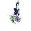
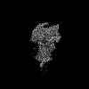
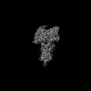
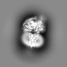
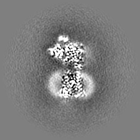
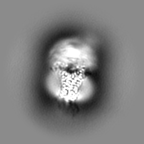
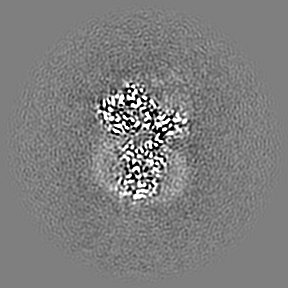
Journal: Structure / Year: 2023
Title: Molecular insights into peptide agonist engagement with the PTH receptor.
Authors: Brian P Cary / Elliot J Gerrard / Matthew J Belousoff / Madeleine M Fletcher / Yan Jiang / Isabella C Russell / Sarah J Piper / Denise Wootten / Patrick M Sexton /
Abstract: The parathyroid hormone (PTH) 1 receptor (PTH1R) is a G protein-coupled receptor (GPCR) that regulates skeletal development and calcium homeostasis. Here, we describe cryo-EM structures of the PTH1R ...The parathyroid hormone (PTH) 1 receptor (PTH1R) is a G protein-coupled receptor (GPCR) that regulates skeletal development and calcium homeostasis. Here, we describe cryo-EM structures of the PTH1R in complex with fragments of the two hormones, PTH and PTH-related protein, the drug abaloparatide, as well as the engineered tool compounds, long-acting PTH (LA-PTH) and the truncated peptide, M-PTH(1-14). We found that the critical N terminus of each agonist engages the transmembrane bundle in a topologically similar fashion, reflecting similarities in measures of Gαs activation. The full-length peptides induce subtly different extracellular domain (ECD) orientations relative to the transmembrane domain. In the structure bound to M-PTH, the ECD is unresolved, demonstrating that the ECD is highly dynamic when unconstrained by a peptide. High resolutions enabled identification of water molecules near peptide and G protein binding sites. Our results illuminate the action of orthosteric agonists of the PTH1R.
History
DepositionDec 22, 2022-
Header (metadata) releaseApr 26, 2023-
Map releaseApr 26, 2023-
UpdateJun 14, 2023-
Current statusJun 14, 2023Processing site: RCSB / Status: Released

-
Structure visualization

Supplemental images

Downloads & links

-
Map

FileDownload / File: emd_29286.map.gz / Format: CCP4 / Size: 91.1 MB / Type: IMAGE STORED AS FLOATING POINT NUMBER (4 BYTES)
AnnotationConsensus map sharpened; rescaled to 0.85 angstrom/px
Voxel sizeX=Y=Z: 0.85 Å
Density
Contour LevelBy AUTHOR: 0.61
Minimum - Maximum-3.5174825 - 4.5491505
Average (Standard dev.)-0.0023783005 (±0.10081797)
SymmetrySpace group: 1
Details

EMDB XML:

Map geometry
Axis orderXYZ
Origin000
Dimensions288288288
Spacing288288288
CellA=B=C: 244.8 Å
α=β=γ: 90.0 °

-
Supplemental data

-
Mask #1

Fileemd_29286_msk_1.map
Projections & Slices
AxesZYX

Projections

Slices (1/2)
Density Histograms

-
Additional map: Consensus map unsharpened; rescaled to 0.85 angstrom/px

Fileemd_29286_additional_1.map
AnnotationConsensus map unsharpened; rescaled to 0.85 angstrom/px
Projections & Slices
AxesZYX

Projections

Slices (1/2)
Density Histograms

-
Additional map: local refinement (receptor) map unsharpened; rescaled to 0.85...

Fileemd_29286_additional_2.map
Annotationlocal refinement (receptor) map unsharpened; rescaled to 0.85 angstrom/px
Projections & Slices
AxesZYX

Projections

Slices (1/2)
Density Histograms

-
Additional map: Consensus map unsharpened; original pixel size

Fileemd_29286_additional_3.map
AnnotationConsensus map unsharpened; original pixel size
Projections & Slices
AxesZYX

Projections

Slices (1/2)
Density Histograms

-
Additional map: Consensus map sharpened; original pixel size

Fileemd_29286_additional_4.map
AnnotationConsensus map sharpened; original pixel size
Projections & Slices
AxesZYX

Projections

Slices (1/2)
Density Histograms

-
Half map: half map

Fileemd_29286_half_map_1.map
Annotationhalf map
Projections & Slices
AxesZYX

Projections

Slices (1/2)
Density Histograms

-
Half map: half map

Fileemd_29286_half_map_2.map
Annotationhalf map
Projections & Slices
AxesZYX

Projections

Slices (1/2)
Density Histograms

-
Sample components

-
Entire : Human PTH1R in complex with M-PTH(1-14) and Gs

EntireName: Human PTH1R in complex with M-PTH(1-14) and GsParathyroid hormone 1 receptor
Components
  • Complex: Human PTH1R in complex with M-PTH(1-14) and GsParathyroid hormone 1 receptor
    • Protein or peptide: Guanine nucleotide-binding protein G(s) subunit alpha isoforms short
    • Protein or peptide: Guanine nucleotide-binding protein G(I)/G(S)/G(T) subunit beta-1
    • Protein or peptide: Guanine nucleotide-binding protein G(I)/G(S)/G(O) subunit gamma-2
    • Protein or peptide: Nanobody35
    • Protein or peptide: M-PTH(1-14)
    • Protein or peptide: Parathyroid hormone/parathyroid hormone-related peptide receptor

-
Supramolecule #1: Human PTH1R in complex with M-PTH(1-14) and Gs

SupramoleculeName: Human PTH1R in complex with M-PTH(1-14) and Gs / type: complex / ID: 1 / Parent: 0 / Macromolecule list: all
Source (natural)Organism: Homo sapiens (human)

-
Macromolecule #1: Guanine nucleotide-binding protein G(s) subunit alpha isoforms short

MacromoleculeName: Guanine nucleotide-binding protein G(s) subunit alpha isoforms short
type: protein_or_peptide / ID: 1 / Number of copies: 1 / Enantiomer: LEVO
Source (natural)Organism: Homo sapiens (human)
Molecular weightTheoretical: 45.683434 KDa
Recombinant expressionOrganism: Trichoplusia ni (cabbage looper)
SequenceString: MGCLGNSKTE DQRNEEKAQR EANKKIEKQL QKDKQVYRAT HRLLLLGAGE SGKNTIVKQM RILHVNGFNG EGGEEDPQAA RSNSDGEKA TKVQDIKNNL KEAIETIVAA MSNLVPPVEL ANPENQFRVD YILSVMNVPD FDFPPEFYEH AKALWEDEGV R ACYERSNE ...String:
MGCLGNSKTE DQRNEEKAQR EANKKIEKQL QKDKQVYRAT HRLLLLGAGE SGKNTIVKQM RILHVNGFNG EGGEEDPQAA RSNSDGEKA TKVQDIKNNL KEAIETIVAA MSNLVPPVEL ANPENQFRVD YILSVMNVPD FDFPPEFYEH AKALWEDEGV R ACYERSNE YQLIDCAQYF LDKIDVIKQA DYVPSDQDLL RCRVLTSGIF ETKFQVDKVN FHMFDVGAQR DERRKWIQCF ND VTAIIFV VASSSYNMVI REDNQTNRLQ AALKLFDSIW NNKWLRDTSV ILFLNKQDLL AEKVLAGKSK IEDYFPEFAR YTT PEDATP EPGEDPRVTR AKYFIRDEFL RISTASGDGR HYCYPHFTCA VDTENIRRVF NDCRDIIQRM HLRQYELL

-
Macromolecule #2: Guanine nucleotide-binding protein G(I)/G(S)/G(T) subunit beta-1

MacromoleculeName: Guanine nucleotide-binding protein G(I)/G(S)/G(T) subunit beta-1
type: protein_or_peptide / ID: 2 / Number of copies: 1 / Enantiomer: LEVO
Source (natural)Organism: Homo sapiens (human)
Molecular weightTheoretical: 37.413863 KDa
Recombinant expressionOrganism: Trichoplusia ni (cabbage looper)
SequenceString: QSELDQLRQE AEQLKNQIRD ARKACADATL SQITNNIDPV GRIQMRTRRT LRGHLAKIYA MHWGTDSRLL VSASQDGKLI IWDSYTTNK VHAIPLRSSW VMTCAYAPSG NYVACGGLDN ICSIYNLKTR EGNVRVSREL AGHTGYLSCC RFLDDNQIVT S SGDTTCAL ...String:
QSELDQLRQE AEQLKNQIRD ARKACADATL SQITNNIDPV GRIQMRTRRT LRGHLAKIYA MHWGTDSRLL VSASQDGKLI IWDSYTTNK VHAIPLRSSW VMTCAYAPSG NYVACGGLDN ICSIYNLKTR EGNVRVSREL AGHTGYLSCC RFLDDNQIVT S SGDTTCAL WDIETGQQTT TFTGHTGDVM SLSLAPDTRL FVSGACDASA KLWDVREGMC RQTFTGHESD INAICFFPNG NA FATGSDD ATCRLFDLRA DQELMTYSHD NIICGITSVS FSKSGRLLLA GYDDFNCNVW DALKADRAGV LAGHDNRVSC LGV TDDGMA VATGSWDSFL KIWN

-
Macromolecule #3: Guanine nucleotide-binding protein G(I)/G(S)/G(O) subunit gamma-2

MacromoleculeName: Guanine nucleotide-binding protein G(I)/G(S)/G(O) subunit gamma-2
type: protein_or_peptide / ID: 3 / Number of copies: 1 / Enantiomer: LEVO
Source (natural)Organism: Homo sapiens (human)
Molecular weightTheoretical: 6.375332 KDa
Recombinant expressionOrganism: Trichoplusia ni (cabbage looper)
SequenceString:
NTASIAQARK LVEQLKMEAN IDRIKVSKAA ADLMAYCEAH AKEDPLLTPV PASENPFR

-
Macromolecule #4: Nanobody35

MacromoleculeName: Nanobody35 / type: protein_or_peptide / ID: 4 / Number of copies: 1 / Enantiomer: LEVO
Source (natural)Organism: Lama glama (llama)
Molecular weightTheoretical: 13.885439 KDa
Recombinant expressionOrganism: Escherichia coli (E. coli)
SequenceString:
QVQLQESGGG LVQPGGSLRL SCAASGFTFS NYKMNWVRQA PGKGLEWVSD ISQSGASISY TGSVKGRFTI SRDNAKNTLY LQMNSLKPE DTAVYYCARC PAPFTRDCFD VTSTTYAYRG QGTQVTVSS

-
Macromolecule #5: M-PTH(1-14)

MacromoleculeName: M-PTH(1-14) / type: protein_or_peptide / ID: 5 / Number of copies: 1 / Enantiomer: LEVO
Source (natural)Organism: synthetic construct (others)
Molecular weightTheoretical: 1.725065 KDa
SequenceString:
(AIB)V(AIB)EIQLMHQ (HRG)AKW

-
Macromolecule #6: Parathyroid hormone/parathyroid hormone-related peptide receptor

MacromoleculeName: Parathyroid hormone/parathyroid hormone-related peptide receptor
type: protein_or_peptide / ID: 6 / Number of copies: 1 / Enantiomer: LEVO
Source (natural)Organism: Homo sapiens (human)
Molecular weightTheoretical: 69.513758 KDa
Recombinant expressionOrganism: Trichoplusia ni (cabbage looper)
SequenceString: MKTIIALSYI FCLVFADYKD DDDLEVLFQG PADDVMTKEE QIFLLHRAQA QCEKRLKEVL QRPASIMESD KGWTSASTSG KPRKDKASG KLYPESEEDK EAPTGSRYRG RPCLPEWDHI LCWPLGAPGE VVAVPCPDYI YDFNHKGHAY RRCDRNGSWE L VPGHNRTW ...String:
MKTIIALSYI FCLVFADYKD DDDLEVLFQG PADDVMTKEE QIFLLHRAQA QCEKRLKEVL QRPASIMESD KGWTSASTSG KPRKDKASG KLYPESEEDK EAPTGSRYRG RPCLPEWDHI LCWPLGAPGE VVAVPCPDYI YDFNHKGHAY RRCDRNGSWE L VPGHNRTW ANYSECVKFL TNETREREVF DRLGMIYTVG YSVSLASLTV AVLILAYFRR LHCTRNYIHM HLFLSFMLRA VS IFVKDAV LYSGATLDEA ERLTEEELRA IAQAPPPPAT AAAGYAGCRV AVTFFLYFLA TNYYWILVEG LYLHSLIFMA FFS EKKYLW GFTVFGWGLP AVFVAVWVSV RATLANTGCW DLSSGNKKWI IQVPILASIV LNFILFINIV RVLATKLRET NAGR CDTRQ QYRKLLKSTL VLMPLFGVHY IVFMATPYTE VSGTLWQVQM HYEMLFNSFQ GFFVAIIYCF CNGEVQAEIK KSWSR WTLA LDFKRKARSG SSSYSYGPMV SHTSVTNVGP RVGLGLPLSP RLLPTATTNG HPQLPGHAKP GTPALETLET TPPAMA APK DDGFLNGSCS GLDEEASGPE RPPALLQEEW ETVMPAGLEV LFQGPHHHHH HHH

-
Experimental details

-
Structure determination

Methodcryo EM
Processingsingle particle reconstruction
Aggregation stateparticle

-
Sample preparation

Concentration3.55 mg/mL
BufferpH: 7.4
Component:
ConcentrationFormulaName
20.0 mMC8H18N2O4SHEPES
100.0 mMNaClSodium chlorideSodium Chloride
2.0 mMMgCl2Magnesium Chloride
0.1 mMC9H15O6PTCEP
0.01 percentC47H88O22LMNG
6e-05 percentC31H50O4CHS

Details: Buffer also contained 25 micromolar M-PTH peptide.
GridModel: UltrAuFoil R1.2/1.3 / Material: GOLD / Pretreatment - Type: GLOW DISCHARGE / Pretreatment - Time: 60 sec. / Pretreatment - Atmosphere: AIR / Details: 20 mA current, negative polarity
VitrificationCryogen name: ETHANE / Chamber humidity: 100 % / Chamber temperature: 277 K / Instrument: FEI VITROBOT MARK IV

-
Electron microscopy

MicroscopeTFS GLACIOS
Electron beamAcceleration voltage: 200 kV / Electron source: FIELD EMISSION GUN
Electron opticsIllumination mode: FLOOD BEAM / Imaging mode: BRIGHT FIELDBright-field microscopy / Cs: 2.7 mm / Nominal defocus max: 1.3 µm / Nominal defocus min: 0.6 µm
Sample stageCooling holder cryogen: NITROGEN
Image recordingFilm or detector model: FEI FALCON IV (4k x 4k) / Number grids imaged: 1 / Number real images: 5797 / Average exposure time: 8.23 sec. / Average electron dose: 49.91 e/Å2

-
Image processing

Particle selectionNumber selected: 1723403
Startup modelType of model: NONE
Initial angle assignmentType: MAXIMUM LIKELIHOOD / Software - Name: cryoSPARC (ver. 3.3.1)
Final angle assignmentType: MAXIMUM LIKELIHOOD / Software - Name: cryoSPARC (ver. 3.3.2)
Final reconstructionApplied symmetry - Point group: C1 (asymmetric) / Resolution.type: BY AUTHOR / Resolution: 3.03 Å / Resolution method: FSC 0.143 CUT-OFF / Software - Name: cryoSPARC (ver. 3.3.2) / Number images used: 189793
FSC plot (resolution estimation)

-
Atomic model buiding 1

Initial model
PDB IDChain

chain_id: R

Output model

PDB-8flt:
Human PTH1R in complex with M-PTH(1-14) and Gs

+
About Yorodumi

-
News

-
Feb 9, 2022. New format data for meta-information of EMDB entries

New format data for meta-information of EMDB entries

  • Version 3 of the EMDB header file is now the official format.
  • The previous official version 1.9 will be removed from the archive.

Related info.:EMDB header

External links:wwPDB to switch to version 3 of the EMDB data model

-
Aug 12, 2020. Covid-19 info

Covid-19 info

URL: https://pdbj.org/emnavi/covid19.php

New page: Covid-19 featured information page in EM Navigator.

Related info.:Covid-19 info / Mar 5, 2020. Novel coronavirus structure data

+
Mar 5, 2020. Novel coronavirus structure data

Novel coronavirus structure data

Related info.:Yorodumi Speices / Aug 12, 2020. Covid-19 info

External links:COVID-19 featured content - PDBj / Molecule of the Month (242):Coronavirus Proteases

+
Jan 31, 2019. EMDB accession codes are about to change! (news from PDBe EMDB page)

EMDB accession codes are about to change! (news from PDBe EMDB page)

  • The allocation of 4 digits for EMDB accession codes will soon come to an end. Whilst these codes will remain in use, new EMDB accession codes will include an additional digit and will expand incrementally as the available range of codes is exhausted. The current 4-digit format prefixed with “EMD-” (i.e. EMD-XXXX) will advance to a 5-digit format (i.e. EMD-XXXXX), and so on. It is currently estimated that the 4-digit codes will be depleted around Spring 2019, at which point the 5-digit format will come into force.
  • The EM Navigator/Yorodumi systems omit the EMD- prefix.

Related info.:Q: What is EMD? / ID/Accession-code notation in Yorodumi/EM Navigator

External links:EMDB Accession Codes are Changing Soon! / Contact to PDBj

+
Jul 12, 2017. Major update of PDB

Major update of PDB

  • wwPDB released updated PDB data conforming to the new PDBx/mmCIF dictionary.
  • This is a major update changing the version number from 4 to 5, and with Remediation, in which all the entries are updated.
  • In this update, many items about electron microscopy experimental information are reorganized (e.g. em_software).
  • Now, EM Navigator and Yorodumi are based on the updated data.

External links:wwPDB Remediation / Enriched Model Files Conforming to OneDep Data Standards Now Available in the PDB FTP Archive

-
Yorodumi

Thousand views of thousand structures

  • Yorodumi is a browser for structure data from EMDB, PDB, SASBDB, etc.
  • This page is also the successor to EM Navigator detail page, and also detail information page/front-end page for Omokage search.
  • The word "yorodu" (or yorozu) is an old Japanese word meaning "ten thousand". "mi" (miru) is to see.

Related info.:EMDB / PDB / SASBDB / Comparison of 3 databanks / Yorodumi Search / Aug 31, 2016. New EM Navigator & Yorodumi / Yorodumi Papers / Jmol/JSmol / Function and homology information / Changes in new EM Navigator and Yorodumi

Read more