- EMDB-28816: Wild type P53 dimer structure from human cancer cells -
+
データを開く
IDまたはキーワード:
読み込み中...
-
基本情報
登録情報
データベース: EMDB / ID: EMD-28816
タイトル
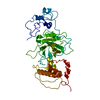
Wild type P53 dimer structure from human cancer cells
マップデータ
P53 dimer structure
試料
細胞: P53 dimer isolated from U87-MG cells
タンパク質・ペプチド: Cellular tumor antigen p53
キーワード
cancer / tumor suppressor / cell cycle / apoptosis / DNA repair / ANTITUMOR PROTEIN
機能・相同性
機能・相同性情報
Loss of function of TP53 in cancer due to loss of tetramerization ability / Regulation of TP53 Expression / signal transduction by p53 class mediator / negative regulation of G1 to G0 transition / regulation of fibroblast apoptotic process / negative regulation of glucose catabolic process to lactate via pyruvate / Transcriptional activation of cell cycle inhibitor p21 / regulation of intrinsic apoptotic signaling pathway by p53 class mediator / Activation of NOXA and translocation to mitochondria / negative regulation of pentose-phosphate shunt ...Loss of function of TP53 in cancer due to loss of tetramerization ability / Regulation of TP53 Expression / signal transduction by p53 class mediator / negative regulation of G1 to G0 transition / regulation of fibroblast apoptotic process / negative regulation of glucose catabolic process to lactate via pyruvate / Transcriptional activation of cell cycle inhibitor p21 / regulation of intrinsic apoptotic signaling pathway by p53 class mediator / Activation of NOXA and translocation to mitochondria / negative regulation of pentose-phosphate shunt / ATP-dependent DNA/DNA annealing activity / negative regulation of helicase activity / regulation of cell cycle G2/M phase transition / intrinsic apoptotic signaling pathway in response to hypoxia / oxidative stress-induced premature senescence / oligodendrocyte apoptotic process / negative regulation of miRNA processing / positive regulation of thymocyte apoptotic process / regulation of tissue remodeling / glucose catabolic process to lactate via pyruvate / positive regulation of mitochondrial membrane permeability / negative regulation of mitophagy / positive regulation of programmed necrotic cell death / circadian behavior / mRNA transcription / bone marrow development / regulation of mitochondrial membrane permeability involved in apoptotic process / histone deacetylase regulator activity / germ cell nucleus / T cell lineage commitment / RUNX3 regulates CDKN1A transcription / regulation of DNA damage response, signal transduction by p53 class mediator / TP53 regulates transcription of additional cell cycle genes whose exact role in the p53 pathway remain uncertain / TP53 Regulates Transcription of Death Receptors and Ligands / Activation of PUMA and translocation to mitochondria / DNA damage response, signal transduction by p53 class mediator resulting in transcription of p21 class mediator / B cell lineage commitment / thymocyte apoptotic process / negative regulation of glial cell proliferation / negative regulation of neuroblast proliferation / Formation of Senescence-Associated Heterochromatin Foci (SAHF) / Regulation of TP53 Activity through Association with Co-factors / mitochondrial DNA repair / positive regulation of cardiac muscle cell apoptotic process / TP53 Regulates Transcription of Caspase Activators and Caspases / ER overload response / negative regulation of DNA replication / positive regulation of release of cytochrome c from mitochondria / TP53 regulates transcription of several additional cell death genes whose specific roles in p53-dependent apoptosis remain uncertain / entrainment of circadian clock by photoperiod / cardiac septum morphogenesis / PI5P Regulates TP53 Acetylation / positive regulation of execution phase of apoptosis / Association of TriC/CCT with target proteins during biosynthesis / necroptotic process / Zygotic genome activation (ZGA) / TP53 Regulates Transcription of Genes Involved in Cytochrome C Release / TFIID-class transcription factor complex binding / rRNA transcription / negative regulation of telomere maintenance via telomerase / SUMOylation of transcription factors / intrinsic apoptotic signaling pathway by p53 class mediator / general transcription initiation factor binding / intrinsic apoptotic signaling pathway in response to endoplasmic reticulum stress / response to X-ray / Transcriptional Regulation by VENTX / DNA damage response, signal transduction by p53 class mediator / Pyroptosis / replicative senescence / mitophagy / cellular response to UV-C / positive regulation of RNA polymerase II transcription preinitiation complex assembly / neuroblast proliferation / negative regulation of reactive oxygen species metabolic process / hematopoietic stem cell differentiation / somitogenesis / embryonic organ development / intrinsic apoptotic signaling pathway in response to DNA damage by p53 class mediator / chromosome organization / T cell proliferation involved in immune response / hematopoietic progenitor cell differentiation / glial cell proliferation / type II interferon-mediated signaling pathway / cis-regulatory region sequence-specific DNA binding / TP53 Regulates Transcription of Genes Involved in G1 Cell Cycle Arrest / cellular response to glucose starvation / viral process / cellular response to actinomycin D / positive regulation of intrinsic apoptotic signaling pathway / core promoter sequence-specific DNA binding / negative regulation of stem cell proliferation / mitotic G1 DNA damage checkpoint signaling / negative regulation of fibroblast proliferation / gastrulation / MDM2/MDM4 family protein binding / tumor necrosis factor-mediated signaling pathway / response to salt stress / cardiac muscle cell apoptotic process / Regulation of TP53 Activity through Acetylation / 14-3-3 protein binding 類似検索 - 分子機能
National Institutes of Health/National Cancer Institute (NIH/NCI)
R01CA193578
米国
National Institutes of Health/National Cancer Institute (NIH/NCI)
R01CA227261
米国
National Institutes of Health/National Cancer Institute (NIH/NCI)
R01CA219700
米国
引用
ジャーナル: Chembiochem / 年: 2022 タイトル: High-Resolution Imaging of Human Cancer Proteins Using Microprocessor Materials. 著者: Maria J Solares / G M Jonaid / William Y Luqiu / Samantha Berry / Janki Khadela / Yanping Liang / Madison C Evans / Kevin J Pridham / William J Dearnaley / Zhi Sheng / Deborah F Kelly / 要旨: Mutations in tumor suppressor genes, such as Tumor Protein 53 (TP53), are heavily implicated in aggressive cancers giving rise to gain- and loss-of-function phenotypes. While individual domains of ...Mutations in tumor suppressor genes, such as Tumor Protein 53 (TP53), are heavily implicated in aggressive cancers giving rise to gain- and loss-of-function phenotypes. While individual domains of the p53 protein have been studied extensively, structural information for full-length p53 remains incomplete. Functionalized microprocessor chips (microchips) with properties amenable to electron microscopy permitted us to visualize complete p53 assemblies for the first time. The new structures revealed p53 in an inactive dimeric state independent of DNA binding. Residues located at the protein-protein interface corresponded with modification sites in cancer-related hot spots. Changes in these regions may amplify the toxic effects of clinical mutations. Taken together, these results contribute advances in technology and imaging approaches to decode native protein models in different states of activation.
pH: 7.5 詳細: 20 mM HEPES (pH 7.5), 140 mM NaCl, 2 mM MgCl2, 2 mM CaCl2, 5 mM imidazole
グリッド
モデル: Homemade / 材質: SILICON NITRIDE 詳細: Silicon nitride chips coated with Ni-NTA layers prior to sample application
凍結
凍結剤: ETHANE / チャンバー内湿度: 90 % / チャンバー内温度: 298 K / 装置: FEI VITROBOT MARK III 詳細: The microchip samples were loaded into a FEI Mark III Vitrobot and flash-frozen into liquid ethane..
詳細
Sample was placed on Silicon nitride chips coated with Ni-NTA layers
-
電子顕微鏡法
顕微鏡
TFS TALOS F200C
撮影
フィルム・検出器のモデル: FEI CETA (4k x 4k) / 撮影したグリッド数: 10 / 実像数: 300 / 平均露光時間: 1.0 sec. / 平均電子線量: 5.0 e/Å2
 ムービー
ムービー コントローラー


 データを開く
 基本情報
基本情報
 マップデータ
 試料
試料 キーワード
キーワード 機能・相同性情報
 Homo sapiens (ヒト)
 データ登録者
 米国, 3件
  引用
引用 構造の表示
構造の表示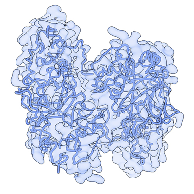
 ダウンロードとリンク
 emd_28816.png
 http://ftp.pdbj.org/pub/emdb/structures/EMD-28816


 Z (Sec.)
 Y (Row.)
 X (Col.)


 試料の構成要素
 解析
解析 電子顕微鏡法
 FIELD EMISSION GUN

