ATP synthase, F0 complex, subunit H / ATP synthase complex subunit h / ATP synthase, F0 complex, subunit J / ATP synthase protein 8, fungal type / ATP synthase, F0 complex, subunit F, mitochondria, fungi / ATP synthase j chain / Fungal ATP synthase protein 8 (A6L) / Mitochondrial F1-F0 ATP synthase subunit F of fungi / : / Fungal epsilon subunit of F1F0-ATP synthase C-terminal domain ...ATP synthase, F0 complex, subunit H / ATP synthase complex subunit h / ATP synthase, F0 complex, subunit J / ATP synthase protein 8, fungal type / ATP synthase, F0 complex, subunit F, mitochondria, fungi / ATP synthase j chain / Fungal ATP synthase protein 8 (A6L) / Mitochondrial F1-F0 ATP synthase subunit F of fungi / : / Fungal epsilon subunit of F1F0-ATP synthase C-terminal domain / ATP synthase, F0 complex, subunit B/MI25 / ATP synthase, F0 complex, subunit B / Mitochondrial ATP synthase B chain precursor (ATP-synt_B) / ATP synthase, F0 complex, subunit D, mitochondrial / ATP synthase D chain, mitochondrial (ATP5H) / ATP synthase, F0 complex, subunit D superfamily, mitochondrial / ATP synthase, F0 complex, subunit A, bacterial/mitochondria / ATP synthase, F1 complex, epsilon subunit, mitochondrial / ATP synthase, F1 complex, epsilon subunit superfamily, mitochondrial / Mitochondrial ATP synthase epsilon chain / ATPase, OSCP/delta subunit, conserved site / ATP synthase delta (OSCP) subunit signature. / F1F0 ATP synthase OSCP/delta subunit, N-terminal domain superfamily / ATP synthase, F0 complex, subunit A / ATP synthase, F0 complex, subunit A, active site / ATP synthase, F0 complex, subunit A superfamily / ATP synthase A chain / ATP synthase a subunit signature. / ATPase, OSCP/delta subunit / ATP synthase delta (OSCP) subunit / ATP synthase, F1 complex, delta/epsilon subunit / ATP synthase, F1 complex, delta/epsilon subunit, N-terminal / F0F1 ATP synthase delta/epsilon subunit, N-terminal / ATP synthase, Delta/Epsilon chain, beta-sandwich domain / ATP synthase, F0 complex, subunit C / F1F0 ATP synthase subunit C superfamily / ATP synthase, F0 complex, subunit C, DCCD-binding site / ATP synthase c subunit signature. / ATP synthase, F1 complex, gamma subunit conserved site / ATP synthase gamma subunit signature. / ATP synthase, F1 complex, beta subunit / ATP synthase, alpha subunit, C-terminal domain superfamily / : / ATP synthase, F1 complex, alpha subunit nucleotide-binding domain / ATP synthase, F1 complex, gamma subunit / ATP synthase, F1 complex, gamma subunit superfamily / ATP synthase / ATP synthase, alpha subunit, C-terminal / ATP synthase, F1 complex, alpha subunit / ATP synthase alpha/beta chain, C terminal domain / V-ATPase proteolipid subunit C-like domain / F/V-ATP synthase subunit C superfamily / ATP synthase subunit C / : / ATPase, F1/V1 complex, beta/alpha subunit, C-terminal / C-terminal domain of V and A type ATP synthase / ATPase, F1/V1/A1 complex, alpha/beta subunit, N-terminal domain superfamily / ATP synthase subunit alpha, N-terminal domain-like superfamily / ATPase, F1/V1/A1 complex, alpha/beta subunit, N-terminal domain / ATP synthase alpha/beta family, beta-barrel domain / ATPase, alpha/beta subunit, nucleotide-binding domain, active site / ATP synthase alpha and beta subunits signature. / ATPase, F1/V1/A1 complex, alpha/beta subunit, nucleotide-binding domain / ATP synthase alpha/beta family, nucleotide-binding domain / ATPases associated with a variety of cellular activities / AAA+ ATPase domain / P-loop containing nucleoside triphosphate hydrolase Similarity search - Domain/homology
ATP synthase subunit beta, mitochondrial / ATP synthase subunit a / ATP synthase protein 8 / ATP synthase subunit 4, mitochondrial / ATP synthase subunit alpha, mitochondrial / ATP synthase subunit 5, mitochondrial / ATP synthase subunit epsilon, mitochondrial / ATP synthase subunit d, mitochondrial / ATP synthase subunit gamma, mitochondrial / ATP synthase subunit 9, mitochondrial ...ATP synthase subunit beta, mitochondrial / ATP synthase subunit a / ATP synthase protein 8 / ATP synthase subunit 4, mitochondrial / ATP synthase subunit alpha, mitochondrial / ATP synthase subunit 5, mitochondrial / ATP synthase subunit epsilon, mitochondrial / ATP synthase subunit d, mitochondrial / ATP synthase subunit gamma, mitochondrial / ATP synthase subunit 9, mitochondrial / ATP synthase subunit J, mitochondrial / ATP synthase subunit f, mitochondrial / ATP synthase subunit delta, mitochondrial / ATP synthase subunit H, mitochondrial Similarity search - Component
Biological species
Saccharomyces cerevisiae (brewer's yeast)
Method
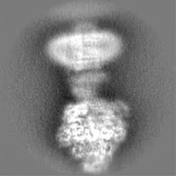
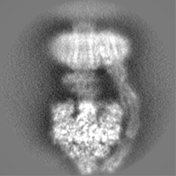
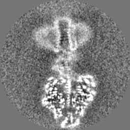
single particle reconstruction / cryo EM / Resolution: 4.0 Å
National Institutes of Health/National Institute of General Medical Sciences (NIH/NIGMS)
United States
Citation
Journal: Nat Struct Mol Biol / Year: 2024 Title: Conformational ensemble of yeast ATP synthase at low pH reveals unique intermediates and plasticity in F-F coupling. Authors: Stuti Sharma / Min Luo / Hiral Patel / David M Mueller / Maofu Liao / Abstract: Mitochondrial adenosine triphosphate (ATP) synthase uses the proton gradient across the inner mitochondrial membrane to synthesize ATP. Structural and single molecule studies conducted mostly at ...Mitochondrial adenosine triphosphate (ATP) synthase uses the proton gradient across the inner mitochondrial membrane to synthesize ATP. Structural and single molecule studies conducted mostly at neutral or basic pH have provided details of the reaction mechanism of ATP synthesis. However, pH of the mitochondrial matrix is slightly acidic during hypoxia and pH-dependent conformational changes in the ATP synthase have been reported. Here we use single-particle cryo-EM to analyze the conformational ensemble of the yeast (Saccharomyces cerevisiae) ATP synthase at pH 6. Of the four conformations resolved in this study, three are reaction intermediates. In addition to canonical catalytic dwell and binding dwell structures, we identify two unique conformations with nearly identical positions of the central rotor but different catalytic site conformations. These structures provide new insights into the catalytic mechanism of the ATP synthase and highlight elastic coupling between the catalytic and proton translocating domains.
In the structure databanks used in Yorodumi, some data are registered as the other names, "COVID-19 virus" and "2019-nCoV". Here are the details of the virus and the list of structure data.
Jan 31, 2019. EMDB accession codes are about to change! (news from PDBe EMDB page)
EMDB accession codes are about to change! (news from PDBe EMDB page)
The allocation of 4 digits for EMDB accession codes will soon come to an end. Whilst these codes will remain in use, new EMDB accession codes will include an additional digit and will expand incrementally as the available range of codes is exhausted. The current 4-digit format prefixed with “EMD-” (i.e. EMD-XXXX) will advance to a 5-digit format (i.e. EMD-XXXXX), and so on. It is currently estimated that the 4-digit codes will be depleted around Spring 2019, at which point the 5-digit format will come into force.
The EM Navigator/Yorodumi systems omit the EMD- prefix.
Related info.:Q: What is EMD? / ID/Accession-code notation in Yorodumi/EM Navigator
Yorodumi is a browser for structure data from EMDB, PDB, SASBDB, etc.
This page is also the successor to EM Navigator detail page, and also detail information page/front-end page for Omokage search.
The word "yorodu" (or yorozu) is an old Japanese word meaning "ten thousand". "mi" (miru) is to see.
Related info.:EMDB / PDB / SASBDB / Comparison of 3 databanks / Yorodumi Search / Aug 31, 2016. New EM Navigator & Yorodumi / Yorodumi Papers / Jmol/JSmol / Function and homology information / Changes in new EM Navigator and Yorodumi
 Movie
Movie Controller


 Open data
 Basic information
 Map data
 Sample
 Keywords
 Function and homology information
 Authors
 United States, 1 items
  Citation

 Structure visualization
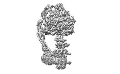
 Downloads & links
 emd_28809.png
 http://ftp.pdbj.org/pub/emdb/structures/EMD-28809


 Z (Sec.)
 Y (Row.)
 X (Col.)


 Sample components

 Processing
 Electron microscopy
 FIELD EMISSION GUN

