 Movie
Movie Controller


+ Open data

- Basic information
| Entry | 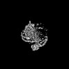 | |||||||||
|---|---|---|---|---|---|---|---|---|---|---|
| Title | cryo-EM structure of TMEM63A in nanodisc | |||||||||
 Map data Map data | ||||||||||
 Sample Sample |
| |||||||||
 Keywords Keywords | ion channel / mechanosensitive / monomeric / MEMBRANE PROTEIN | |||||||||
| Function / homology |  Function and homology information Function and homology informationsurfactant secretion / osmolarity-sensing monoatomic cation channel activity / mechanosensitive monoatomic ion channel activity / calcium-activated cation channel activity / tertiary granule membrane / centriolar satellite / specific granule membrane / early endosome membrane / nucleic acid binding / lysosomal membrane ...surfactant secretion / osmolarity-sensing monoatomic cation channel activity / mechanosensitive monoatomic ion channel activity / calcium-activated cation channel activity / tertiary granule membrane / centriolar satellite / specific granule membrane / early endosome membrane / nucleic acid binding / lysosomal membrane / intracellular membrane-bounded organelle / Neutrophil degranulation / extracellular exosome / plasma membrane Similarity search - Function | |||||||||
| Biological species |  Homo sapiens (human) Homo sapiens (human) | |||||||||
| Method | single particle reconstruction / cryo EM / Resolution: 3.8 Å | |||||||||
 Authors Authors | Zheng W / Fu TM / Holt JR | |||||||||
| Funding support |  United States, 1 items United States, 1 items
| |||||||||
 Citation Citation | Journal: Neuron / Year: 2023 Title: TMEM63 proteins function as monomeric high-threshold mechanosensitive ion channels. Authors: Wang Zheng / Shaun Rawson / Zhangfei Shen / Elakkiya Tamilselvan / Harper E Smith / Julia Halford / Chen Shen / Swetha E Murthy / Maximilian H Ulbrich / Marcos Sotomayor / Tian-Min Fu / Jeffrey R Holt /  Abstract: OSCA/TMEM63s form mechanically activated (MA) ion channels in plants and animals, respectively. OSCAs and related TMEM16s and transmembrane channel-like (TMC) proteins form homodimers with two pores. ...OSCA/TMEM63s form mechanically activated (MA) ion channels in plants and animals, respectively. OSCAs and related TMEM16s and transmembrane channel-like (TMC) proteins form homodimers with two pores. Here, we uncover an unanticipated monomeric configuration of TMEM63 proteins. Structures of TMEM63A and TMEM63B (referred to as TMEM63s) revealed a single highly restricted pore. Functional analyses demonstrated that TMEM63s are bona fide mechanosensitive ion channels, characterized by small conductance and high thresholds. TMEM63s possess evolutionary variations in the intracellular linker IL2, which mediates dimerization in OSCAs. Replacement of OSCA1.2 IL2 with TMEM63A IL2 or mutations to key variable residues resulted in monomeric OSCA1.2 and MA currents with significantly higher thresholds. Structural analyses revealed substantial conformational differences in the mechano-sensing domain IL2 and gating helix TM6 between TMEM63s and OSCA1.2. Our studies reveal that mechanosensitivity in OSCA/TMEM63 channels is affected by oligomerization and suggest gating mechanisms that may be shared by OSCA/TMEM63, TMEM16, and TMC channels. | |||||||||
| History |
|
- Structure visualization
| Supplemental images |
|---|

- Downloads & links
-EMDB archive
| Map data | emd_28153.map.gz | 66 MB | EMDB map data format | |
|---|---|---|---|---|
| Header (meta data) | emd-28153-v30.xmlemd-28153.xml | 17.3 KB 17.3 KB | Display Display | EMDB header |
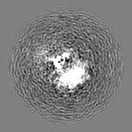
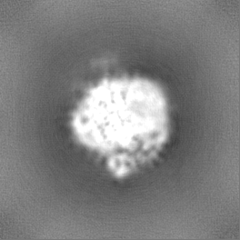
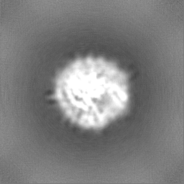
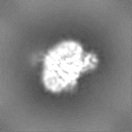
| Images |  emd_28153.png emd_28153.png | 31.4 KB | ||
| Filedesc metadata | emd-28153.cif.gz | 6 KB | ||
| Others | emd_28153_half_map_1.map.gzemd_28153_half_map_2.map.gz | 65 MB 65 MB | ||
| Archive directory |  http://ftp.pdbj.org/pub/emdb/structures/EMD-28153ftp://ftp.pdbj.org/pub/emdb/structures/EMD-28153 http://ftp.pdbj.org/pub/emdb/structures/EMD-28153ftp://ftp.pdbj.org/pub/emdb/structures/EMD-28153 | HTTPS FTP |
-Validation report
| Summary document | emd_28153_validation.pdf.gz | 761.7 KB | Display | EMDB validaton report |
|---|---|---|---|---|
| Full document | emd_28153_full_validation.pdf.gz | 761.3 KB | Display | |
| Data in XML | emd_28153_validation.xml.gz | 12.5 KB | Display | |
| Data in CIF | emd_28153_validation.cif.gz | 14.6 KB | Display | |
| Arichive directory | https://ftp.pdbj.org/pub/emdb/validation_reports/EMD-28153ftp://ftp.pdbj.org/pub/emdb/validation_reports/EMD-28153 | HTTPS FTP |
-Related structure data
| Related structure data | 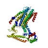 8ehwMC 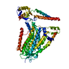 8ehxC M: atomic model generated by this map C: citing same article ( |
|---|---|
| Similar structure data |
-Links
| EMDB pages | EMDB (EBI/PDBe) / EMDataResource |
|---|
-Map
| File | Download / File: emd_28153.map.gz / Format: CCP4 / Size: 70.2 MB / Type: IMAGE STORED AS FLOATING POINT NUMBER (4 BYTES) | ||||||||||||||||||||||||||||||||||||
|---|---|---|---|---|---|---|---|---|---|---|---|---|---|---|---|---|---|---|---|---|---|---|---|---|---|---|---|---|---|---|---|---|---|---|---|---|---|
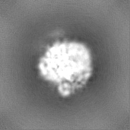
| Projections & slices | Image control
Images are generated by Spider. | ||||||||||||||||||||||||||||||||||||
| Voxel size | X=Y=Z: 0.825 Å | ||||||||||||||||||||||||||||||||||||
| Density |
| ||||||||||||||||||||||||||||||||||||
| Symmetry | Space group: 1 | ||||||||||||||||||||||||||||||||||||
| Details | EMDB XML:
|
 Z (Sec.)
 Y (Row.)
 X (Col.)


-Supplemental data
-Half map: #2
| File | emd_28153_half_map_1.map | ||||||||||||
|---|---|---|---|---|---|---|---|---|---|---|---|---|---|
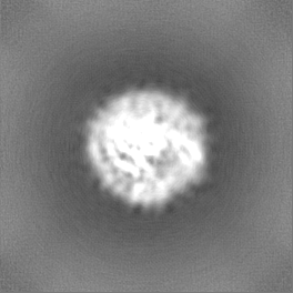
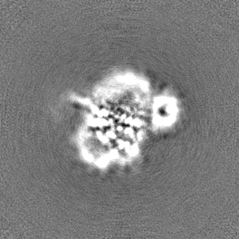
| Projections & Slices |
| ||||||||||||
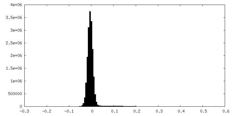
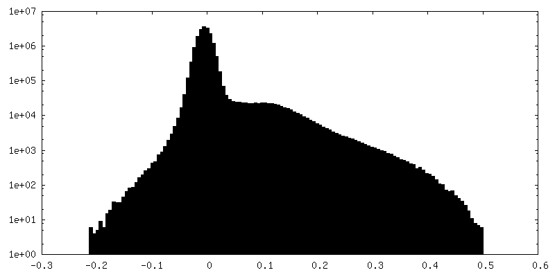
| Density Histograms |


-Half map: #1
| File | emd_28153_half_map_2.map | ||||||||||||
|---|---|---|---|---|---|---|---|---|---|---|---|---|---|
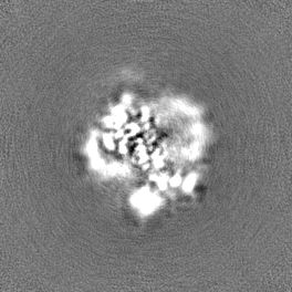
| Projections & Slices |
| ||||||||||||
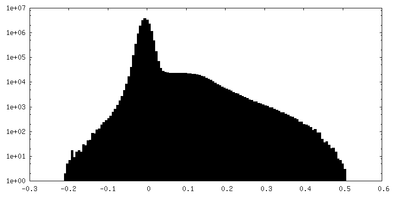
| Density Histograms |


- Sample components
-Entire : TMEM63A purified protein in nanodisc
| Entire | Name: TMEM63A purified protein in nanodisc |
|---|---|
| Components |
|
-Supramolecule #1: TMEM63A purified protein in nanodisc
| Supramolecule | Name: TMEM63A purified protein in nanodisc / type: complex / ID: 1 / Parent: 0 / Macromolecule list: all |
|---|---|
| Source (natural) | Organism: Homo sapiens (human) |
| Molecular weight | Theoretical: 110 KDa |
-Macromolecule #1: CSC1-like protein 1
| Macromolecule | Name: CSC1-like protein 1 / type: protein_or_peptide / ID: 1 / Number of copies: 1 / Enantiomer: LEVO |
|---|---|
| Source (natural) | Organism: Homo sapiens (human) |
| Molecular weight | Theoretical: 93.200992 KDa |
| Recombinant expression | Organism: Homo sapiens (human) |
| Sequence | String: MMDSPFLELW QSKAVSIREQ LGLGDRPNDS YCYNSAKNST VLQGVTFGGI PTVLLIDVSC FLFLILVFSI IRRRFWDYGR IALVSEADS ESRFQRLSST SSSGQQDFEN ELGCCPWLTA IFRLHDDQIL EWCGEDAIHY LSFQRHIIFL LVVVSFLSLC V ILPVNLSG ...String: MMDSPFLELW QSKAVSIREQ LGLGDRPNDS YCYNSAKNST VLQGVTFGGI PTVLLIDVSC FLFLILVFSI IRRRFWDYGR IALVSEADS ESRFQRLSST SSSGQQDFEN ELGCCPWLTA IFRLHDDQIL EWCGEDAIHY LSFQRHIIFL LVVVSFLSLC V ILPVNLSG DLLDKDPYSF GRTTIANLQT DNDLLWLHTI FAVIYLFLTV GFMRHHTQSI KYKEENLVRR TLFITGLPRD AR KETVESH FRDAYPTCEV VDVQLCYNVA KLIYLCKEKK KTEKSLTYYT NLQVKTGQRT LINPKPCGQF CCCEVLGCEW EDA ISYYTR MKDRLLERIT EEERHVQDQP LGMAFVTFQE KSMATYILKD FNACKCQSLQ CKGEPQPSSH SRELYTSKWT VTFA ADPED ICWKNLSIQG LRWWLQWLGI NFTLFLGLFF LTTPSIILST MDKFNVTKPI HALNNPIISQ FFPTLLLWSF SALLP SIVY YSTLLESHWT KSGENQIMMT KVYIFLIFMV LILPSLGLTS LDFFFRWLFD KTSSEASIRL ECVFLPDQGA FFVNYV IAS AFIGNGMELL RLPGLILYTF RMIMAKTAAD RRNVKQNQAF QYEFGAMYAW MLCVFTVIVA YSITCPIIAP FGLIYIL LK HMVDRHNLYF VYLPAKLEKG IHFAAVNQAL AAPILCLFWL YFFSFLRLGM KAPATLFTFL VLLLTILVCL AHTCFGCF K HLSPLNYKTE EPASDKGSEA EAHMPPPFTP YVPRILNGLA SERTALSPQQ QQQQTYGAIH NISGTIPGQC LAQSATGSV AAAPQEATRL EVLFQ UniProtKB: CSC1-like protein 1 |
-Experimental details
-Structure determination
| Method | cryo EM |
|---|---|
 Processing Processing | single particle reconstruction |
| Aggregation state | particle |
-Sample preparation
| Concentration | 2 mg/mL | |||||||||
|---|---|---|---|---|---|---|---|---|---|---|
| Buffer | pH: 7.4 Component:
| |||||||||
| Grid | Model: Quantifoil R1.2/1.3 / Material: GOLD / Mesh: 400 / Support film - Material: CARBON / Support film - topology: HOLEY / Pretreatment - Type: GLOW DISCHARGE / Pretreatment - Time: 60 sec. | |||||||||
| Vitrification | Cryogen name: ETHANE / Chamber humidity: 100 % / Chamber temperature: 279.15 K / Instrument: FEI VITROBOT MARK IV |
- Electron microscopy
| Microscope | FEI TITAN KRIOS |
|---|---|
| Specialist optics | Energy filter - Slit width: 20 eV |
| Image recording | Film or detector model: GATAN K3 (6k x 4k) / Average exposure time: 2.497 sec. / Average electron dose: 50.7 e/Å2 |
| Electron beam | Acceleration voltage: 300 kV / Electron source:  FIELD EMISSION GUN FIELD EMISSION GUN |
| Electron optics | C2 aperture diameter: 50.0 µm / Illumination mode: FLOOD BEAM / Imaging mode: BRIGHT FIELD / Cs: 2.7 mm / Nominal defocus max: 2.0 µm / Nominal defocus min: 1.0 µm / Nominal magnification: 105000 |
| Sample stage | Specimen holder model: FEI TITAN KRIOS AUTOGRID HOLDER / Cooling holder cryogen: NITROGEN |
| Experimental equipment |  Model: Titan Krios / Image courtesy: FEI Company |
-Image processing
| Startup model | Type of model: NONE |
|---|---|
| Final reconstruction | Applied symmetry - Point group: C1 (asymmetric) / Algorithm: BACK PROJECTION / Resolution.type: BY AUTHOR / Resolution: 3.8 Å / Resolution method: FSC 0.143 CUT-OFF / Software - Name: cryoSPARC / Number images used: 64399 |
| Initial angle assignment | Type: MAXIMUM LIKELIHOOD / Software - Name: cryoSPARC (ver. 3.3.2) |
| Final angle assignment | Type: MAXIMUM LIKELIHOOD / Software - Name: cryoSPARC (ver. 3.3.2) |
-Atomic model buiding 1
| Refinement | Space: REAL / Protocol: AB INITIO MODEL |
|---|---|
| Output model | PDB-8ehw: |
