Movie
Movie Controller


+ Open data

- Basic information
| Entry |  | |||||||||
|---|---|---|---|---|---|---|---|---|---|---|
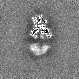
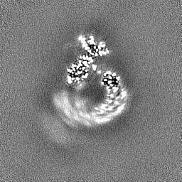
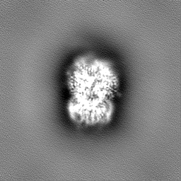
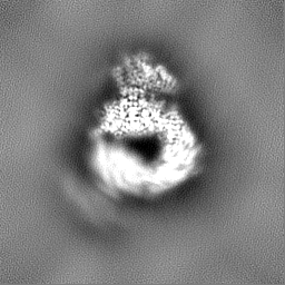
| Title | Structure of focused PtuA(dimer) and PtuB(monomer) complex | |||||||||
 Map data Map data | ||||||||||
 Sample Sample |
| |||||||||
 Keywords Keywords | PtuA / IMMUNE SYSTEM | |||||||||
| Function / homology | Retron Ec78 putative HNH endonuclease-like / TIGR02646 family protein Function and homology information Function and homology information | |||||||||
| Biological species |  | |||||||||
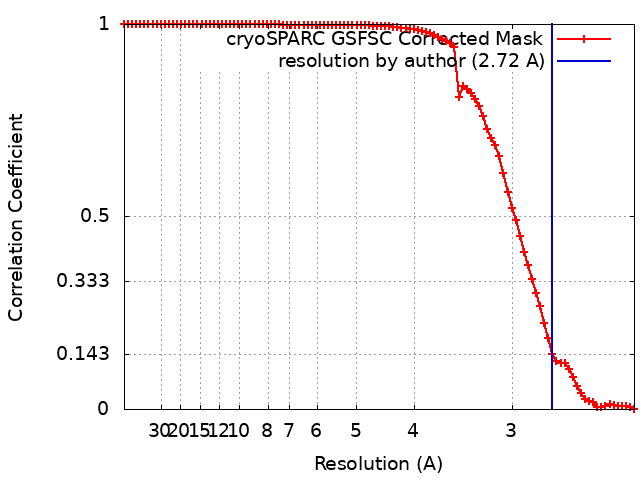
| Method | single particle reconstruction / cryo EM / Resolution: 2.72 Å | |||||||||
 Authors Authors | Shen ZF / Fu TM | |||||||||
| Funding support | 1 items
| |||||||||
 Citation Citation | Journal: Nat Struct Mol Biol / Year: 2024 Title: PtuA and PtuB assemble into an inflammasome-like oligomer for anti-phage defense. Authors: Yuanyuan Li / Zhangfei Shen / Mengyuan Zhang / Xiao-Yuan Yang / Sean P Cleary / Jiale Xie / Ila A Marathe / Marius Kostelic / Jacelyn Greenwald / Anthony D Rish / Vicki H Wysocki / Chong ...Authors: Yuanyuan Li / Zhangfei Shen / Mengyuan Zhang / Xiao-Yuan Yang / Sean P Cleary / Jiale Xie / Ila A Marathe / Marius Kostelic / Jacelyn Greenwald / Anthony D Rish / Vicki H Wysocki / Chong Chen / Qiang Chen / Tian-Min Fu / Yamei Yu /   Abstract: Escherichia coli Septu system, an anti-phage defense system, comprises two components: PtuA and PtuB. PtuA contains an ATPase domain, while PtuB is predicted to function as a nuclease. Here we show ...Escherichia coli Septu system, an anti-phage defense system, comprises two components: PtuA and PtuB. PtuA contains an ATPase domain, while PtuB is predicted to function as a nuclease. Here we show that PtuA and PtuB form a stable complex with a 6:2 stoichiometry. Cryo-electron microscopy structure of PtuAB reveals a distinctive horseshoe-like configuration. PtuA adopts a hexameric arrangement, organized as an asymmetric trimer of dimers, contrasting the ring-like structure by other ATPases. Notably, the three pairs of PtuA dimers assume distinct conformations and fulfill unique roles in recruiting PtuB. Our functional assays have further illuminated the importance of the oligomeric assembly of PtuAB in anti-phage defense. Moreover, we have uncovered that ATP molecules can directly bind to PtuA and inhibit the activities of PtuAB. Together, the assembly and function of the Septu system shed light on understanding other ATPase-containing systems in bacterial immunity. | |||||||||
| History |
|
- Structure visualization
| Supplemental images |
|---|

- Downloads & links
-EMDB archive
| Map data | emd_28048.map.gz | 59.7 MB | EMDB map data format | |
|---|---|---|---|---|
| Header (meta data) | emd-28048-v30.xmlemd-28048.xml | 20 KB 20 KB | Display Display | EMDB header |
| FSC (resolution estimation) | emd_28048_fsc.xml | 8.4 KB | Display | FSC data file |
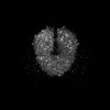
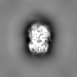
| Images | 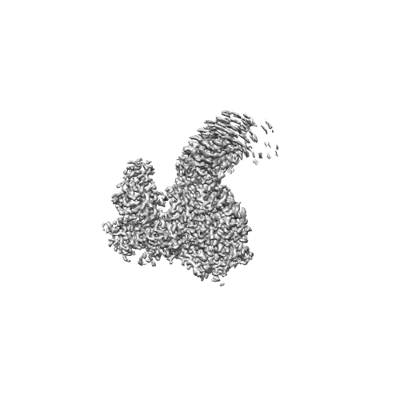 emd_28048.png emd_28048.png | 41.6 KB | ||
| Masks | emd_28048_msk_1.map | 64 MB | Mask map | |
| Filedesc metadata | emd-28048.cif.gz | 6.5 KB | ||
| Others | emd_28048_half_map_1.map.gzemd_28048_half_map_2.map.gz | 59.4 MB 59.4 MB | ||
| Archive directory |  http://ftp.pdbj.org/pub/emdb/structures/EMD-28048ftp://ftp.pdbj.org/pub/emdb/structures/EMD-28048 http://ftp.pdbj.org/pub/emdb/structures/EMD-28048ftp://ftp.pdbj.org/pub/emdb/structures/EMD-28048 | HTTPS FTP |
-Related structure data
| Related structure data | 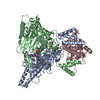 8ee7MC 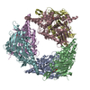 8ee4C 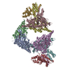 8eeaC 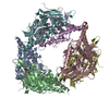 8suxC C: citing same article ( M: atomic model generated by this map |
|---|---|
| Similar structure data |

-Links
| EMDB pages | EMDB (EBI/PDBe) / EMDataResource |
|---|
-Map
| File | Download / File: emd_28048.map.gz / Format: CCP4 / Size: 64 MB / Type: IMAGE STORED AS FLOATING POINT NUMBER (4 BYTES) | ||||||||||||||||||||||||||||||||||||
|---|---|---|---|---|---|---|---|---|---|---|---|---|---|---|---|---|---|---|---|---|---|---|---|---|---|---|---|---|---|---|---|---|---|---|---|---|---|
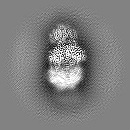
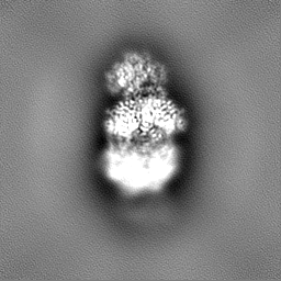
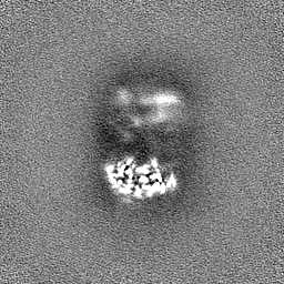
| Projections & slices | Image control
Images are generated by Spider. | ||||||||||||||||||||||||||||||||||||
| Voxel size | X=Y=Z: 1.12 Å | ||||||||||||||||||||||||||||||||||||
| Density |
| ||||||||||||||||||||||||||||||||||||
| Symmetry | Space group: 1 | ||||||||||||||||||||||||||||||||||||
| Details | EMDB XML:
|
 Z (Sec.)
 Y (Row.)
 X (Col.)


-Supplemental data
-Mask #1
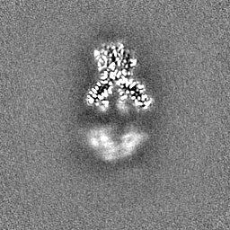
| File | emd_28048_msk_1.map | ||||||||||||
|---|---|---|---|---|---|---|---|---|---|---|---|---|---|
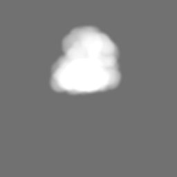
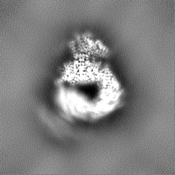
| Projections & Slices |
| ||||||||||||
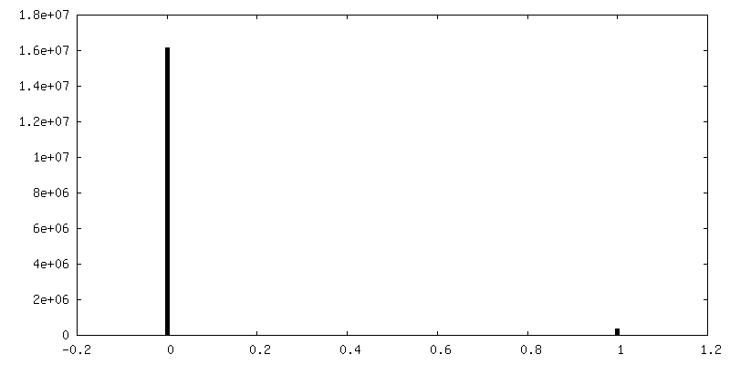
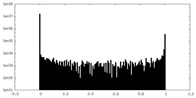
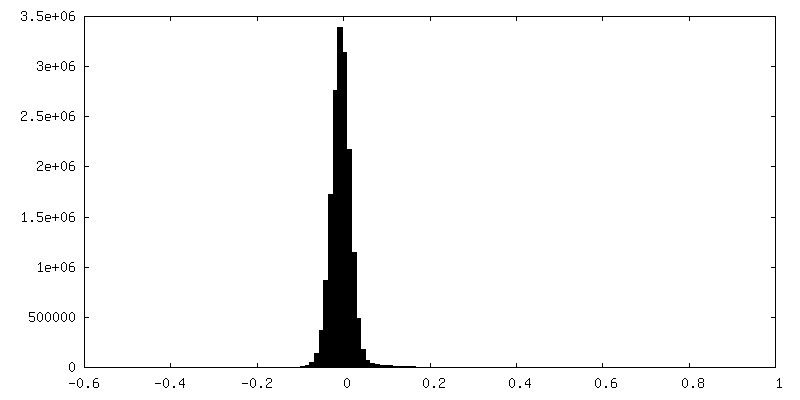
| Density Histograms |


-Half map: #2
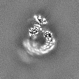
| File | emd_28048_half_map_1.map | ||||||||||||
|---|---|---|---|---|---|---|---|---|---|---|---|---|---|
| Projections & Slices |
| ||||||||||||
| Density Histograms |


-Half map: #1
| File | emd_28048_half_map_2.map | ||||||||||||
|---|---|---|---|---|---|---|---|---|---|---|---|---|---|
| Projections & Slices |
| ||||||||||||
| Density Histograms |

- Sample components
-Entire : 6 ptuA form as a complex
| Entire | Name: 6 ptuA form as a complex |
|---|---|
| Components |
|
-Supramolecule #1: 6 ptuA form as a complex
| Supramolecule | Name: 6 ptuA form as a complex / type: complex / ID: 1 / Parent: 0 / Macromolecule list: #1 |
|---|---|
| Source (natural) | Organism: |
-Macromolecule #1: PtuA
| Macromolecule | Name: PtuA / type: protein_or_peptide / ID: 1 / Number of copies: 2 / Enantiomer: LEVO |
|---|---|
| Source (natural) | Organism: |
| Molecular weight | Theoretical: 53.189656 KDa |
| Recombinant expression | Organism: |
| Sequence | String: MRIDKLSLLN FRCFKQLDIT FDEHITILVA PNGAGKTTVL DAVRLALFPF IRGFDASLYV KDKSLAIRTE DLRLIYRQEA LNMEMSSPA KITATGEWAS GKTATWMLDK RGEQPPHEDK MAAQLTRWGE QLQKRVREEH SLQQVELPLM LYLGTARLWY Q ERYEKQPT ...String: MRIDKLSLLN FRCFKQLDIT FDEHITILVA PNGAGKTTVL DAVRLALFPF IRGFDASLYV KDKSLAIRTE DLRLIYRQEA LNMEMSSPA KITATGEWAS GKTATWMLDK RGEQPPHEDK MAAQLTRWGE QLQKRVREEH SLQQVELPLM LYLGTARLWY Q ERYEKQPT EQRLDNSAFS RLSGYDDCLS ATSNYKQFEQ WYSWLWLSYR EHQITQLESP SAKLKEGVRV QRMKEAIQAI QQ AINCLTQ QVTGWHDLEY SASHNQQLVM SHPQYGKIPL SQLSDGLRNA VAMVADIAFR CVKLNPHLQN DAALKTQGIV LID EVDMFL HPAWQQQIIQ SLRSAFPQIQ FIVTTHSPQV LSTVKRESIR LLEQDENGNG KALMPLGATY GEPSNDVLQS VMGV DPQPA VKEKADLQKL TGWVDQGKYD EPKTQQLMVA LEVALGEKHP QLQRLQRSIA RQRLLKGKAQ |
-Macromolecule #2: PtuB
| Macromolecule | Name: PtuB / type: protein_or_peptide / ID: 2 / Number of copies: 1 / Enantiomer: LEVO |
|---|---|
| Source (natural) | Organism: |
| Molecular weight | Theoretical: 28.136291 KDa |
| Recombinant expression | Organism: |
| Sequence | String: MRHVIKTQLG TVALLTAHEN PPQDADQSTR RWRNFRRDKA AVMVQLINEQ YHLCCYSEIR SDLRGLGYHI EHVENKSQHP ERTFDYQNL AASALDSGEN GGLSSLKGKN AFGGHAQGKQ DVVDMAKFIH CHIRDCSRYF AYLSDGRIVP ADELNAQETE N AQYTIDLL ...String: MRHVIKTQLG TVALLTAHEN PPQDADQSTR RWRNFRRDKA AVMVQLINEQ YHLCCYSEIR SDLRGLGYHI EHVENKSQHP ERTFDYQNL AASALDSGEN GGLSSLKGKN AFGGHAQGKQ DVVDMAKFIH CHIRDCSRYF AYLSDGRIVP ADELNAQETE N AQYTIDLL NLNSGFLQTE RRNHWEELEQ LFDEHIEKDW DLQQLLQLDL VSTPDHKLHE FFSITRQFFQ QEAEQVLQSH AP ALI UniProtKB: TIGR02646 family protein |
-Macromolecule #3: ADENOSINE-5'-TRIPHOSPHATE
| Macromolecule | Name: ADENOSINE-5'-TRIPHOSPHATE / type: ligand / ID: 3 / Number of copies: 1 / Formula: ATP |
|---|---|
| Molecular weight | Theoretical: 507.181 Da |
| Chemical component information |  ChemComp-ATP: |
-Experimental details
-Structure determination
| Method | cryo EM |
|---|---|
 Processing Processing | single particle reconstruction |
| Aggregation state | particle |
-Sample preparation
| Concentration | 1.2 mg/mL |
|---|---|
| Buffer | pH: 7.5 |
| Vitrification | Cryogen name: ETHANE |
- Electron microscopy
| Microscope | TFS KRIOS |
|---|---|
| Image recording | Film or detector model: GATAN K3 (6k x 4k) / Average electron dose: 50.0 e/Å2 |
| Electron beam | Acceleration voltage: 300 kV / Electron source:  FIELD EMISSION GUN FIELD EMISSION GUN |
| Electron optics | Illumination mode: FLOOD BEAM / Imaging mode: BRIGHT FIELD / Cs: 2.7 mm / Nominal defocus max: 2.0 µm / Nominal defocus min: 0.5 µm |
| Experimental equipment |  Model: Titan Krios / Image courtesy: FEI Company |