 Movie
Movie Controller


+ Open data

- Basic information
| Entry |  | |||||||||
|---|---|---|---|---|---|---|---|---|---|---|
| Title | E. coli 50S ribosome bound to compound SAB002 | |||||||||
 Map data Map data | ||||||||||
 Sample Sample |
| |||||||||
 Keywords Keywords | E. coli ribosome / streptogramin A / streptogramin B / antibiotics / RIBOSOME-ANTIBIOTIC complex | |||||||||
| Function / homology |  Function and homology information Function and homology informationDnaA-L2 complex / negative regulation of DNA-templated DNA replication initiation / cytosolic ribosome assembly / ribosome assembly / assembly of large subunit precursor of preribosome / large ribosomal subunit / transferase activity / ribosomal large subunit assembly / large ribosomal subunit rRNA binding / cytosolic large ribosomal subunit ...DnaA-L2 complex / negative regulation of DNA-templated DNA replication initiation / cytosolic ribosome assembly / ribosome assembly / assembly of large subunit precursor of preribosome / large ribosomal subunit / transferase activity / ribosomal large subunit assembly / large ribosomal subunit rRNA binding / cytosolic large ribosomal subunit / cytoplasmic translation / rRNA binding / structural constituent of ribosome / ribosome / translation / ribonucleoprotein complex / response to antibiotic / RNA binding / zinc ion binding / cytoplasm / cytosol Similarity search - Function | |||||||||
| Biological species |  | |||||||||
| Method | single particle reconstruction / cryo EM / Resolution: 2.27 Å | |||||||||
 Authors Authors | Pellegrino J / Lee DJ / Fraser JS / Seiple IB | |||||||||
| Funding support |  United States, 1 items United States, 1 items
| |||||||||
 Citation Citation | Journal: To Be Published Title: Potential for clicked streptogramin analogs Authors: Takahashi Y / Pellegrino J / Lee DJ / Fraser JS / Seiple IB | |||||||||
| History |
|
- Structure visualization
| Supplemental images |
|---|

- Downloads & links
-EMDB archive
| Map data | emd_27857.map.gz | 474.1 MB | EMDB map data format | |
|---|---|---|---|---|
| Header (meta data) | emd-27857-v30.xmlemd-27857.xml | 30.4 KB 30.4 KB | Display Display | EMDB header |
| Images | 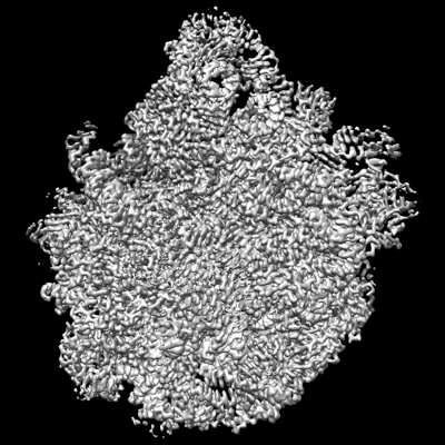 emd_27857.png emd_27857.png | 176.2 KB | ||
| Filedesc metadata | emd-27857.cif.gz | 8.9 KB | ||
| Others | emd_27857_half_map_1.map.gzemd_27857_half_map_2.map.gz | 112.6 MB 112.6 MB | ||
| Archive directory |  http://ftp.pdbj.org/pub/emdb/structures/EMD-27857ftp://ftp.pdbj.org/pub/emdb/structures/EMD-27857 http://ftp.pdbj.org/pub/emdb/structures/EMD-27857ftp://ftp.pdbj.org/pub/emdb/structures/EMD-27857 | HTTPS FTP |
-Related structure data
| Related structure data |  8e35MC  8e30C  8e32C  8e33C  8e36C C: citing same article ( M: atomic model generated by this map |
|---|---|
| Similar structure data |


-Links
| EMDB pages | EMDB (EBI/PDBe) / EMDataResource |
|---|---|
| Related items in Molecule of the Month |


-Map
| File | Download / File: emd_27857.map.gz / Format: CCP4 / Size: 512 MB / Type: IMAGE STORED AS FLOATING POINT NUMBER (4 BYTES) | ||||||||||||||||||||||||||||||||||||
|---|---|---|---|---|---|---|---|---|---|---|---|---|---|---|---|---|---|---|---|---|---|---|---|---|---|---|---|---|---|---|---|---|---|---|---|---|---|
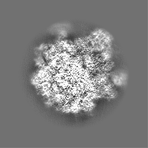
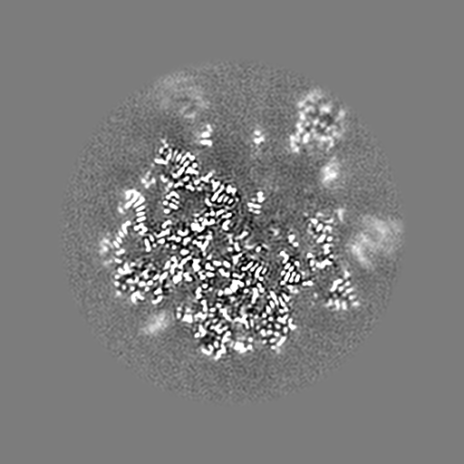
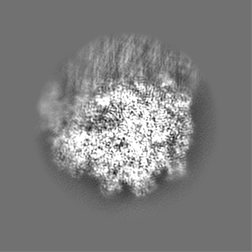
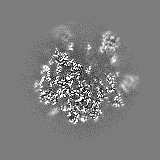
| Projections & slices | Image control
Images are generated by Spider. | ||||||||||||||||||||||||||||||||||||
| Voxel size | X=Y=Z: 0.665 Å | ||||||||||||||||||||||||||||||||||||
| Density |
| ||||||||||||||||||||||||||||||||||||
| Symmetry | Space group: 1 | ||||||||||||||||||||||||||||||||||||
| Details | EMDB XML:
|
 Z (Sec.)
 Y (Row.)
 X (Col.)


-Supplemental data
-Half map: #2
| File | emd_27857_half_map_1.map | ||||||||||||
|---|---|---|---|---|---|---|---|---|---|---|---|---|---|
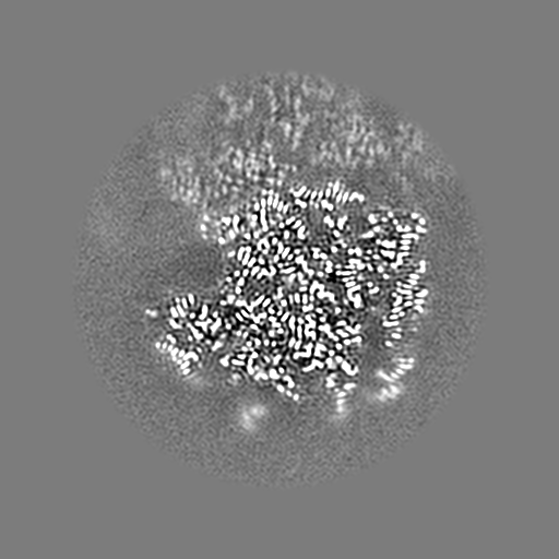
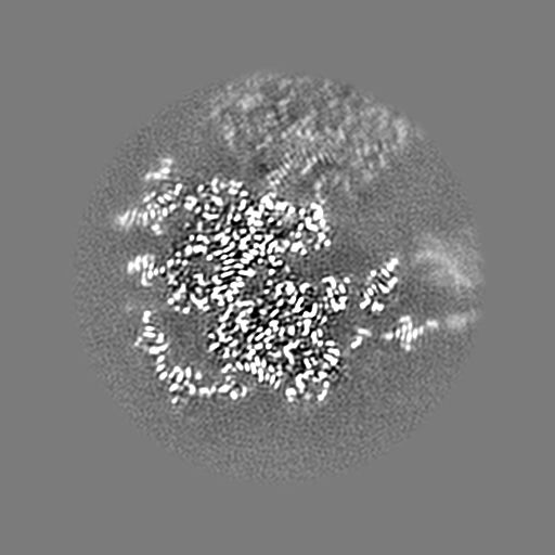
| Projections & Slices |
| ||||||||||||
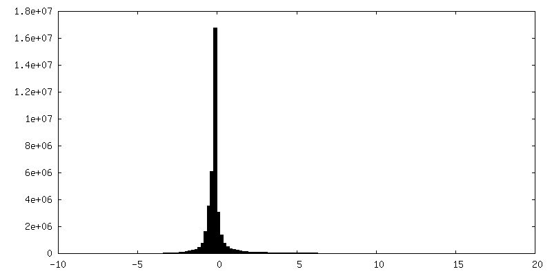
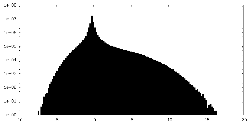
| Density Histograms |


-Half map: #1
| File | emd_27857_half_map_2.map | ||||||||||||
|---|---|---|---|---|---|---|---|---|---|---|---|---|---|
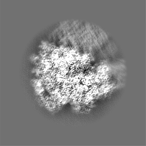
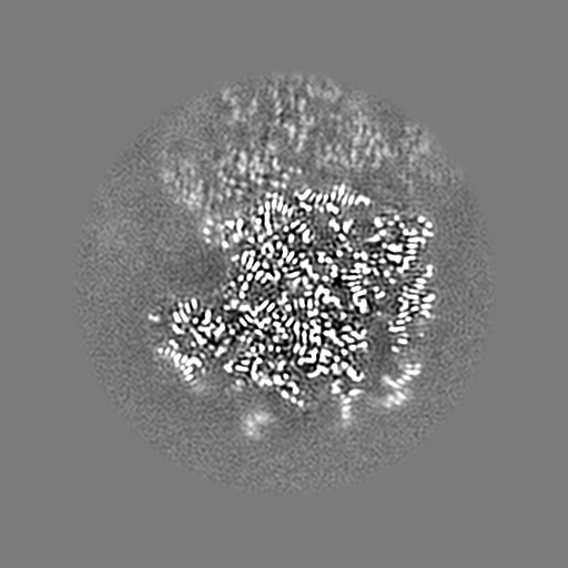
| Projections & Slices |
| ||||||||||||
| Density Histograms |


- Sample components
+Entire : 50S E. coli ribosome
+Supramolecule #1: 50S E. coli ribosome
+Macromolecule #1: 23S ribosomal RNA
+Macromolecule #2: 5S ribosomal RNA
+Macromolecule #3: 50S ribosomal protein L2
+Macromolecule #4: 50S ribosomal protein L15
+Macromolecule #5: 50S ribosomal protein L4
+Macromolecule #6: 50S ribosomal protein L3
+Macromolecule #7: 50S ribosomal protein L13
+Macromolecule #8: 50S ribosomal protein L22
+Macromolecule #9: 50S ribosomal protein L32
+Macromolecule #10: 50S ribosomal protein L34
+Macromolecule #11: N-{(4R,6R,9S,10R,13S,15aS,17R,20R,22S,24aS)-22-benzyl-6-[3-(4-{[(...

+Macromolecule #12: water

-Experimental details
-Structure determination
| Method | cryo EM |
|---|---|
 Processing Processing | single particle reconstruction |
| Aggregation state | particle |
-Sample preparation
| Buffer | pH: 7.5 |
|---|---|
| Vitrification | Cryogen name: ETHANE |
- Electron microscopy
| Microscope | FEI TITAN KRIOS |
|---|---|
| Image recording | Film or detector model: GATAN K3 BIOQUANTUM (6k x 4k) / Average electron dose: 47.2 e/Å2 |
| Electron beam | Acceleration voltage: 300 kV / Electron source:  FIELD EMISSION GUN FIELD EMISSION GUN |
| Electron optics | Illumination mode: FLOOD BEAM / Imaging mode: BRIGHT FIELD / Nominal defocus max: 1.6 µm / Nominal defocus min: 0.6 µm |
| Experimental equipment |  Model: Titan Krios / Image courtesy: FEI Company |
-Image processing
| Startup model | Type of model: NONE |
|---|---|
| Final reconstruction | Resolution.type: BY AUTHOR / Resolution: 2.27 Å / Resolution method: FSC 0.143 CUT-OFF / Number images used: 537105 |
| Initial angle assignment | Type: PROJECTION MATCHING |
| Final angle assignment | Type: PROJECTION MATCHING |
