 Movie
Movie Controller


+ Open data

- Basic information
| Entry |  | |||||||||
|---|---|---|---|---|---|---|---|---|---|---|
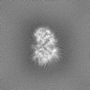
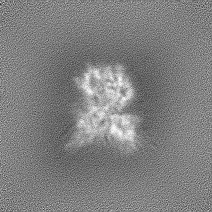
| Title | RNA-free Human Dis3L2 | |||||||||
 Map data Map data | ||||||||||
 Sample Sample |
| |||||||||
 Keywords Keywords | 3'-5' exonuclease / RNA-free exonuclease / human exonuclease / RNA BIND PROTEIN-RNA complex / RNA BINDING PROTEIN-RNA complex | |||||||||
| Function / homology |  Function and homology information Function and homology informationpolyuridylation-dependent mRNA catabolic process / miRNA catabolic process / mitotic sister chromatid separation / Z-decay: degradation of maternal mRNAs by zygotically expressed factors / poly(U) RNA binding / stem cell population maintenance / mRNA catabolic process / nuclear-transcribed mRNA catabolic process / RNA nuclease activity / P-body ...polyuridylation-dependent mRNA catabolic process / miRNA catabolic process / mitotic sister chromatid separation / Z-decay: degradation of maternal mRNAs by zygotically expressed factors / poly(U) RNA binding / stem cell population maintenance / mRNA catabolic process / nuclear-transcribed mRNA catabolic process / RNA nuclease activity / P-body / mitotic cell cycle / Hydrolases; Acting on ester bonds; Exoribonucleases producing 5'-phosphomonoesters / 3'-5'-RNA exonuclease activity / negative regulation of cell population proliferation / cell division / magnesium ion binding / cytoplasm Similarity search - Function | |||||||||
| Biological species |  Homo sapiens (human) Homo sapiens (human) | |||||||||
| Method | single particle reconstruction / cryo EM / Resolution: 3.4 Å | |||||||||
 Authors Authors | Meze K / Thomas DR / Joshua-Tor L | |||||||||
| Funding support |  United States, 2 items United States, 2 items
| |||||||||
 Citation Citation | Journal: Nat Struct Mol Biol / Year: 2023 Title: A shape-shifting nuclease unravels structured RNA. Authors: Katarina Meze / Armend Axhemi / Dennis R Thomas / Ahmet Doymaz / Leemor Joshua-Tor / Abstract: RNA turnover pathways ensure appropriate gene expression levels by eliminating unwanted transcripts. Dis3-like 2 (Dis3L2) is a 3'-5' exoribonuclease that plays a critical role in human development. ...RNA turnover pathways ensure appropriate gene expression levels by eliminating unwanted transcripts. Dis3-like 2 (Dis3L2) is a 3'-5' exoribonuclease that plays a critical role in human development. Dis3L2 independently degrades structured substrates, including coding and noncoding 3' uridylated RNAs. While the basis for Dis3L2's substrate recognition has been well characterized, the mechanism of structured RNA degradation by this family of enzymes is unknown. We characterized the discrete steps of the degradation cycle by determining cryogenic electron microscopy structures representing snapshots along the RNA turnover pathway and measuring kinetic parameters for RNA processing. We discovered a dramatic conformational change that is triggered by double-stranded RNA (dsRNA), repositioning two cold shock domains by 70 Å. This movement exposes a trihelix linker region, which acts as a wedge to separate the two RNA strands. Furthermore, we show that the trihelix linker is critical for dsRNA, but not single-stranded RNA, degradation. These findings reveal the conformational plasticity of Dis3L2 and detail a mechanism of structured RNA degradation. | |||||||||
| History |
|
- Structure visualization
| Supplemental images |
|---|

- Downloads & links
-EMDB archive
| Map data | emd_27827.map.gz | 96.9 MB | EMDB map data format | |
|---|---|---|---|---|
| Header (meta data) | emd-27827-v30.xmlemd-27827.xml | 19.9 KB 19.9 KB | Display Display | EMDB header |
| Images | 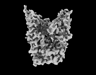 emd_27827.png emd_27827.png | 49.7 KB | ||
| Filedesc metadata | emd-27827.cif.gz | 6.7 KB | ||
| Others | emd_27827_half_map_1.map.gzemd_27827_half_map_2.map.gz | 95.2 MB 95.2 MB | ||
| Archive directory |  http://ftp.pdbj.org/pub/emdb/structures/EMD-27827ftp://ftp.pdbj.org/pub/emdb/structures/EMD-27827 http://ftp.pdbj.org/pub/emdb/structures/EMD-27827ftp://ftp.pdbj.org/pub/emdb/structures/EMD-27827 | HTTPS FTP |
-Related structure data
| Related structure data |  8e27MC  8e28C  8e29C  8e2aC M: atomic model generated by this map C: citing same article ( |
|---|---|
| Similar structure data |

-Links
| EMDB pages | EMDB (EBI/PDBe) / EMDataResource |
|---|
-Map
| File | Download / File: emd_27827.map.gz / Format: CCP4 / Size: 103 MB / Type: IMAGE STORED AS FLOATING POINT NUMBER (4 BYTES) | ||||||||||||||||||||||||||||||||||||
|---|---|---|---|---|---|---|---|---|---|---|---|---|---|---|---|---|---|---|---|---|---|---|---|---|---|---|---|---|---|---|---|---|---|---|---|---|---|
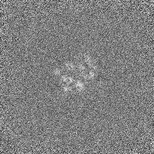
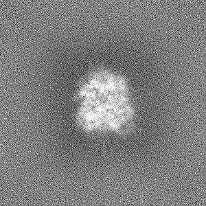
| Projections & slices | Image control
Images are generated by Spider. | ||||||||||||||||||||||||||||||||||||
| Voxel size | X=Y=Z: 0.66 Å | ||||||||||||||||||||||||||||||||||||
| Density |
| ||||||||||||||||||||||||||||||||||||
| Symmetry | Space group: 1 | ||||||||||||||||||||||||||||||||||||
| Details | EMDB XML:
|
 Z (Sec.)
 Y (Row.)
 X (Col.)


-Supplemental data
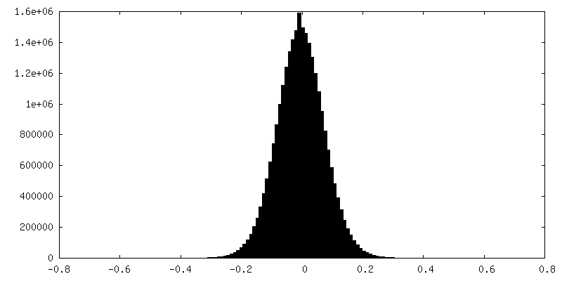
-Half map: #2
| File | emd_27827_half_map_1.map | ||||||||||||
|---|---|---|---|---|---|---|---|---|---|---|---|---|---|
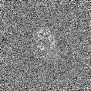
| Projections & Slices |
| ||||||||||||
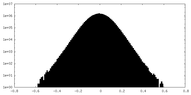
| Density Histograms |


-Half map: #1
| File | emd_27827_half_map_2.map | ||||||||||||
|---|---|---|---|---|---|---|---|---|---|---|---|---|---|
| Projections & Slices |
| ||||||||||||
| Density Histograms |

- Sample components
-Entire : Inactive RNA-free HsDis3L2 (D391N)
| Entire | Name: Inactive RNA-free HsDis3L2 (D391N) |
|---|---|
| Components |
|
-Supramolecule #1: Inactive RNA-free HsDis3L2 (D391N)
| Supramolecule | Name: Inactive RNA-free HsDis3L2 (D391N) / type: complex / ID: 1 / Parent: 0 / Macromolecule list: all |
|---|---|
| Source (natural) | Organism: Homo sapiens (human) |
| Molecular weight | Theoretical: 100 KDa |
-Macromolecule #1: DIS3-like exonuclease 2
| Macromolecule | Name: DIS3-like exonuclease 2 / type: protein_or_peptide / ID: 1 / Number of copies: 1 / Enantiomer: LEVO EC number: Hydrolases; Acting on ester bonds; Exoribonucleases producing 5'-phosphomonoesters |
|---|---|
| Source (natural) | Organism: Homo sapiens (human) |
| Molecular weight | Theoretical: 96.54332 KDa |
| Recombinant expression | Organism:   Spodoptera frugiperda (fall armyworm) Spodoptera frugiperda (fall armyworm) |
| Sequence | String: MSHPDYRMNL RPLGTPRGVS AVAGPHDIGA SPGDKKSKNR STRGKKKSIF ETYMSKEDVS EGLKRGTLIQ GVLRINPKKF HEAFIPSPD GDRDIFIDGV VARNRALNGD LVVVKLLPEE HWKVVKPESN DKETEAAYES DIPEELCGHH LPQQSLKSYN D SPDVIVEA ...String: MSHPDYRMNL RPLGTPRGVS AVAGPHDIGA SPGDKKSKNR STRGKKKSIF ETYMSKEDVS EGLKRGTLIQ GVLRINPKKF HEAFIPSPD GDRDIFIDGV VARNRALNGD LVVVKLLPEE HWKVVKPESN DKETEAAYES DIPEELCGHH LPQQSLKSYN D SPDVIVEA QFDGSDSEDG HGITQNVLVD GVKKLSVCVS EKGREDGDAP VTKDETTCIS QDTRALSEKS LQRSAKVVYI LE KKHSRAA TGFLKLLADK NSELFRKYAL FSPSDHRVPR IYVPLKDCPQ DFVARPKDYA NTLFICRIVD WKEDCNFALG QLA KSLGQA GEIEPETEGI LTEYGVDFSD FSSEVLECLP QGLPWTIPPE EFSKRRDLRK DCIFTIDPST ARDLNDALSC KPLA DGNFK VGVHIADVSY FVPEGSDLDK VAAERATSVY LVQKVVPMLP RLLCEELCSL NPMSDKLTFS VIWTLTPEGK ILDEW FGRT IIRSCTKLSY EHAQSMIESP TEKIPAKELP PISPEHSSEE VHQAVLNLHG IAKQLRQQRF VDGALRLDQL KLAFTL DHE TGLPQGCHIY EYRESNKLVE EFMLLANMAV AHKIHRAFPE QALLRRHPPP QTRMLSDLVE FCDQMGLPVD FSSAGAL NK SLTQTFGDDK YSLARKEVLT NMCSRPMQMA LYFCSGLLQD PAQFRHYALN VPLYTHFTSP IRRFADVLVH RLLAAALG Y RERLDMAPDT LQKQADHCND RRMASKRVQE LSTSLFFAVL VKESGPLESE AMVMGILKQA FDVLVLRYGV QKRIYCNAL ALRSHHFQKV GKKPELTLVW EPEDMEQEPA QQVITIFSLV EVVLQAESTA LKYSAILK UniProtKB: DIS3-like exonuclease 2 |
-Experimental details
-Structure determination
| Method | cryo EM |
|---|---|
 Processing Processing | single particle reconstruction |
| Aggregation state | particle |
-Sample preparation
| Concentration | 0.3 mg/mL | ||||||||||||
|---|---|---|---|---|---|---|---|---|---|---|---|---|---|
| Buffer | pH: 7.5 Component:
| ||||||||||||
| Grid | Model: UltrAuFoil R1.2/1.3 / Material: GOLD / Mesh: 300 / Support film - Material: GOLD / Support film - topology: HOLEY ARRAY / Pretreatment - Type: GLOW DISCHARGE / Pretreatment - Time: 30 sec. / Pretreatment - Atmosphere: AIR / Pretreatment - Pressure: 0.042 kPa | ||||||||||||
| Vitrification | Cryogen name: ETHANE / Chamber humidity: 90 % / Chamber temperature: 295 K / Instrument: FEI VITROBOT MARK IV |
- Electron microscopy
| Microscope | FEI TITAN KRIOS |
|---|---|
| Temperature | Min: 80.0 K / Max: 80.0 K |
| Image recording | Film or detector model: GATAN K2 QUANTUM (4k x 4k) / Detector mode: COUNTING / Digitization - Dimensions - Width: 3840 pixel / Digitization - Dimensions - Height: 3712 pixel / Digitization - Frames/image: 1-25 / Number grids imaged: 1 / Number real images: 4095 / Average exposure time: 5.0 sec. / Average electron dose: 51.75 e/Å2 |
| Electron beam | Acceleration voltage: 300 kV / Electron source:  FIELD EMISSION GUN FIELD EMISSION GUN |
| Electron optics | C2 aperture diameter: 70.0 µm / Calibrated defocus max: 2.8000000000000003 µm / Calibrated defocus min: 0.5 µm / Illumination mode: FLOOD BEAM / Imaging mode: BRIGHT FIELD / Cs: 2.7 mm / Nominal defocus max: 2.6 µm / Nominal defocus min: 0.7000000000000001 µm / Nominal magnification: 160000 |
| Sample stage | Specimen holder model: FEI TITAN KRIOS AUTOGRID HOLDER / Cooling holder cryogen: NITROGEN |
| Experimental equipment |  Model: Titan Krios / Image courtesy: FEI Company |
+Image processing
-Atomic model buiding 1
| Initial model | PDB ID: Chain - Chain ID: A / Chain - Source name: PDB / Chain - Initial model type: experimental model |
|---|---|
| Details | Chain A of PDB 4PMW (MmDis3L2) was fit into the map via rigid body fitting. Residues were then mutated to match the human protein and manual building done to correct for differences. Next rounds of refinement and building were done in PHENIX and Coot, respectively, until the final structure was obtained. |
| Refinement | Space: REAL / Protocol: FLEXIBLE FIT |
| Output model | PDB-8e27: |

