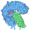
+ Open data
- Basic information
| Entry |  | |||||||||||||||
|---|---|---|---|---|---|---|---|---|---|---|---|---|---|---|---|---|
| Title | Symmetry expansion of dimeric LRRK1 | |||||||||||||||
 Map data Map data | ||||||||||||||||
 Sample Sample |
| |||||||||||||||
 Keywords Keywords | dimer / TRANSFERASE | |||||||||||||||
| Function / homology |  Function and homology information Function and homology informationosteoclast development / positive regulation of intracellular signal transduction / bone resorption / positive regulation of canonical Wnt signaling pathway / non-specific serine/threonine protein kinase / intracellular signal transduction / protein serine kinase activity / protein serine/threonine kinase activity / GTP binding / mitochondrion ...osteoclast development / positive regulation of intracellular signal transduction / bone resorption / positive regulation of canonical Wnt signaling pathway / non-specific serine/threonine protein kinase / intracellular signal transduction / protein serine kinase activity / protein serine/threonine kinase activity / GTP binding / mitochondrion / ATP binding / metal ion binding / identical protein binding / plasma membrane / cytosol Similarity search - Function | |||||||||||||||
| Biological species |  Homo sapiens (human) Homo sapiens (human) | |||||||||||||||
| Method | single particle reconstruction / cryo EM / Resolution: 4.3 Å | |||||||||||||||
 Authors Authors | Reimer JM / Lin YX / Leschziner AE | |||||||||||||||
| Funding support |  United States, 4 items United States, 4 items
| |||||||||||||||
 Citation Citation |  Journal: Nat Struct Mol Biol / Year: 2023 Journal: Nat Struct Mol Biol / Year: 2023Title: Structure of LRRK1 and mechanisms of autoinhibition and activation. Authors: Janice M Reimer / Andrea M Dickey / Yu Xuan Lin / Robert G Abrisch / Sebastian Mathea / Deep Chatterjee / Elizabeth J Fay / Stefan Knapp / Matthew D Daugherty / Samara L Reck-Peterson / Andres E Leschziner /   Abstract: Leucine Rich Repeat Kinase 1 and 2 (LRRK1 and LRRK2) are homologs in the ROCO family of proteins in humans. Despite their shared domain architecture and involvement in intracellular trafficking, ...Leucine Rich Repeat Kinase 1 and 2 (LRRK1 and LRRK2) are homologs in the ROCO family of proteins in humans. Despite their shared domain architecture and involvement in intracellular trafficking, their disease associations are strikingly different: LRRK2 is involved in familial Parkinson's disease while LRRK1 is linked to bone diseases. Furthermore, Parkinson's disease-linked mutations in LRRK2 are typically autosomal dominant gain-of-function while those in LRRK1 are autosomal recessive loss-of-function. Here, to understand these differences, we solved cryo-EM structures of LRRK1 in its monomeric and dimeric forms. Both differ from the corresponding LRRK2 structures. Unlike LRRK2, which is sterically autoinhibited as a monomer, LRRK1 is sterically autoinhibited in a dimer-dependent manner. LRRK1 has an additional level of autoinhibition that prevents activation of the kinase and is absent in LRRK2. Finally, we place the structural signatures of LRRK1 and LRRK2 in the context of the evolution of the LRRK family of proteins. | |||||||||||||||
| History |
|
- Structure visualization
| Supplemental images |
|---|
- Downloads & links
-EMDB archive
| Map data |  emd_27818.map.gz emd_27818.map.gz | 86.1 MB |  EMDB map data format EMDB map data format | |
|---|---|---|---|---|
| Header (meta data) |  emd-27818-v30.xml emd-27818-v30.xml emd-27818.xml emd-27818.xml | 17.2 KB 17.2 KB | Display Display |  EMDB header EMDB header |
| FSC (resolution estimation) |  emd_27818_fsc.xml emd_27818_fsc.xml | 10.8 KB | Display |  FSC data file FSC data file |
| Images |  emd_27818.png emd_27818.png | 50.3 KB | ||
| Filedesc metadata |  emd-27818.cif.gz emd-27818.cif.gz | 6.7 KB | ||
| Others |  emd_27818_half_map_1.map.gz emd_27818_half_map_1.map.gz emd_27818_half_map_2.map.gz emd_27818_half_map_2.map.gz | 84.4 MB 84.4 MB | ||
| Archive directory |  http://ftp.pdbj.org/pub/emdb/structures/EMD-27818 http://ftp.pdbj.org/pub/emdb/structures/EMD-27818 ftp://ftp.pdbj.org/pub/emdb/structures/EMD-27818 ftp://ftp.pdbj.org/pub/emdb/structures/EMD-27818 | HTTPS FTP |
-Validation report
| Summary document |  emd_27818_validation.pdf.gz emd_27818_validation.pdf.gz | 1.1 MB | Display |  EMDB validaton report EMDB validaton report |
|---|---|---|---|---|
| Full document |  emd_27818_full_validation.pdf.gz emd_27818_full_validation.pdf.gz | 1.1 MB | Display | |
| Data in XML |  emd_27818_validation.xml.gz emd_27818_validation.xml.gz | 17.5 KB | Display | |
| Data in CIF |  emd_27818_validation.cif.gz emd_27818_validation.cif.gz | 22 KB | Display | |
| Arichive directory |  https://ftp.pdbj.org/pub/emdb/validation_reports/EMD-27818 https://ftp.pdbj.org/pub/emdb/validation_reports/EMD-27818 ftp://ftp.pdbj.org/pub/emdb/validation_reports/EMD-27818 ftp://ftp.pdbj.org/pub/emdb/validation_reports/EMD-27818 | HTTPS FTP |
-Related structure data
| Related structure data |  8e06MC  8e04C  8e05C C: citing same article ( M: atomic model generated by this map |
|---|---|
| Similar structure data | Similarity search - Function & homology  F&H Search F&H Search |
- Links
Links
| EMDB pages |  EMDB (EBI/PDBe) / EMDB (EBI/PDBe) /  EMDataResource EMDataResource |
|---|---|
| Related items in Molecule of the Month |
- Map
Map
| File |  Download / File: emd_27818.map.gz / Format: CCP4 / Size: 91.1 MB / Type: IMAGE STORED AS FLOATING POINT NUMBER (4 BYTES) Download / File: emd_27818.map.gz / Format: CCP4 / Size: 91.1 MB / Type: IMAGE STORED AS FLOATING POINT NUMBER (4 BYTES) | ||||||||||||||||||||||||||||||||||||
|---|---|---|---|---|---|---|---|---|---|---|---|---|---|---|---|---|---|---|---|---|---|---|---|---|---|---|---|---|---|---|---|---|---|---|---|---|---|
| Projections & slices | Image control
Images are generated by Spider. | ||||||||||||||||||||||||||||||||||||
| Voxel size | X=Y=Z: 1.16 Å | ||||||||||||||||||||||||||||||||||||
| Density |
| ||||||||||||||||||||||||||||||||||||
| Symmetry | Space group: 1 | ||||||||||||||||||||||||||||||||||||
| Details | EMDB XML:
|
-Supplemental data
-Half map: #2
| File | emd_27818_half_map_1.map | ||||||||||||
|---|---|---|---|---|---|---|---|---|---|---|---|---|---|
| Projections & Slices |
| ||||||||||||
| Density Histograms |
-Half map: #1
| File | emd_27818_half_map_2.map | ||||||||||||
|---|---|---|---|---|---|---|---|---|---|---|---|---|---|
| Projections & Slices |
| ||||||||||||
| Density Histograms |
- Sample components
-Entire : Symmetry expansion of dimeric LRRK1
| Entire | Name: Symmetry expansion of dimeric LRRK1 |
|---|---|
| Components |
|
-Supramolecule #1: Symmetry expansion of dimeric LRRK1
| Supramolecule | Name: Symmetry expansion of dimeric LRRK1 / type: complex / ID: 1 / Parent: 0 / Macromolecule list: #1 |
|---|---|
| Source (natural) | Organism:  Homo sapiens (human) Homo sapiens (human) |
-Macromolecule #1: Leucine-rich repeat serine/threonine-protein kinase 1
| Macromolecule | Name: Leucine-rich repeat serine/threonine-protein kinase 1 / type: protein_or_peptide / ID: 1 / Number of copies: 1 / Enantiomer: LEVO / EC number: non-specific serine/threonine protein kinase |
|---|---|
| Source (natural) | Organism:  Homo sapiens (human) Homo sapiens (human) |
| Molecular weight | Theoretical: 225.739219 KDa |
| Recombinant expression | Organism:  |
| Sequence | String: SMAGMSQRPP SMYWCVGPEE SAVCPERAME TLNGAGDTGG KPSTRGGDPA ARSRRTEGIR AAYRRGDRGG ARDLLEEACD QCASQLEKG QLLSIPAAYG DLEMVRYLLS KRLVELPTEP TDDNPAVVAA YFGHTAVVQE LLESLPGPCS PQRLLNWMLA L ACQRGHLG ...String: SMAGMSQRPP SMYWCVGPEE SAVCPERAME TLNGAGDTGG KPSTRGGDPA ARSRRTEGIR AAYRRGDRGG ARDLLEEACD QCASQLEKG QLLSIPAAYG DLEMVRYLLS KRLVELPTEP TDDNPAVVAA YFGHTAVVQE LLESLPGPCS PQRLLNWMLA L ACQRGHLG VVKLLVLTHG ADPESYAVRK NEFPVIVRLP LYAAIKSGNE DIAIFLLRHG AYFCSYILLD SPDPSKHLLR KY FIEASPL PSSYPGKTAL RVKWSHLRLP WVDLDWLIDI SCQITELDLS ANCLATLPSV IPWGLINLRK LNLSDNHLGE LPG VQSSDE IICSRLLEID ISSNKLSHLP PGFLHLSKLQ KLTASKNCLE KLFEEENATN WIGLRKLQEL DISDNKLTEL PALF LHSFK SLNSLNVSRN NLKVFPDPWA CPLKCCKASR NALECLPDKM AVFWKNHLKD VDFSENALKE VPLGLFQLDA LMFLR LQGN QLAALPPQEK WTCRQLKTLD LSRNQLGKNE DGLKTKRIAF FTTRGRQRSG TEAASVLEFP AFLSESLEVL CLNDNH LDT VPPSVCLLKS LSELYLGNNP GLRELPPELG QLGNLWQLDT EDLTISNVPA EIQKEGPKAM LSYLRAQLRK AEKCKLM KM IIVGPPRQGK STLLEILQTG RAPQVVHGEA TIRTTKWELQ RPAGSRAKVE SVEFNVWDIG GPASMATVNQ CFFTDKAL Y VVVWNLALGE EAVANLQFWL LNIEAKAPNA VVLVVGTHLD LIEAKFRVER IATLRAYVLA LCRSPSGSRA TGFPDITFK HLHEISCKSL EGQEGLRQLI FHVTCSMKDV GSTIGCQRLA GRLIPRSYLS LQEAVLAEQQ RRSRDDDVQY LTDRQLEQLV EQTPDNDIK DYEDLQSAIS FLIETGTLLH FPDTSHGLRN LYFLDPIWLS ECLQRIFNIK GSRSVAKNGV IRAEDLRMLL V GTGFTQQT EEQYFQFLAK FEIALPVAND SYLLPHLLPS KPGLDTHGMR HPTANTIQRV FKMSFVPVGF WQRFIARMLI SL AEMDLQL FENKKNTKSR NRKVTIYSFT GNQRNRCSTF RVKRNQTIYW QEGLLVTFDG GYLSVESSDV NWKKKKSGGM KIV CQSEVR DFSAMAFITD HVNSLIDQWF PALTATESDG TPLMEQYVPC PVCETAWAQH TDPSEKSEDV QYFDMEDCVL TAIE RDFIS CPRHPDLPVP LQELVPELFM TDFPARLFLE NSKLEHSEDE GSVLGQGGSG TVIYRARYQG QPVAVKRFHI KKFKN FANV PADTMLRHLR ATDAMKNFSE FRQEASMLHA LQHPCIVALI GISIHPLCFA LELAPLSSLN TVLSENARDS SFIPLG HML TQKIAYQIAS GLAYLHKKNI IFCDLKSDNI LVWSLDVKEH INIKLSDYGI SRQSFHEGAL GVEGTPGYQA PEIRPRI VY DEKVDMFSYG MVLYELLSGQ RPALGHHQLQ IAKKLSKGIR PVLGQPEEVQ FRRLQALMME CWDTKPEKRP LALSVVSQ M KDPTFATFMY ELCCGKQTAF FSSQGQEYTV VFWDGKEESR NYTVVNTEKG LMEVQRMCCP GMKVSCQLQV QRSLWTATE DQKIYIYTLK GMCPLNTPQQ ALDTPAVVTC FLAVPVIKKN SYLVLAGLAD GLVAVFPVVR GTPKDSCSYL CSHTANRSKF SIADEDARQ NPYPVKAMEV VNSGSEVWYS NGPGLLVIDC ASLEICRRLE PYMAPSMVTS VVCSSEGRGE EVVWCLDDKA N SLVMYHST TYQLCARYFC GVPSPLRDMF PVRPLDTEPP AASHTANPKV PEGDSIADVS IMYSEELGTQ ILIHQESLTD YC SMSSYSS SPPRQAARSP SSLPSSPASS SSVPFSTDCE DSDMLHTPGA ASDRSEHDLT PMDGETFSQH LQAVKILAVR DLI WVPRRG GDVIVIGLEK DSGAQRGRVI AVLKARELTP HGVLVDAAVV AKDTVVCTFE NENTEWCLAV WRGWGAREFD IFYQ SYEEL GRLEACTRKR R UniProtKB: Leucine-rich repeat serine/threonine-protein kinase 1 |
-Macromolecule #2: GUANOSINE-5'-DIPHOSPHATE
| Macromolecule | Name: GUANOSINE-5'-DIPHOSPHATE / type: ligand / ID: 2 / Number of copies: 1 / Formula: GDP |
|---|---|
| Molecular weight | Theoretical: 443.201 Da |
| Chemical component information |  ChemComp-GDP: |
-Experimental details
-Structure determination
| Method | cryo EM |
|---|---|
 Processing Processing | single particle reconstruction |
| Aggregation state | particle |
- Sample preparation
| Buffer | pH: 7.4 |
|---|---|
| Vitrification | Cryogen name: ETHANE |
- Electron microscopy
| Microscope | FEI TALOS ARCTICA |
|---|---|
| Image recording | Film or detector model: GATAN K2 SUMMIT (4k x 4k) / Average electron dose: 55.0 e/Å2 |
| Electron beam | Acceleration voltage: 200 kV / Electron source:  FIELD EMISSION GUN FIELD EMISSION GUN |
| Electron optics | Illumination mode: FLOOD BEAM / Imaging mode: BRIGHT FIELD / Nominal defocus max: 2.124 µm / Nominal defocus min: 1.2630000000000001 µm |
| Experimental equipment |  Model: Talos Arctica / Image courtesy: FEI Company |
 Movie
Movie Controller


 Z (Sec.)
 Y (Row.)
 X (Col.)


