 Movie
Movie Controller

[English] 日本語
 Yorodumi
- EMDB-27637: Structure of EBOV GP lacking the mucin-like domain with 2.1.1D5 s... -
+ Open data

- Basic information
| Entry |  | |||||||||
|---|---|---|---|---|---|---|---|---|---|---|
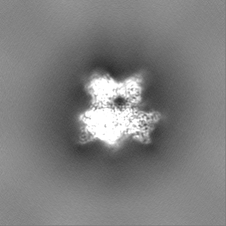
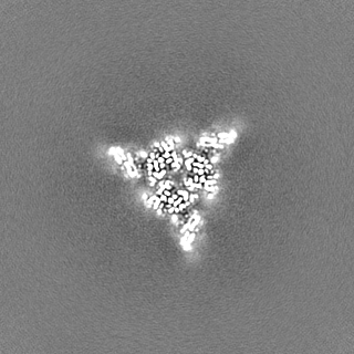
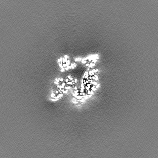
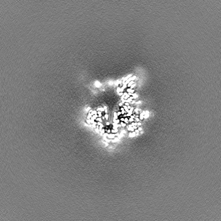
| Title | Structure of EBOV GP lacking the mucin-like domain with 2.1.1D5 scFv and 6D6 scFv bound | |||||||||
 Map data Map data | Structure of EBOV GP lacking the mucin-like domain with 2.1.1D5 scFv and 6D6 scFv bound | |||||||||
 Sample Sample |
| |||||||||
 Keywords Keywords | VIRAL PROTEIN / glycoprotein / immune system / antibody / Ebola virus / VIRAL PROTEIN-Immune System complex | |||||||||
| Function / homology |  Function and homology information Function and homology informationhost cell endoplasmic reticulum / viral budding from plasma membrane / clathrin-dependent endocytosis of virus by host cell / suppression by virus of host tetherin activity / host cell cytoplasm / entry receptor-mediated virion attachment to host cell / symbiont-mediated suppression of host innate immune response / symbiont entry into host cell / membrane raft / fusion of virus membrane with host endosome membrane ...host cell endoplasmic reticulum / viral budding from plasma membrane / clathrin-dependent endocytosis of virus by host cell / suppression by virus of host tetherin activity / host cell cytoplasm / entry receptor-mediated virion attachment to host cell / symbiont-mediated suppression of host innate immune response / symbiont entry into host cell / membrane raft / fusion of virus membrane with host endosome membrane / lipid binding / viral envelope / host cell plasma membrane / virion membrane / extracellular region / identical protein binding / membrane Similarity search - Function | |||||||||
| Biological species |   Ebola virus - Mayinga, Zaire, 1976 / Ebola virus - Mayinga, Zaire, 1976 /  Homo sapiens (human) / Homo sapiens (human) /  | |||||||||
| Method | single particle reconstruction / cryo EM / Resolution: 2.53 Å | |||||||||
 Authors Authors | Yu X / Saphire EO | |||||||||
| Funding support |  United States, 2 items United States, 2 items
| |||||||||
 Citation Citation | Journal: Cell Rep / Year: 2023 Title: The evolution and determinants of neutralization of potent head-binding antibodies against Ebola virus. Authors: Xiaoying Yu / Kathryn M Hastie / Carl W Davis / Ruben Diaz Avalos / Dewight Williams / Diptiben Parekh / Sean Hui / Colin Mann / Chitra Hariharan / Ayato Takada / Rafi Ahmed / Erica Ollmann Saphire /  Abstract: Monoclonal antibodies against the Ebola virus (EBOV) surface glycoprotein are effective treatments for EBOV disease. Antibodies targeting the EBOV glycoprotein (GP) head epitope have potent ...Monoclonal antibodies against the Ebola virus (EBOV) surface glycoprotein are effective treatments for EBOV disease. Antibodies targeting the EBOV glycoprotein (GP) head epitope have potent neutralization and Fc effector function activity and thus are of high interest as therapeutics and for vaccine design. Here we focus on the head-binding antibodies 1A2 and 1D5, which have been identified previously in a longitudinal study of survivors of EBOV infection. 1A2 and 1D5 have the same heavy- and light-chain germlines despite being isolated from different individuals and at different time points after recovery from infection. Cryoelectron microscopy analysis of each antibody in complex with the EBOV surface GP reveals key amino acid substitutions in 1A2 that contribute to greater affinity, improved neutralization potency, and enhanced breadth as well as two strategies for antibody evolution from a common site. | |||||||||
| History |
|
- Structure visualization
| Supplemental images |
|---|

- Downloads & links
-EMDB archive
| Map data | emd_27637.map.gz | 61.9 MB | EMDB map data format | |
|---|---|---|---|---|
| Header (meta data) | emd-27637-v30.xmlemd-27637.xml | 19 KB 19 KB | Display Display | EMDB header |
| Images | 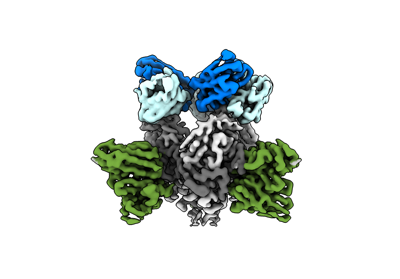 emd_27637.png emd_27637.png | 57 KB | ||
| Filedesc metadata | emd-27637.cif.gz | 6.2 KB | ||
| Others | emd_27637_half_map_1.map.gzemd_27637_half_map_2.map.gz | 115.8 MB 115.8 MB | ||
| Archive directory |  http://ftp.pdbj.org/pub/emdb/structures/EMD-27637ftp://ftp.pdbj.org/pub/emdb/structures/EMD-27637 http://ftp.pdbj.org/pub/emdb/structures/EMD-27637ftp://ftp.pdbj.org/pub/emdb/structures/EMD-27637 | HTTPS FTP |
-Validation report
| Summary document | emd_27637_validation.pdf.gz | 689.6 KB | Display | EMDB validaton report |
|---|---|---|---|---|
| Full document | emd_27637_full_validation.pdf.gz | 689.2 KB | Display | |
| Data in XML | emd_27637_validation.xml.gz | 14 KB | Display | |
| Data in CIF | emd_27637_validation.cif.gz | 16.6 KB | Display | |
| Arichive directory | https://ftp.pdbj.org/pub/emdb/validation_reports/EMD-27637ftp://ftp.pdbj.org/pub/emdb/validation_reports/EMD-27637 | HTTPS FTP |
-Related structure data
| Related structure data |  8dplMC  8dpmC M: atomic model generated by this map C: citing same article ( |
|---|---|
| Similar structure data |

-Links
| EMDB pages | EMDB (EBI/PDBe) / EMDataResource |
|---|---|
| Related items in Molecule of the Month |


-Map
| File | Download / File: emd_27637.map.gz / Format: CCP4 / Size: 125 MB / Type: IMAGE STORED AS FLOATING POINT NUMBER (4 BYTES) | ||||||||||||||||||||
|---|---|---|---|---|---|---|---|---|---|---|---|---|---|---|---|---|---|---|---|---|---|
| Annotation | Structure of EBOV GP lacking the mucin-like domain with 2.1.1D5 scFv and 6D6 scFv bound | ||||||||||||||||||||
| Voxel size | X=Y=Z: 0.99 Å | ||||||||||||||||||||
| Density |
| ||||||||||||||||||||
| Symmetry | Space group: 1 | ||||||||||||||||||||
| Details | EMDB XML:
|
-Supplemental data
-Half map: Half Map 1
| File | emd_27637_half_map_1.map | ||||||||||||
|---|---|---|---|---|---|---|---|---|---|---|---|---|---|
| Annotation | Half Map 1 | ||||||||||||
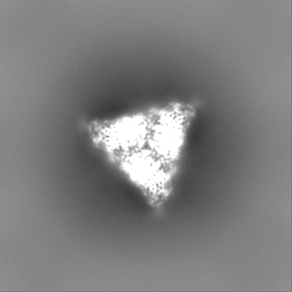
| Projections & Slices |
| ||||||||||||
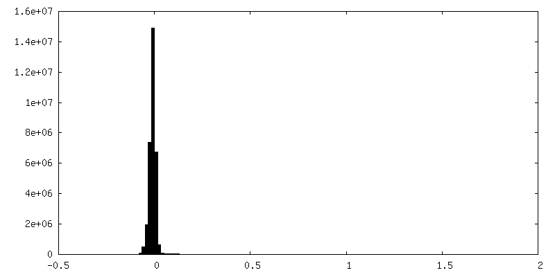
| Density Histograms |
 Z
Z Y
Y X
X


-Half map: Half Map 2
| File | emd_27637_half_map_2.map | ||||||||||||
|---|---|---|---|---|---|---|---|---|---|---|---|---|---|
| Annotation | Half Map 2 | ||||||||||||
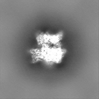
| Projections & Slices |
| ||||||||||||
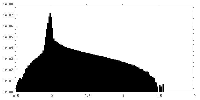
| Density Histograms |
- Sample components
-Entire : Structure of EBOV GP in complex with 2.1.1D5 scFv and 6D6 scFv
| Entire | Name: Structure of EBOV GP in complex with 2.1.1D5 scFv and 6D6 scFv |
|---|---|
| Components |
|
-Supramolecule #1: Structure of EBOV GP in complex with 2.1.1D5 scFv and 6D6 scFv
| Supramolecule | Name: Structure of EBOV GP in complex with 2.1.1D5 scFv and 6D6 scFv type: complex / ID: 1 / Parent: 0 / Macromolecule list: #1-#5 |
|---|---|
| Source (natural) | Organism: Ebola virus - Mayinga, Zaire, 1976 |
-Macromolecule #1: 2.1.1D5 heavy chain variable domain
| Macromolecule | Name: 2.1.1D5 heavy chain variable domain / type: protein_or_peptide / ID: 1 / Number of copies: 3 / Enantiomer: LEVO |
|---|---|
| Source (natural) | Organism: Homo sapiens (human) |
| Molecular weight | Theoretical: 13.057619 KDa |
| Recombinant expression | Organism:  |
| Sequence | String: EVQLVESGGG LVKPGGSLRL SCAASGFTFS NAWMNWVRQA PGKGLEWVGR IKSKTDGGAA DYAAPVKGRF TISRDDSKNT LYLQMNSLK TEDTAVYFCT TVYRYNYDSV WGQGTLVTVS S |
-Macromolecule #2: 2.1.1D5 light chain variable domain
| Macromolecule | Name: 2.1.1D5 light chain variable domain / type: protein_or_peptide / ID: 2 / Number of copies: 3 / Enantiomer: LEVO |
|---|---|
| Source (natural) | Organism: Homo sapiens (human) |
| Molecular weight | Theoretical: 11.738831 KDa |
| Recombinant expression | Organism: |
| Sequence | String: QSVLTQPPSV SGAPGQRVTI SCTGSSSNIG AGYDVYWYQQ LPGTAPKLLI YGNSNRPSGV PDRFSGSKSG TSASLAITGL QAEDEADYY CQSFDSSLRD SWVFGGGTKL TVL |
-Macromolecule #3: 6D6 single-chain variable fragment
| Macromolecule | Name: 6D6 single-chain variable fragment / type: protein_or_peptide / ID: 3 / Number of copies: 3 / Enantiomer: LEVO |
|---|---|
| Source (natural) | Organism: |
| Molecular weight | Theoretical: 25.853459 KDa |
| Recombinant expression | Organism: |
| Sequence | String: QVQLQQSGTE LVKPGASVKL SCKASGYTFT SYWMHWVKQR PGQGLEWIGE INPRNGRTDF SEKFKSKATL TVDTSSSTAF IQLSSLTSE DSAVYYCARW GYYGSSDYWG QGTALTVSSG TGGSGGGGSG GGGSGGGASD IVVTQSHKFM STSVGDRVSI T CKASQDVS ...String: QVQLQQSGTE LVKPGASVKL SCKASGYTFT SYWMHWVKQR PGQGLEWIGE INPRNGRTDF SEKFKSKATL TVDTSSSTAF IQLSSLTSE DSAVYYCARW GYYGSSDYWG QGTALTVSSG TGGSGGGGSG GGGSGGGASD IVVTQSHKFM STSVGDRVSI T CKASQDVS VAVAWYQQKT GQSPKLLIYS ASYRITGVPD RFTGSGSGTD FTFTISSVQA EDMAVYYCQQ HYSTPPWTFG GG TKL |
-Macromolecule #4: Glycoprotein GP1
| Macromolecule | Name: Glycoprotein GP1 / type: protein_or_peptide / ID: 4 / Number of copies: 3 / Enantiomer: LEVO |
|---|---|
| Source (natural) | Organism: Ebola virus - Mayinga, Zaire, 1976 / Strain: Mayinga-76 |
| Molecular weight | Theoretical: 31.297158 KDa |
| Recombinant expression | Organism: |
| Sequence | String: IPLGVIHNST LQVSDVDKLV CRDKLSSTNQ LRSVGLNLEG NGVATDVPSA TKRWGFRSGV PPKVVNYEAG EWAENCYNLE IKKPDGSEC LPAAPDGIRG FPRCRYVHKV SGTGPCAGDF AFHKEGAFFL YDRLASTVIY RGTTFAEGVV AFLILPQAKK D FFSSHPLR ...String: IPLGVIHNST LQVSDVDKLV CRDKLSSTNQ LRSVGLNLEG NGVATDVPSA TKRWGFRSGV PPKVVNYEAG EWAENCYNLE IKKPDGSEC LPAAPDGIRG FPRCRYVHKV SGTGPCAGDF AFHKEGAFFL YDRLASTVIY RGTTFAEGVV AFLILPQAKK D FFSSHPLR EPVNATEDPS SGYYSTTIRY QATGFGTNET EYLFEVDNLT YVQLESRFTP QFLLQLNETI YTSGKRSNTT GK LIWKVNP EIDTTIGEWA FWETKKNLTR KIRSEELSFT VVS UniProtKB: Envelope glycoprotein |
-Macromolecule #5: Glycoprotein GP2
| Macromolecule | Name: Glycoprotein GP2 / type: protein_or_peptide / ID: 5 / Number of copies: 3 / Enantiomer: LEVO |
|---|---|
| Source (natural) | Organism: Ebola virus - Mayinga, Zaire, 1976 |
| Molecular weight | Theoretical: 15.335362 KDa |
| Recombinant expression | Organism: |
| Sequence | String: EAIVNAQPKC NPNLHYWTTQ DEGAAIGLAW IPYFGPAAEG IYTEGLMHNQ DGLICGLRQL ANETTQALQL FLRATTELRT FSILNRKAI DFLLQRWGGT CHILGPDCCI EPHDWTKNIT DKIDQIIHDF VDKTLPD UniProtKB: Envelope glycoprotein |
-Macromolecule #7: 2-acetamido-2-deoxy-beta-D-glucopyranose
| Macromolecule | Name: 2-acetamido-2-deoxy-beta-D-glucopyranose / type: ligand / ID: 7 / Number of copies: 3 / Formula: NAG |
|---|---|
| Molecular weight | Theoretical: 221.208 Da |
| Chemical component information |  ChemComp-NAG: |
-Experimental details
-Structure determination
| Method | cryo EM |
|---|---|
 Processing Processing | single particle reconstruction |
| Aggregation state | particle |
-Sample preparation
| Buffer | pH: 7.4 / Details: TBS buffer pH 7.4 |
|---|---|
| Vitrification | Cryogen name: ETHANE |
- Electron microscopy
| Microscope | FEI TITAN KRIOS |
|---|---|
| Image recording | Film or detector model: GATAN K3 (6k x 4k) / Average electron dose: 50.0 e/Å2 |
| Electron beam | Acceleration voltage: 300 kV / Electron source:  FIELD EMISSION GUN FIELD EMISSION GUN |
| Electron optics | Illumination mode: FLOOD BEAM / Imaging mode: BRIGHT FIELD / Nominal defocus max: 2.5 µm / Nominal defocus min: 1.0 µm |
| Experimental equipment |  Model: Titan Krios / Image courtesy: FEI Company |
-Image processing
| Startup model | Type of model: INSILICO MODEL |
|---|---|
| Final reconstruction | Resolution.type: BY AUTHOR / Resolution: 2.53 Å / Resolution method: FSC 0.143 CUT-OFF / Number images used: 610008 |
| Initial angle assignment | Type: MAXIMUM LIKELIHOOD |
| Final angle assignment | Type: MAXIMUM LIKELIHOOD |
