 Movie
Movie Controller


+ Open data

- Basic information
| Entry |  | |||||||||
|---|---|---|---|---|---|---|---|---|---|---|
| Title | Cryo-EM of the OmcZ nanowires from Geobacter sulfurreducens | |||||||||
 Map data Map data | cryo-EM of OmcZ filament | |||||||||
 Sample Sample |
| |||||||||
 Keywords Keywords | helical symmetry / cytochrome nanowire / filament / ELECTRON TRANSPORT / PROTEIN FIBRIL | |||||||||
| Function / homology | IPT/TIG domain / Multiheme cytochrome superfamily / IPT domain / Immunoglobulin E-set / Immunoglobulin-like fold / Cytochrome c Function and homology information Function and homology information | |||||||||
| Biological species |  Geobacter sulfurreducens (bacteria) Geobacter sulfurreducens (bacteria) | |||||||||
| Method | helical reconstruction / cryo EM / Resolution: 4.2 Å | |||||||||
 Authors Authors | Wang F / Chan CH / Mustafa K / Hochbaum AI / Bond DR / Egelman EH | |||||||||
| Funding support |  United States, 2 items United States, 2 items
| |||||||||
 Citation Citation | Journal: Elife / Year: 2022 Title: Structure of OmcZ filaments suggests extracellular cytochrome polymers evolved independently multiple times. Authors: Fengbin Wang / Chi Ho Chan / Victor Suciu / Khawla Mustafa / Madeline Ammend / Dong Si / Allon I Hochbaum / Edward H Egelman / Daniel R Bond / Abstract: While early genetic and low-resolution structural observations suggested that extracellular conductive filaments on metal-reducing organisms such as were composed of type IV pili, it has now been ...While early genetic and low-resolution structural observations suggested that extracellular conductive filaments on metal-reducing organisms such as were composed of type IV pili, it has now been established that bacterial -type cytochromes can polymerize to form extracellular filaments capable of long-range electron transport. Atomic structures exist for two such cytochrome filaments, formed from the hexaheme cytochrome OmcS and the tetraheme cytochrome OmcE. Due to the highly conserved heme packing within the central OmcS and OmcE cores, and shared pattern of heme coordination between subunits, it has been suggested that these polymers have a common origin. We have now used cryo-electron microscopy (cryo-EM) to determine the structure of a third extracellular filament, formed from the octaheme cytochrome, OmcZ. In contrast to the linear heme chains in OmcS and OmcE from the same organism, the packing of hemes, heme:heme angles, and between-subunit heme coordination is quite different in OmcZ. A branched heme arrangement within OmcZ leads to a highly surface exposed heme in every subunit, which may account for the formation of conductive biofilm networks, and explain the higher measured conductivity of OmcZ filaments. This new structural evidence suggests that conductive cytochrome polymers arose independently on more than one occasion from different ancestral multiheme proteins. | |||||||||
| History |
|
- Structure visualization
| Supplemental images |
|---|

- Downloads & links
-EMDB archive
| Map data | emd_27266.map.gz | 37.1 MB | EMDB map data format | |
|---|---|---|---|---|
| Header (meta data) | emd-27266-v30.xmlemd-27266.xml | 16.7 KB 16.7 KB | Display Display | EMDB header |
| Images |  emd_27266.png emd_27266.png | 92.4 KB | ||
| Filedesc metadata | emd-27266.cif.gz | 5.8 KB | ||
| Others | emd_27266_half_map_1.map.gzemd_27266_half_map_2.map.gz | 200.3 MB 200.3 MB | ||
| Archive directory |  http://ftp.pdbj.org/pub/emdb/structures/EMD-27266ftp://ftp.pdbj.org/pub/emdb/structures/EMD-27266 http://ftp.pdbj.org/pub/emdb/structures/EMD-27266ftp://ftp.pdbj.org/pub/emdb/structures/EMD-27266 | HTTPS FTP |
-Related structure data
| Related structure data |  8d9mMC M: atomic model generated by this map C: citing same article ( |
|---|---|
| Similar structure data |
-Links
| EMDB pages | EMDB (EBI/PDBe) / EMDataResource |
|---|---|
| Related items in Molecule of the Month |


-Map
| File | Download / File: emd_27266.map.gz / Format: CCP4 / Size: 216 MB / Type: IMAGE STORED AS FLOATING POINT NUMBER (4 BYTES) | ||||||||||||||||||||||||||||||||||||
|---|---|---|---|---|---|---|---|---|---|---|---|---|---|---|---|---|---|---|---|---|---|---|---|---|---|---|---|---|---|---|---|---|---|---|---|---|---|
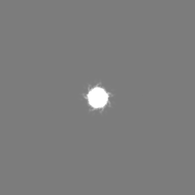
| Annotation | cryo-EM of OmcZ filament | ||||||||||||||||||||||||||||||||||||
| Projections & slices | Image control
Images are generated by Spider. | ||||||||||||||||||||||||||||||||||||
| Voxel size | X=Y=Z: 1.08 Å | ||||||||||||||||||||||||||||||||||||
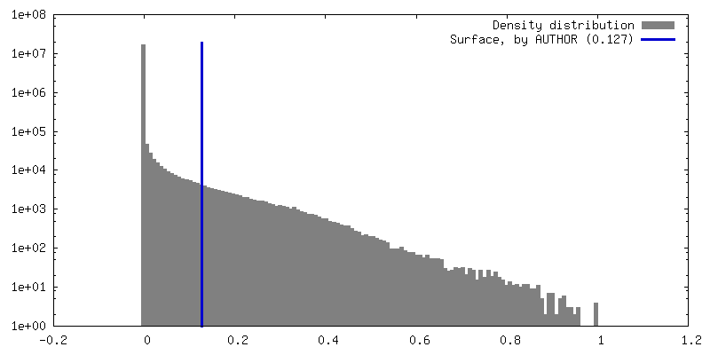
| Density |
| ||||||||||||||||||||||||||||||||||||
| Symmetry | Space group: 1 | ||||||||||||||||||||||||||||||||||||
| Details | EMDB XML:
|
 Z (Sec.)
 Y (Row.)
 X (Col.)


-Supplemental data
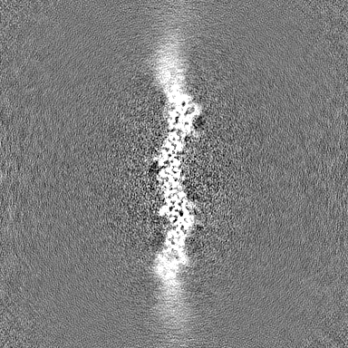
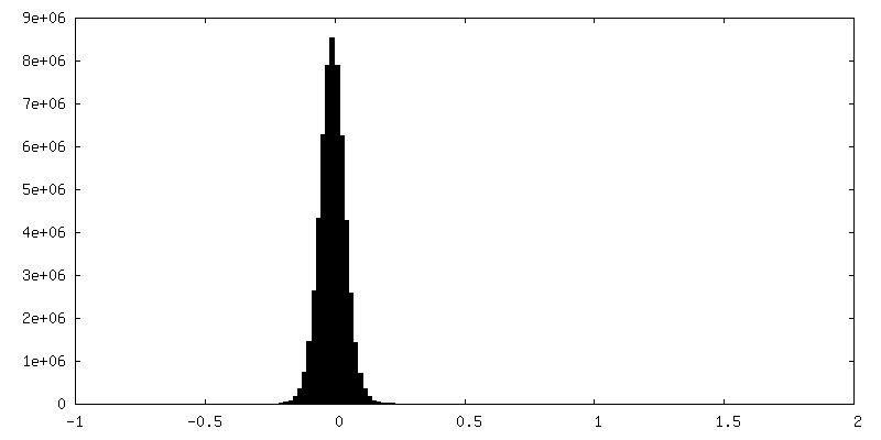
-Half map: half map A
| File | emd_27266_half_map_1.map | ||||||||||||
|---|---|---|---|---|---|---|---|---|---|---|---|---|---|
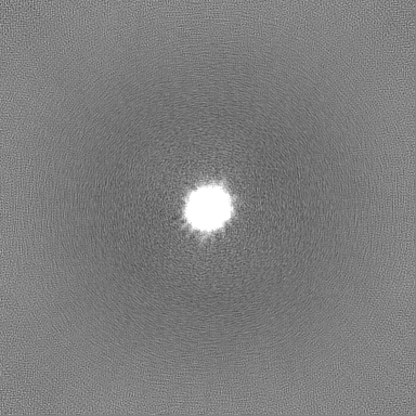
| Annotation | half map A | ||||||||||||
| Projections & Slices |
| ||||||||||||
| Density Histograms |


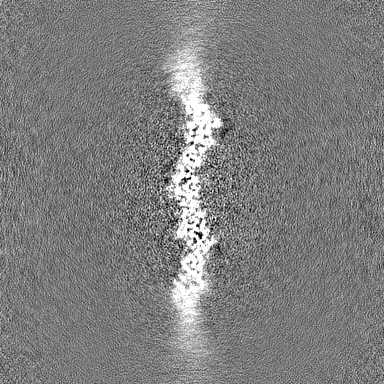
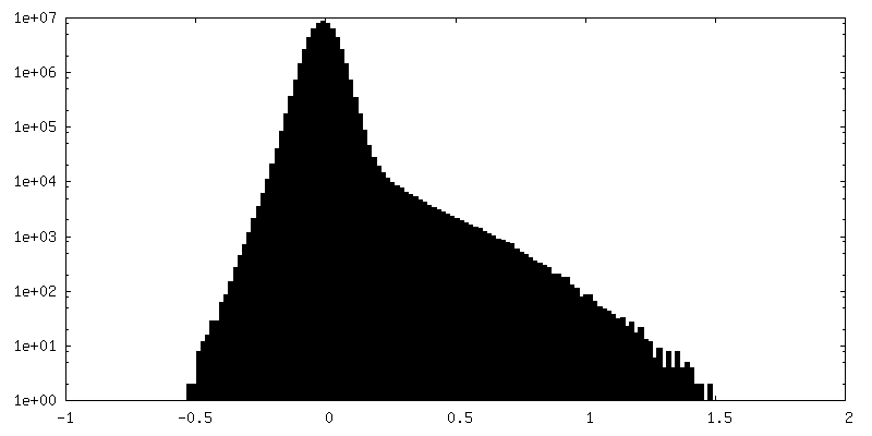
-Half map: half map B
| File | emd_27266_half_map_2.map | ||||||||||||
|---|---|---|---|---|---|---|---|---|---|---|---|---|---|
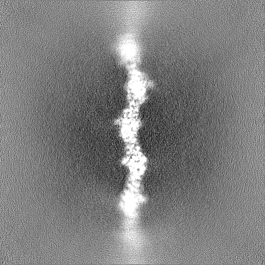
| Annotation | half map B | ||||||||||||
| Projections & Slices |
| ||||||||||||
| Density Histograms |


- Sample components
-Entire : Filament of OmcZ protein
| Entire | Name: Filament of OmcZ protein |
|---|---|
| Components |
|
-Supramolecule #1: Filament of OmcZ protein
| Supramolecule | Name: Filament of OmcZ protein / type: complex / ID: 1 / Parent: 0 / Macromolecule list: #1 |
|---|---|
| Source (natural) | Organism: Geobacter sulfurreducens (bacteria) |
-Macromolecule #1: Cytochrome c
| Macromolecule | Name: Cytochrome c / type: protein_or_peptide / ID: 1 / Number of copies: 1 / Enantiomer: LEVO |
|---|---|
| Source (natural) | Organism: Geobacter sulfurreducens (bacteria) / Strain: ATCC 51573 / DSM 12127 / PCA |
| Molecular weight | Theoretical: 49.424559 KDa |
| Sequence | String: MKKKVLIGAS LAAVVLTGAA MVGAAVPPPP VNQFLGIYDT KFPNLTKADC LECHVSDTVL VQQHHALINT VTPPASCINT SGTVPPTLA TGCHVMVPDG SGGFTFQDFR NCFNCHTQTP HHTSPAAVAK DCKYCHGNFI DNPLDGHYIP TYSASSVTPM P SGRSVTAT ...String: MKKKVLIGAS LAAVVLTGAA MVGAAVPPPP VNQFLGIYDT KFPNLTKADC LECHVSDTVL VQQHHALINT VTPPASCINT SGTVPPTLA TGCHVMVPDG SGGFTFQDFR NCFNCHTQTP HHTSPAAVAK DCKYCHGNFI DNPLDGHYIP TYSASSVTPM P SGRSVTAT DGNVVIVQGC EACHQAAPNA IDPKTNTVRP IFSNQDTHHG TGITDCNLCH NTSSNVPIRQ CEVCHGVNSL HN IQKDSPN AANLGTVKPG LEDLGWGHIG NNWDCQGCHW SWFGNSSPYT NATVPAINGQ SSYTVTAGKE AVLTIVGSSF VNV GPDGVT TYQPTVALVS GSTSLTLTPF SVTESEIKVS VPALVEGVYE LRITKANKVS NLAKLTVAPA RIIASATLAT GKTL TITGT GFGPAPSSEY DAGIGVYAGT TQANVISWSD TKVVATSPDF ATNGYVTVKT INGPLSGKIL AAPKKVKR UniProtKB: Cytochrome c |
-Macromolecule #2: HEME C
| Macromolecule | Name: HEME C / type: ligand / ID: 2 / Number of copies: 8 / Formula: HEC |
|---|---|
| Molecular weight | Theoretical: 618.503 Da |
| Chemical component information |  ChemComp-HEC: |
-Experimental details
-Structure determination
| Method | cryo EM |
|---|---|
 Processing Processing | helical reconstruction |
| Aggregation state | filament |
-Sample preparation
| Buffer | pH: 10.5 |
|---|---|
| Vitrification | Cryogen name: ETHANE |
- Electron microscopy
| Microscope | FEI TITAN KRIOS |
|---|---|
| Image recording | Film or detector model: GATAN K3 (6k x 4k) / Average electron dose: 48.0 e/Å2 |
| Electron beam | Acceleration voltage: 300 kV / Electron source:  FIELD EMISSION GUN FIELD EMISSION GUN |
| Electron optics | Illumination mode: FLOOD BEAM / Imaging mode: BRIGHT FIELD / Nominal defocus max: 2.0 µm / Nominal defocus min: 1.0 µm |
| Experimental equipment |  Model: Titan Krios / Image courtesy: FEI Company |
-Image processing
| Final reconstruction | Applied symmetry - Helical parameters - Δz: 58.1 Å Applied symmetry - Helical parameters - Δ&Phi: -158.2 ° Applied symmetry - Helical parameters - Axial symmetry: C1 (asymmetric) Resolution.type: BY AUTHOR / Resolution: 4.2 Å / Resolution method: FSC 0.143 CUT-OFF / Number images used: 92170 |
|---|---|
| CTF correction | Type: PHASE FLIPPING AND AMPLITUDE CORRECTION |
| Startup model | Type of model: NONE |
| Final angle assignment | Type: NOT APPLICABLE |
