Movie
Movie Controller

[English] 日本語
 Yorodumi
- EMDB-27251: Yeast mitochondrial small subunit assembly intermediate (State 3) -
+ Open data

- Basic information
| Entry |  | |||||||||
|---|---|---|---|---|---|---|---|---|---|---|
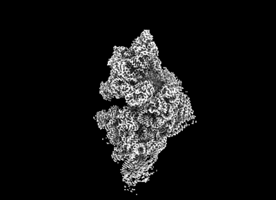
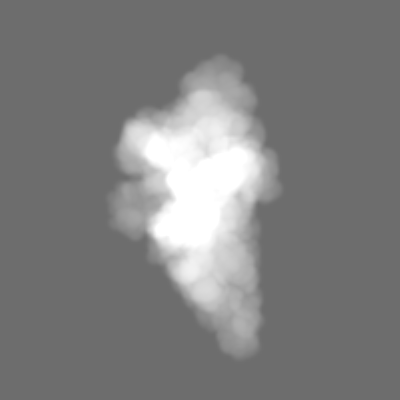
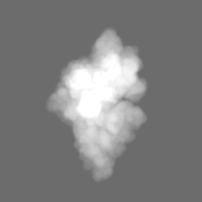
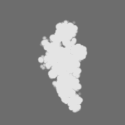
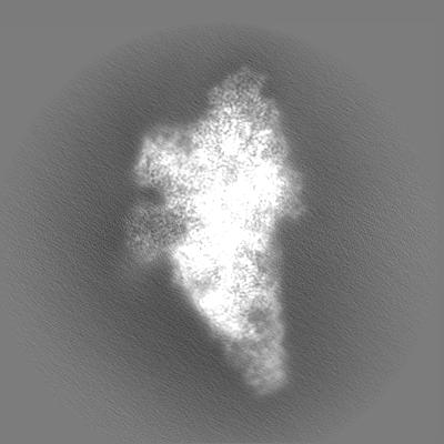
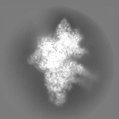
| Title | Yeast mitochondrial small subunit assembly intermediate (State 3) | |||||||||
 Map data Map data | ||||||||||
 Sample Sample |
| |||||||||
| Function / homology |  Function and homology information Function and homology informationBranched-chain amino acid catabolism / 3-hydroxyisobutyryl-CoA hydrolase / 3-hydroxyisobutyryl-CoA hydrolase activity / mitochondrial translational initiation / valine catabolic process / Mitochondrial protein degradation / mitochondrial small ribosomal subunit / mitochondrial ribosome / sporulation resulting in formation of a cellular spore / mitochondrial translation ...Branched-chain amino acid catabolism / 3-hydroxyisobutyryl-CoA hydrolase / 3-hydroxyisobutyryl-CoA hydrolase activity / mitochondrial translational initiation / valine catabolic process / Mitochondrial protein degradation / mitochondrial small ribosomal subunit / mitochondrial ribosome / sporulation resulting in formation of a cellular spore / mitochondrial translation / superoxide dismutase activity / methyltransferase activity / ribosomal small subunit biogenesis / ribosomal small subunit assembly / small ribosomal subunit / small ribosomal subunit rRNA binding / methylation / cytosolic small ribosomal subunit / mitochondrial inner membrane / rRNA binding / hydrolase activity / ribosome / structural constituent of ribosome / translation / mRNA binding / GTP binding / mitochondrion / RNA binding / ATP binding / metal ion binding / cytoplasm / cytosol Similarity search - Function | |||||||||
| Biological species |  | |||||||||
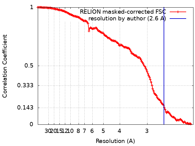
| Method | single particle reconstruction / cryo EM / Resolution: 2.6 Å | |||||||||
 Authors Authors | Burnside C / Harper N / Klinge S | |||||||||
| Funding support |  United States, 2 items United States, 2 items
| |||||||||
 Citation Citation | Journal: Nature / Year: 2023 Title: Principles of mitoribosomal small subunit assembly in eukaryotes. Authors: Nathan J Harper / Chloe Burnside / Sebastian Klinge / Abstract: Mitochondrial ribosomes (mitoribosomes) synthesize proteins encoded within the mitochondrial genome that are assembled into oxidative phosphorylation complexes. Thus, mitoribosome biogenesis is ...Mitochondrial ribosomes (mitoribosomes) synthesize proteins encoded within the mitochondrial genome that are assembled into oxidative phosphorylation complexes. Thus, mitoribosome biogenesis is essential for ATP production and cellular metabolism. Here we used cryo-electron microscopy to determine nine structures of native yeast and human mitoribosomal small subunit assembly intermediates, illuminating the mechanistic basis for how GTPases are used to control early steps of decoding centre formation, how initial rRNA folding and processing events are mediated, and how mitoribosomal proteins have active roles during assembly. Furthermore, this series of intermediates from two species with divergent mitoribosomal architecture uncovers both conserved principles and species-specific adaptations that govern the maturation of mitoribosomal small subunits in eukaryotes. By revealing the dynamic interplay between assembly factors, mitoribosomal proteins and rRNA that are required to generate functional subunits, our structural analysis provides a vignette for how molecular complexity and diversity can evolve in large ribonucleoprotein assemblies. | |||||||||
| History |
|
- Structure visualization
| Supplemental images |
|---|
- Downloads & links
-EMDB archive
| Map data | emd_27251.map.gz | 228.8 MB | EMDB map data format | |
|---|---|---|---|---|
| Header (meta data) | emd-27251-v30.xmlemd-27251.xml | 54.8 KB 54.8 KB | Display Display | EMDB header |
| FSC (resolution estimation) | emd_27251_fsc.xml | 14.1 KB | Display | FSC data file |
| Images |  emd_27251.png emd_27251.png | 51.2 KB | ||
| Masks | emd_27251_msk_1.map | 244.1 MB | Mask map | |
| Others | emd_27251_half_map_1.map.gzemd_27251_half_map_2.map.gz | 200.7 MB 200.1 MB | ||
| Archive directory |  http://ftp.pdbj.org/pub/emdb/structures/EMD-27251ftp://ftp.pdbj.org/pub/emdb/structures/EMD-27251 http://ftp.pdbj.org/pub/emdb/structures/EMD-27251ftp://ftp.pdbj.org/pub/emdb/structures/EMD-27251 | HTTPS FTP |
-Validation report
| Summary document | emd_27251_validation.pdf.gz | 826.5 KB | Display | EMDB validaton report |
|---|---|---|---|---|
| Full document | emd_27251_full_validation.pdf.gz | 826.1 KB | Display | |
| Data in XML | emd_27251_validation.xml.gz | 22.1 KB | Display | |
| Data in CIF | emd_27251_validation.cif.gz | 29.1 KB | Display | |
| Arichive directory | https://ftp.pdbj.org/pub/emdb/validation_reports/EMD-27251ftp://ftp.pdbj.org/pub/emdb/validation_reports/EMD-27251 | HTTPS FTP |
-Related structure data
| Related structure data |  8d8lMC  8cspC  8csqC  8csrC  8cssC  8cstC  8csuC  8d8jC  8d8kC M: atomic model generated by this map C: citing same article ( |
|---|---|
| Similar structure data |


-Links
| EMDB pages | EMDB (EBI/PDBe) / EMDataResource |
|---|---|
| Related items in Molecule of the Month |


-Map
| File | Download / File: emd_27251.map.gz / Format: CCP4 / Size: 244.1 MB / Type: IMAGE STORED AS FLOATING POINT NUMBER (4 BYTES) | ||||||||||||||||||||
|---|---|---|---|---|---|---|---|---|---|---|---|---|---|---|---|---|---|---|---|---|---|
| Voxel size | X=Y=Z: 1.057 Å | ||||||||||||||||||||
| Density |
| ||||||||||||||||||||
| Symmetry | Space group: 1 | ||||||||||||||||||||
| Details | EMDB XML:
|
-Supplemental data
-Mask #1
| File | emd_27251_msk_1.map | ||||||||||||
|---|---|---|---|---|---|---|---|---|---|---|---|---|---|
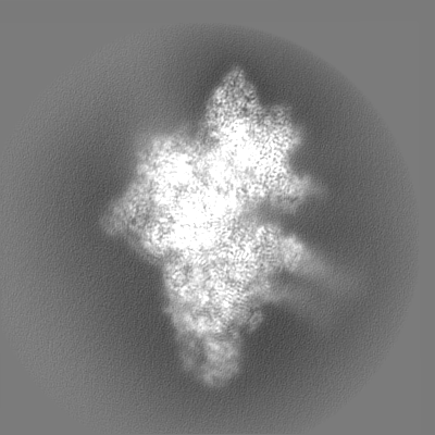
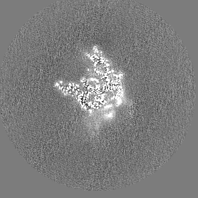
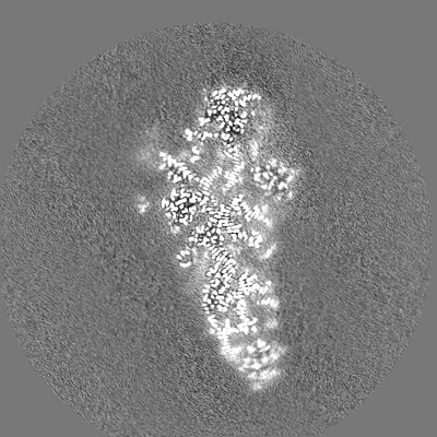
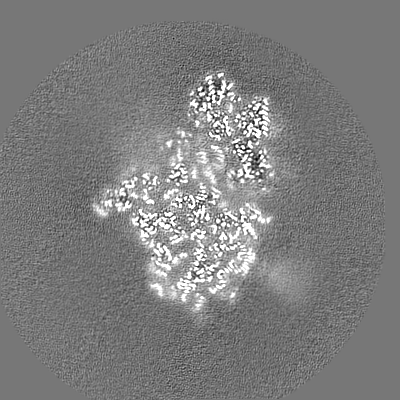
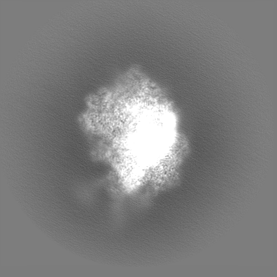
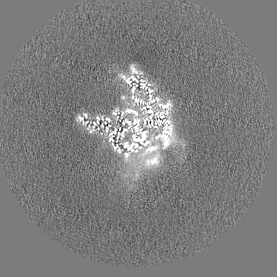
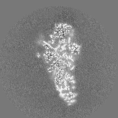
| Projections & Slices |
| ||||||||||||
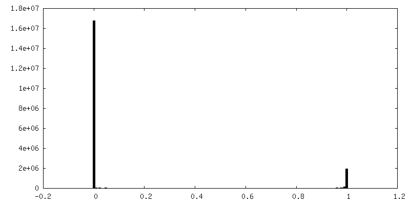
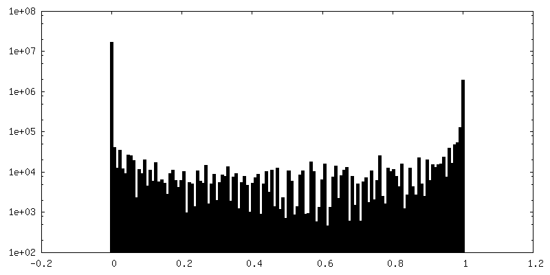
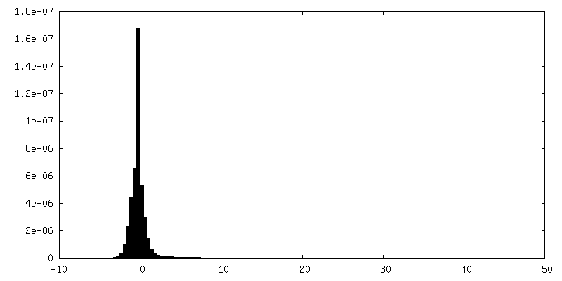
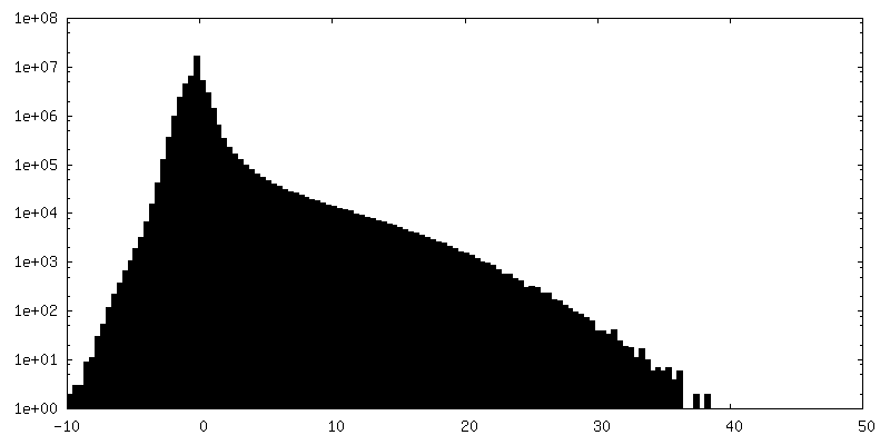
| Density Histograms |
 Z
Z Y
Y X
X


-Half map: #2
| File | emd_27251_half_map_1.map | ||||||||||||
|---|---|---|---|---|---|---|---|---|---|---|---|---|---|
| Projections & Slices |
| ||||||||||||
| Density Histograms |


-Half map: #1
| File | emd_27251_half_map_2.map | ||||||||||||
|---|---|---|---|---|---|---|---|---|---|---|---|---|---|
| Projections & Slices |
| ||||||||||||
| Density Histograms |

- Sample components
+Entire : Yeast mitochondrial small subunit assembly intermediate (State 3)
+Supramolecule #1: Yeast mitochondrial small subunit assembly intermediate (State 3)
+Macromolecule #1: Probable S-adenosyl-L-methionine-dependent RNA methyltransferase ...
+Macromolecule #2: 37S ribosomal protein MRP13, mitochondrial
+Macromolecule #3: Ribosomal protein VAR1, mitochondrial
+Macromolecule #4: 37S ribosomal protein SWS2, mitochondrial
+Macromolecule #5: 37S ribosomal protein MRP2, mitochondrial
+Macromolecule #6: 37S ribosomal protein S28, mitochondrial
+Macromolecule #7: 37S ribosomal protein S16, mitochondrial
+Macromolecule #8: 37S ribosomal protein S17, mitochondrial
+Macromolecule #9: 37S ribosomal protein RSM18, mitochondrial
+Macromolecule #10: 37S ribosomal protein S19, mitochondrial
+Macromolecule #11: 37S ribosomal protein MRP21, mitochondrial
+Macromolecule #12: 37S ribosomal protein S25, mitochondrial
+Macromolecule #13: 37S ribosomal protein PET123, mitochondrial
+Macromolecule #14: 37S ribosomal protein S23, mitochondrial
+Macromolecule #15: Mitochondrial 37S ribosomal protein S27
+Macromolecule #16: 37S ribosomal protein S24, mitochondrial
+Macromolecule #17: 37S ribosomal protein MRP10, mitochondrial
+Macromolecule #18: 37S ribosomal protein MRP51, mitochondrial
+Macromolecule #19: 37S ribosomal protein MRP4, mitochondrial
+Macromolecule #20: Protein FYV4, mitochondrial
+Macromolecule #21: 37S ribosomal protein S26, mitochondrial
+Macromolecule #22: 37S ribosomal protein NAM9, mitochondrial
+Macromolecule #23: 37S ribosomal protein MRP1, mitochondrial
+Macromolecule #24: 37S ribosomal protein S5, mitochondrial
+Macromolecule #25: 37S ribosomal protein MRP17, mitochondrial
+Macromolecule #26: 37S ribosomal protein S35, mitochondrial
+Macromolecule #27: 37S ribosomal protein S7, mitochondrial
+Macromolecule #28: 37S ribosomal protein S8, mitochondrial
+Macromolecule #29: 3-hydroxyisobutyryl-CoA hydrolase, mitochondrial
+Macromolecule #30: 37S ribosomal protein S9, mitochondrial
+Macromolecule #32: 37S ribosomal protein S10, mitochondrial
+Macromolecule #33: 37S ribosomal protein S18, mitochondrial
+Macromolecule #34: unknown protein sequence
+Macromolecule #35: 37S ribosomal protein S12, mitochondrial
+Macromolecule #31: 15S ribosomal RNA
+Macromolecule #36: IRON/SULFUR CLUSTER

+Macromolecule #37: ADENOSINE-5'-TRIPHOSPHATE

+Macromolecule #38: MAGNESIUM ION
+Macromolecule #39: water

-Experimental details
-Structure determination
| Method | cryo EM |
|---|---|
 Processing Processing | single particle reconstruction |
| Aggregation state | particle |
-Sample preparation
| Buffer | pH: 7.5 |
|---|---|
| Vitrification | Cryogen name: ETHANE / Instrument: FEI VITROBOT MARK IV |
- Electron microscopy
| Microscope | FEI TITAN KRIOS |
|---|---|
| Image recording | Film or detector model: GATAN K3 (6k x 4k) / Number grids imaged: 1 / Number real images: 14111 / Average electron dose: 61.73 e/Å2 |
| Electron beam | Acceleration voltage: 300 kV / Electron source:  FIELD EMISSION GUN FIELD EMISSION GUN |
| Electron optics | Calibrated magnification: 64000 / Illumination mode: FLOOD BEAM / Imaging mode: BRIGHT FIELD / Nominal defocus max: 2.5 µm / Nominal defocus min: 1.0 µm |
| Sample stage | Specimen holder model: FEI TITAN KRIOS AUTOGRID HOLDER |
| Experimental equipment |  Model: Titan Krios / Image courtesy: FEI Company |