 Movie
Movie Controller

[English] 日本語
 Yorodumi
- EMDB-26481: Cryo-EM Structure of Bl_Man38A nucleophile mutant in complex with... -
+ Open data

- Basic information
| Entry |  | |||||||||
|---|---|---|---|---|---|---|---|---|---|---|
| Title | Cryo-EM Structure of Bl_Man38A nucleophile mutant in complex with mannose at 2.7 A | |||||||||
 Map data Map data | bl1327 mut mannose 2.71 A | |||||||||
 Sample Sample |
| |||||||||
 Keywords Keywords | n-glycan / probiotic / a-mannosidase / gh38 / HYDROLASE | |||||||||
| Function / homology |  Function and homology information Function and homology informationalpha-mannosidase activity / mannose metabolic process / oligosaccharide catabolic process / carbohydrate binding / metal ion binding Similarity search - Function | |||||||||
| Biological species |  Bifidobacterium longum (bacteria) Bifidobacterium longum (bacteria) | |||||||||
| Method | single particle reconstruction / cryo EM / Resolution: 2.7 Å | |||||||||
 Authors Authors | Santos CR / Cordeiro RL / Domingues MN / Borges AC / de Farias MA / Van Heel M / Murakami MT / Portugal RV | |||||||||
| Funding support |  Brazil, 2 items Brazil, 2 items
| |||||||||
 Citation Citation | Journal: Nat.Chem.Biol. / Year: 2022 Title: Cryo-EM Structure of Bl_Man38A nucleophile mutant in complex with mannose at 2.7 A Authors: Santos CR / Cordeiro RL / Domingues MN / Borges AC / de Farias MA / Van Heel M / Murakami MT / Portugal RV | |||||||||
| History |
|
- Structure visualization
| Supplemental images |
|---|

- Downloads & links
-EMDB archive
| Map data | emd_26481.map.gz | 676.6 MB | EMDB map data format | |
|---|---|---|---|---|
| Header (meta data) | emd-26481-v30.xmlemd-26481.xml | 12.9 KB 12.9 KB | Display Display | EMDB header |
| Images | 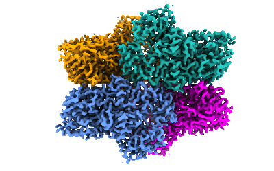 emd_26481.png emd_26481.png | 90.4 KB | ||
| Filedesc metadata | emd-26481.cif.gz | 6.1 KB | ||
| Archive directory |  http://ftp.pdbj.org/pub/emdb/structures/EMD-26481ftp://ftp.pdbj.org/pub/emdb/structures/EMD-26481 http://ftp.pdbj.org/pub/emdb/structures/EMD-26481ftp://ftp.pdbj.org/pub/emdb/structures/EMD-26481 | HTTPS FTP |
-Validation report
| Summary document | emd_26481_validation.pdf.gz | 691.5 KB | Display | EMDB validaton report |
|---|---|---|---|---|
| Full document | emd_26481_full_validation.pdf.gz | 691.1 KB | Display | |
| Data in XML | emd_26481_validation.xml.gz | 8.8 KB | Display | |
| Data in CIF | emd_26481_validation.cif.gz | 10.1 KB | Display | |
| Arichive directory | https://ftp.pdbj.org/pub/emdb/validation_reports/EMD-26481ftp://ftp.pdbj.org/pub/emdb/validation_reports/EMD-26481 | HTTPS FTP |
-Related structure data
| Related structure data |  7ufuMC M: atomic model generated by this map C: citing same article ( |
|---|---|
| Similar structure data |
-Links
| EMDB pages | EMDB (EBI/PDBe) / EMDataResource |
|---|---|
| Related items in Molecule of the Month |


-Map
| File | Download / File: emd_26481.map.gz / Format: CCP4 / Size: 729 MB / Type: IMAGE STORED AS FLOATING POINT NUMBER (4 BYTES) | ||||||||||||||||||||||||||||||||||||
|---|---|---|---|---|---|---|---|---|---|---|---|---|---|---|---|---|---|---|---|---|---|---|---|---|---|---|---|---|---|---|---|---|---|---|---|---|---|
| Annotation | bl1327 mut mannose 2.71 A | ||||||||||||||||||||||||||||||||||||
| Projections & slices | Image control
Images are generated by Spider. | ||||||||||||||||||||||||||||||||||||
| Voxel size | X=Y=Z: 0.67 Å | ||||||||||||||||||||||||||||||||||||
| Density |
| ||||||||||||||||||||||||||||||||||||
| Symmetry | Space group: 1 | ||||||||||||||||||||||||||||||||||||
| Details | EMDB XML:
|
 Z (Sec.)
 Y (Row.)
 X (Col.)


-Supplemental data
- Sample components
-Entire : Tetramer of nucleophile mutant version of Bl1_Man38A in complex w...
| Entire | Name: Tetramer of nucleophile mutant version of Bl1_Man38A in complex with mannose |
|---|---|
| Components |
|
-Supramolecule #1: Tetramer of nucleophile mutant version of Bl1_Man38A in complex w...
| Supramolecule | Name: Tetramer of nucleophile mutant version of Bl1_Man38A in complex with mannose type: complex / ID: 1 / Parent: 0 / Macromolecule list: #1 |
|---|---|
| Source (natural) | Organism: Bifidobacterium longum (bacteria) |
| Molecular weight | Theoretical: 471 KDa |
-Macromolecule #1: Alpha-mannosidase
| Macromolecule | Name: Alpha-mannosidase / type: protein_or_peptide / ID: 1 / Number of copies: 4 / Enantiomer: LEVO |
|---|---|
| Source (natural) | Organism: Bifidobacterium longum (bacteria) / Strain: NCC 2705 |
| Molecular weight | Theoretical: 117.943125 KDa |
| Recombinant expression | Organism: |
| Sequence | String: MGSSHHHHHH SSGLVPRGSH MASMFLKPEQ QLERCRRIVR QRVDPHIHPS IAQLTVESYD IPGEPMPSDE FFAKLDRGDI DFKPFMLGS EWGTTWGTVW FRLTGTVPAG YPKGKPLELI LDLGWYPHSC GGHIEGLVYR ADGTAIKAVH PLNYWVPFMD A EGNAQVPV ...String: MGSSHHHHHH SSGLVPRGSH MASMFLKPEQ QLERCRRIVR QRVDPHIHPS IAQLTVESYD IPGEPMPSDE FFAKLDRGDI DFKPFMLGS EWGTTWGTVW FRLTGTVPAG YPKGKPLELI LDLGWYPHSC GGHIEGLVYR ADGTAIKAVH PLNYWVPFMD A EGNAQVPV AEDGSFTLYL EAASNPLLLG VPPFIETELG DHATGKPDEP YVFKSADLAE FDERYENYSV DLDVVSSLME FA DKQSPRY WQLAKALQRS LNAYDERNPE SVEAARAVLA GVLAKPANAS AMNVSAIGHA HIDSAWLWPV RETRRKVART VSN ALALMD ADPDFKYAMS SAQQYAWLEE DHPDIFKRMK RRIEEGRFIP VGGMWVEADG MLPAGESLIR QIAYGRKYFK EHLG VEPKG VWLPASFGYT GAWPQIARRA GYEWFLTQKI SWNDTTKFPH HSFMWEGIDG SRIFTHFPPA DTYAAWCKVQ ELDYA EKNF QDKDLSDRSL LLFGFGDGGG GPTRNMMEHL HRYENLEGVS KVSIEEPNDF FDKAHQQLAE NAGPEMPVWK GELYLE LHR GTLTSQQDMK RGCRQEESLL RTVEYLGAAA VLSDPEYVYP REELDRIWKT LLLNQFHDIL PGSAIAWVHR EAREDYR RD LKRLAEIAQD MCAVLRKANP QADLLAEARI SQFRNDGASW HANRINEPTD ALSVLTQTLD NGRVLLANGV LSVTIEAD G TISSLLDEEH GRELVPAGTR LGQYELLRDE PAVWDAWEIE RESLLMANAV TGSIESVNTE NGAAQVHVHT ADGDTVITT TITLRPGSHT LDFHADIDWH ERERFLKVDL PLGIVADQAT YDCQYGLIRR PIVKNTASDE AKYESSTNRF AIIGDAGYAA AVINGSVYG SDASPIAGNA AEGRDSGTMF RLSLLSAPTF PDPRTDIGSH EFDWSVVADA TVDRALDAAG VLNAPVLHDV P DITPLASI ESVNGTVVLD WMKLADDGSG DLIVRAYEAA GGQADAMLHV CPALAGASVH ETNVLEGDDL AADLPVALQD GR QNAEGAT LHFGPFQLAT LRITR UniProtKB: Alpha-mannosidase |
-Macromolecule #2: ZINC ION
| Macromolecule | Name: ZINC ION / type: ligand / ID: 2 / Number of copies: 4 / Formula: ZN |
|---|---|
| Molecular weight | Theoretical: 65.409 Da |
-Macromolecule #3: alpha-D-mannopyranose
| Macromolecule | Name: alpha-D-mannopyranose / type: ligand / ID: 3 / Number of copies: 4 / Formula: MAN |
|---|---|
| Molecular weight | Theoretical: 180.156 Da |
| Chemical component information |  ChemComp-MAN: |
-Experimental details
-Structure determination
| Method | cryo EM |
|---|---|
 Processing Processing | single particle reconstruction |
| Aggregation state | particle |
-Sample preparation
| Concentration | 2.2 mg/mL |
|---|---|
| Buffer | pH: 7.5 Details: 150 mM sodium chloride, 20 mM sodium phosphate, pH 7.5 |
| Grid | Model: Quantifoil R2/2 / Material: COPPER / Mesh: 200 / Support film - Material: CARBON / Support film - topology: HOLEY ARRAY / Pretreatment - Type: GLOW DISCHARGE / Pretreatment - Time: 50 sec. |
| Vitrification | Cryogen name: ETHANE / Chamber humidity: 100 % / Chamber temperature: 277 K / Instrument: FEI VITROBOT MARK IV |
- Electron microscopy
| Microscope | FEI TITAN KRIOS |
|---|---|
| Image recording | Film or detector model: FEI FALCON III (4k x 4k) / Detector mode: COUNTING / Average electron dose: 1.5 e/Å2 |
| Electron beam | Acceleration voltage: 300 kV / Electron source:  FIELD EMISSION GUN FIELD EMISSION GUN |
| Electron optics | Illumination mode: FLOOD BEAM / Imaging mode: BRIGHT FIELD / Cs: 0.01 mm |
| Sample stage | Specimen holder model: FEI TITAN KRIOS AUTOGRID HOLDER / Cooling holder cryogen: NITROGEN |
| Experimental equipment |  Model: Titan Krios / Image courtesy: FEI Company |
-Image processing
| Startup model | Type of model: NONE |
|---|---|
| Final reconstruction | Applied symmetry - Point group: D2 (2x2 fold dihedral) / Resolution.type: BY AUTHOR / Resolution: 2.7 Å / Resolution method: FSC 0.143 CUT-OFF / Software - Name: cisTEM / Number images used: 16231 |
| Initial angle assignment | Type: RANDOM ASSIGNMENT / Software - Name: cisTEM |
| Final angle assignment | Type: MAXIMUM LIKELIHOOD / Software - Name: cisTEM |
