 Movie
Movie Controller


+ Open data

- Basic information
| Entry | Database: EMDB / ID: EMD-23331 | |||||||||
|---|---|---|---|---|---|---|---|---|---|---|
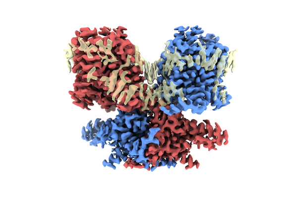
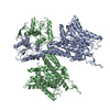
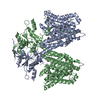
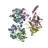
| Title | Structure of human Prestin in nanodisc in the presence of NaCl | |||||||||
 Map data Map data | Prestin in nanodisc in the presence of NaCl | |||||||||
 Sample Sample |
| |||||||||
 Keywords Keywords | transporter family / membrane protein / lipid interaction | |||||||||
| Function / homology |  Function and homology information Function and homology informationlateral wall of outer hair cell / oxalate transport / fructose transmembrane transport / response to salicylic acid / oxalate transmembrane transporter activity / sulfate transmembrane transporter activity / secondary active sulfate transmembrane transporter activity / sulfate transmembrane transport / response to thyroid hormone / response to potassium ion ...lateral wall of outer hair cell / oxalate transport / fructose transmembrane transport / response to salicylic acid / oxalate transmembrane transporter activity / sulfate transmembrane transporter activity / secondary active sulfate transmembrane transporter activity / sulfate transmembrane transport / response to thyroid hormone / response to potassium ion / negative regulation of monoatomic ion transmembrane transport / chloride:bicarbonate antiporter activity / response to salt / response to auditory stimulus / bicarbonate transport / Sensory processing of sound by outer hair cells of the cochlea / positive regulation of cell motility / chloride transport / spectrin binding / cochlea development / lateral plasma membrane / positive regulation of cell size / response to ischemia / chloride transmembrane transport / regulation of membrane potential / sensory perception of sound / regulation of cell shape / basolateral plasma membrane / response to xenobiotic stimulus / protein homodimerization activity / plasma membrane Similarity search - Function | |||||||||
| Biological species |  Homo sapiens (human) Homo sapiens (human) | |||||||||
| Method | single particle reconstruction / cryo EM / Resolution: 2.7 Å | |||||||||
 Authors Authors | Ge J / Gouaux E | |||||||||
| Funding support |  United States, 1 items United States, 1 items
| |||||||||
 Citation Citation | Journal: Cell / Year: 2021 Title: Molecular mechanism of prestin electromotive signal amplification. Authors: Jingpeng Ge / Johannes Elferich / Sepehr Dehghani-Ghahnaviyeh / Zhiyu Zhao / Marc Meadows / Henrique von Gersdorff / Emad Tajkhorshid / Eric Gouaux / Abstract: Hearing involves two fundamental processes: mechano-electrical transduction and signal amplification. Despite decades of studies, the molecular bases for both remain elusive. Here, we show how ...Hearing involves two fundamental processes: mechano-electrical transduction and signal amplification. Despite decades of studies, the molecular bases for both remain elusive. Here, we show how prestin, the electromotive molecule of outer hair cells (OHCs) that senses both voltage and membrane tension, mediates signal amplification by coupling conformational changes to alterations in membrane surface area. Cryoelectron microscopy (cryo-EM) structures of human prestin bound with chloride or salicylate at a common "anion site" adopt contracted or expanded states, respectively. Prestin is ensconced within a perimeter of well-ordered lipids, through which it induces dramatic deformation in the membrane and couples protein conformational changes to the bulk membrane. Together with computational studies, we illustrate how the anion site is allosterically coupled to changes in the transmembrane domain cross-sectional area and the surrounding membrane. These studies provide insight into OHC electromotility by providing a structure-based mechanism of the membrane motor prestin. | |||||||||
| History |
|
- Structure visualization
| Movie |
Movie viewer |
|---|---|
| Structure viewer | EM map: SurfViewMolmilJmol/JSmol |
| Supplemental images |
- Downloads & links
-EMDB archive
| Map data | emd_23331.map.gz | 59.3 MB | EMDB map data format | |
|---|---|---|---|---|
| Header (meta data) | emd-23331-v30.xmlemd-23331.xml | 15 KB 15 KB | Display Display | EMDB header |
| Images |  emd_23331.png emd_23331.png | 167.7 KB | ||
| Filedesc metadata | emd-23331.cif.gz | 6.1 KB | ||
| Archive directory |  http://ftp.pdbj.org/pub/emdb/structures/EMD-23331ftp://ftp.pdbj.org/pub/emdb/structures/EMD-23331 http://ftp.pdbj.org/pub/emdb/structures/EMD-23331ftp://ftp.pdbj.org/pub/emdb/structures/EMD-23331 | HTTPS FTP |
-Related structure data
| Related structure data |  7lgwMC  7lguC  7lh2C  7lh3C M: atomic model generated by this map C: citing same article ( |
|---|---|
| Similar structure data |


-Links
| EMDB pages | EMDB (EBI/PDBe) / EMDataResource |
|---|---|
| Related items in Molecule of the Month |


-Map
| File | Download / File: emd_23331.map.gz / Format: CCP4 / Size: 64 MB / Type: IMAGE STORED AS FLOATING POINT NUMBER (4 BYTES) | ||||||||||||||||||||||||||||||||||||||||||||||||||||||||||||||||||||
|---|---|---|---|---|---|---|---|---|---|---|---|---|---|---|---|---|---|---|---|---|---|---|---|---|---|---|---|---|---|---|---|---|---|---|---|---|---|---|---|---|---|---|---|---|---|---|---|---|---|---|---|---|---|---|---|---|---|---|---|---|---|---|---|---|---|---|---|---|---|
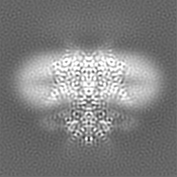
| Annotation | Prestin in nanodisc in the presence of NaCl | ||||||||||||||||||||||||||||||||||||||||||||||||||||||||||||||||||||
| Projections & slices | Image control
Images are generated by Spider. | ||||||||||||||||||||||||||||||||||||||||||||||||||||||||||||||||||||
| Voxel size | X=Y=Z: 0.6507 Å | ||||||||||||||||||||||||||||||||||||||||||||||||||||||||||||||||||||
| Density |
| ||||||||||||||||||||||||||||||||||||||||||||||||||||||||||||||||||||
| Symmetry | Space group: 1 | ||||||||||||||||||||||||||||||||||||||||||||||||||||||||||||||||||||
| Details | EMDB XML:
CCP4 map header:
| ||||||||||||||||||||||||||||||||||||||||||||||||||||||||||||||||||||
 Z (Sec.)
 Y (Row.)
 X (Col.)


-Supplemental data
- Sample components
+Entire : Protein structure bound with substrates and lipids
+Supramolecule #1: Protein structure bound with substrates and lipids
+Macromolecule #1: Prestin
+Macromolecule #2: CHLORIDE ION
+Macromolecule #3: CHOLESTEROL

+Macromolecule #4: N-OCTANE

+Macromolecule #5: DECANE

+Macromolecule #6: DODECANE

+Macromolecule #7: HEXANE
+Macromolecule #8: HEPTANE
+Macromolecule #9: TETRADECANE

+Macromolecule #10: water

-Experimental details
-Structure determination
| Method | cryo EM |
|---|---|
 Processing Processing | single particle reconstruction |
| Aggregation state | particle |
-Sample preparation
| Buffer | pH: 8 |
|---|---|
| Vitrification | Cryogen name: ETHANE / Chamber humidity: 95 % / Chamber temperature: 281 K / Instrument: FEI VITROBOT MARK IV |
- Electron microscopy
| Microscope | FEI TITAN KRIOS |
|---|---|
| Image recording | Film or detector model: GATAN K3 (6k x 4k) / Average electron dose: 50.0 e/Å2 |
| Electron beam | Acceleration voltage: 300 kV / Electron source:  FIELD EMISSION GUN FIELD EMISSION GUN |
| Electron optics | Illumination mode: FLOOD BEAM / Imaging mode: BRIGHT FIELD |
| Experimental equipment |  Model: Titan Krios / Image courtesy: FEI Company |
