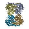
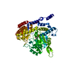
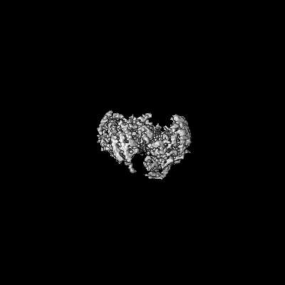
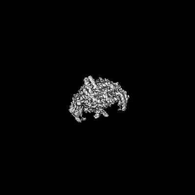
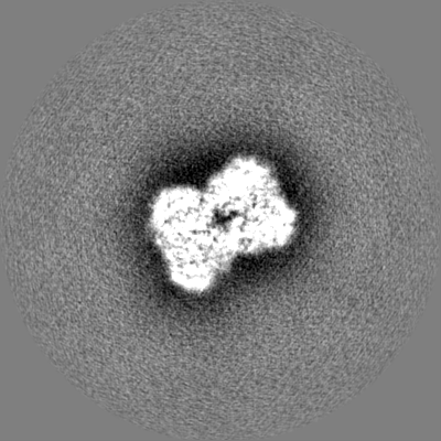
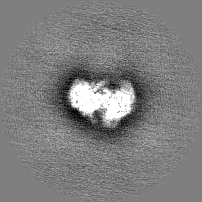
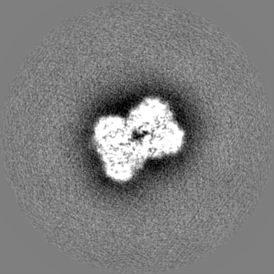
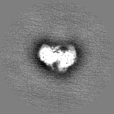
Journal: Mol Cell / Year: 2024 Title: Structure-based mechanism of riboregulation of the metabolic enzyme SHMT1. Authors: Sharon Spizzichino / Federica Di Fonzo / Chiara Marabelli / Angela Tramonti / Antonio Chaves-Sanjuan / Alessia Parroni / Giovanna Boumis / Francesca Romana Liberati / Alessio Paone / Linda ...Authors: Sharon Spizzichino / Federica Di Fonzo / Chiara Marabelli / Angela Tramonti / Antonio Chaves-Sanjuan / Alessia Parroni / Giovanna Boumis / Francesca Romana Liberati / Alessio Paone / Linda Celeste Montemiglio / Matteo Ardini / Arjen J Jakobi / Alok Bharadwaj / Paolo Swuec / Gian Gaetano Tartaglia / Alessandro Paiardini / Roberto Contestabile / Antonello Mai / Dante Rotili / Francesco Fiorentino / Alberto Macone / Alessandra Giorgi / Giancarlo Tria / Serena Rinaldo / Martino Bolognesi / Giorgio Giardina / Francesca Cutruzzolà / Abstract: RNA can directly control protein activity in a process called riboregulation; only a few mechanisms of riboregulation have been described in detail, none of which have been characterized on ...RNA can directly control protein activity in a process called riboregulation; only a few mechanisms of riboregulation have been described in detail, none of which have been characterized on structural grounds. Here, we present a comprehensive structural, functional, and phylogenetic analysis of riboregulation of cytosolic serine hydroxymethyltransferase (SHMT1), the enzyme interconverting serine and glycine in one-carbon metabolism. We have determined the cryoelectron microscopy (cryo-EM) structure of human SHMT1 in its free- and RNA-bound states, and we show that the RNA modulator competes with polyglutamylated folates and acts as an allosteric switch, selectively altering the enzyme's reactivity vs. serine. In addition, we identify the tetrameric assembly and a flap structural motif as key structural elements necessary for binding of RNA to eukaryotic SHMT1. The results presented here suggest that riboregulation may have played a role in evolution of eukaryotic SHMT1 and in compartmentalization of one-carbon metabolism. Our findings provide insights for RNA-based therapeutic strategies targeting this cancer-linked metabolic pathway.
Cryogen name: ETHANE / Chamber humidity: 100 % / Chamber temperature: 277 K / Instrument: FEI VITROBOT MARK IV / Details: blotted for 4 seconds before plunging.
-
Electron microscopy
Microscope
FEI TALOS ARCTICA
Image recording
Film or detector model: FEI FALCON III (4k x 4k) / Detector mode: COUNTING / Number grids imaged: 1 / Number real images: 5450 / Average electron dose: 40.0 e/Å2
Electron beam
Acceleration voltage: 200 kV / Electron source: FIELD EMISSION GUN
In the structure databanks used in Yorodumi, some data are registered as the other names, "COVID-19 virus" and "2019-nCoV". Here are the details of the virus and the list of structure data.
Jan 31, 2019. EMDB accession codes are about to change! (news from PDBe EMDB page)
EMDB accession codes are about to change! (news from PDBe EMDB page)
The allocation of 4 digits for EMDB accession codes will soon come to an end. Whilst these codes will remain in use, new EMDB accession codes will include an additional digit and will expand incrementally as the available range of codes is exhausted. The current 4-digit format prefixed with “EMD-” (i.e. EMD-XXXX) will advance to a 5-digit format (i.e. EMD-XXXXX), and so on. It is currently estimated that the 4-digit codes will be depleted around Spring 2019, at which point the 5-digit format will come into force.
The EM Navigator/Yorodumi systems omit the EMD- prefix.
Related info.:Q: What is EMD? / ID/Accession-code notation in Yorodumi/EM Navigator
Yorodumi is a browser for structure data from EMDB, PDB, SASBDB, etc.
This page is also the successor to EM Navigator detail page, and also detail information page/front-end page for Omokage search.
The word "yorodu" (or yorozu) is an old Japanese word meaning "ten thousand". "mi" (miru) is to see.
Related info.:EMDB / PDB / SASBDB / Comparison of 3 databanks / Yorodumi Search / Aug 31, 2016. New EM Navigator & Yorodumi / Yorodumi Papers / Jmol/JSmol / Function and homology information / Changes in new EM Navigator and Yorodumi
 Movie
Movie Controller


 Open data
 Basic information
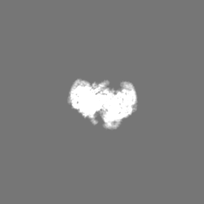
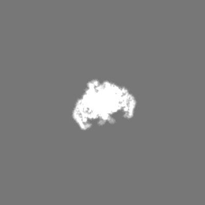
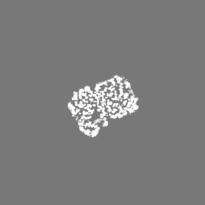
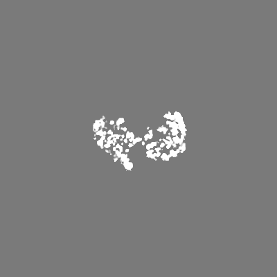
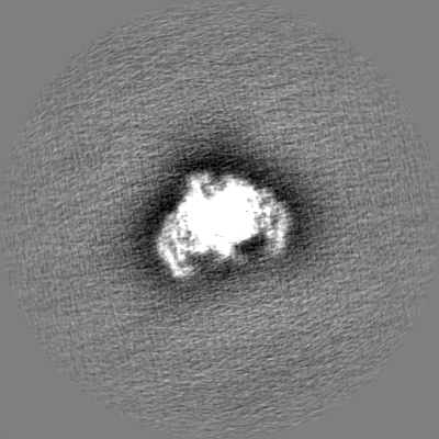
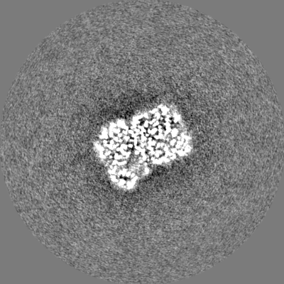
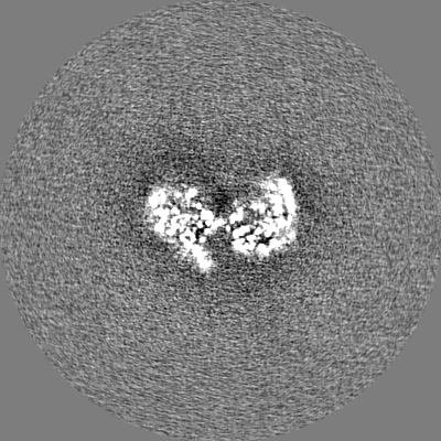
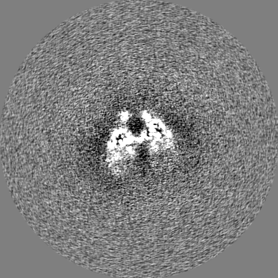
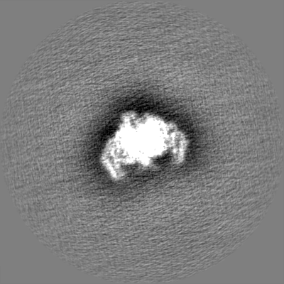
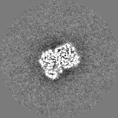
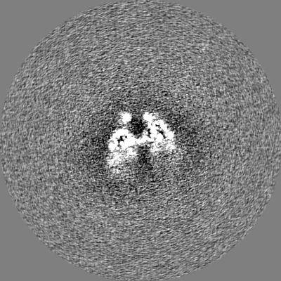
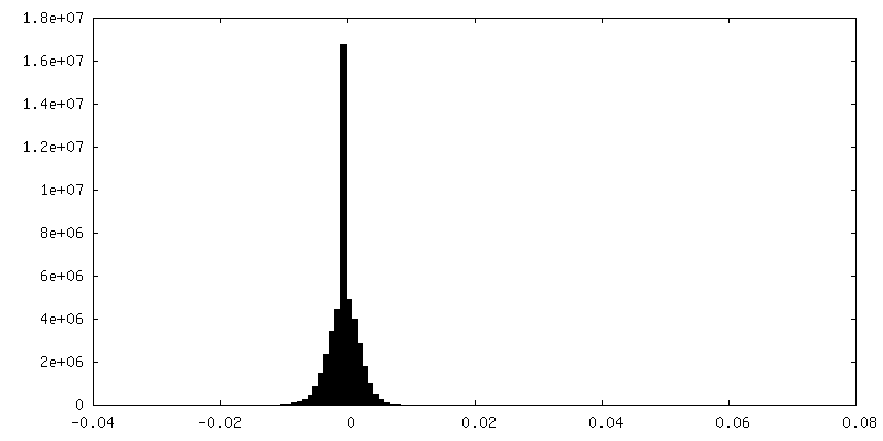
 Map data
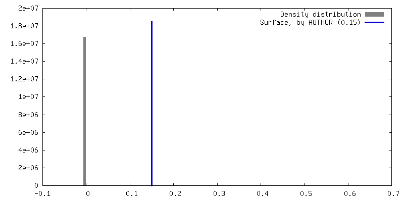
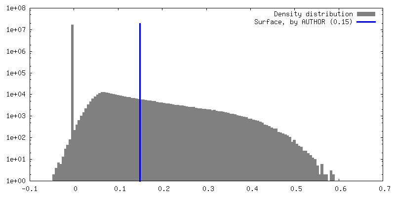
 Sample
 Keywords
 Function and homology information
 Homo sapiens (human)
 Authors
 Italy, 2 items
  Citation
 Structure visualization
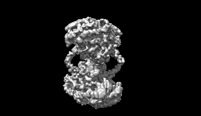
 Downloads & links
 emd_18973.png
 http://ftp.pdbj.org/pub/emdb/structures/EMD-18973
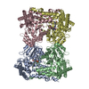


 Z (Sec.)
 Y (Row.)
 X (Col.)


 Sample components
 Processing
 Electron microscopy
 FIELD EMISSION GUN