- EMDB-18268: P301S Tau Filaments from the Brains of PS19 Transgenic Mouse Line -
+
Open data
ID or keywords:
Loading...
-
Basic information
Entry
Database: EMDB / ID: EMD-18268
Title
P301S Tau Filaments from the Brains of PS19 Transgenic Mouse Line
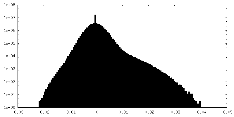
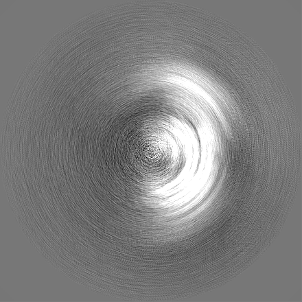
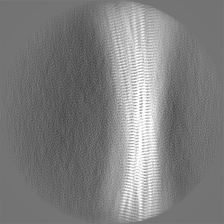
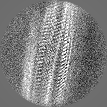
Map data
Sample
Tissue: P301S Tau Protein Filament (PS19)
Protein or peptide: Microtubule-associated protein tau
Keywords
P301S tau / Frontotemporal dementia and parkinsonism linked to chromosome 17 / Transgenic mice / Electron cryo-microscopy / PROTEIN FIBRIL
Function / homology
Function and homology information
plus-end-directed organelle transport along microtubule / histone-dependent DNA binding / negative regulation of protein localization to mitochondrion / neurofibrillary tangle / microtubule lateral binding / axonal transport / tubulin complex / positive regulation of protein localization to synapse / phosphatidylinositol bisphosphate binding / generation of neurons ...plus-end-directed organelle transport along microtubule / histone-dependent DNA binding / negative regulation of protein localization to mitochondrion / neurofibrillary tangle / microtubule lateral binding / axonal transport / tubulin complex / positive regulation of protein localization to synapse / phosphatidylinositol bisphosphate binding / generation of neurons / rRNA metabolic process / axonal transport of mitochondrion / regulation of mitochondrial fission / axon development / regulation of microtubule-based movement / intracellular distribution of mitochondria / regulation of chromosome organization / central nervous system neuron development / minor groove of adenine-thymine-rich DNA binding / lipoprotein particle binding / microtubule polymerization / negative regulation of mitochondrial membrane potential / regulation of microtubule polymerization / dynactin binding / apolipoprotein binding / protein polymerization / main axon / Caspase-mediated cleavage of cytoskeletal proteins / regulation of microtubule polymerization or depolymerization / negative regulation of mitochondrial fission / axolemma / glial cell projection / neurofibrillary tangle assembly / positive regulation of axon extension / regulation of cellular response to heat / positive regulation of microtubule polymerization / positive regulation of protein localization / Activation of AMPK downstream of NMDARs / positive regulation of superoxide anion generation / cellular response to brain-derived neurotrophic factor stimulus / regulation of long-term synaptic depression / supramolecular fiber organization / cytoplasmic microtubule organization / regulation of calcium-mediated signaling / axon cytoplasm / somatodendritic compartment / synapse assembly / phosphatidylinositol binding / nuclear periphery / astrocyte activation / enzyme inhibitor activity / protein phosphatase 2A binding / stress granule assembly / regulation of microtubule cytoskeleton organization / regulation of autophagy / cellular response to reactive oxygen species / microglial cell activation / cellular response to nerve growth factor stimulus / Hsp90 protein binding / protein homooligomerization / SH3 domain binding / PKR-mediated signaling / synapse organization / regulation of synaptic plasticity / response to lead ion / microtubule cytoskeleton organization / memory / neuron projection development / cytoplasmic ribonucleoprotein granule / cell-cell signaling / single-stranded DNA binding / cellular response to heat / growth cone / protein-folding chaperone binding / microtubule cytoskeleton / actin binding / cell body / double-stranded DNA binding / sequence-specific DNA binding / microtubule binding / amyloid fibril formation / dendritic spine / microtubule / protein-macromolecule adaptor activity / learning or memory / neuron projection / membrane raft / negative regulation of gene expression / axon / neuronal cell body / DNA damage response / dendrite / protein kinase binding / enzyme binding / mitochondrion / DNA binding / RNA binding / extracellular region / identical protein binding / nucleus Similarity search - Function
Microtubule-associated protein Tau / Microtubule associated protein, tubulin-binding repeat / Tau and MAP protein, tubulin-binding repeat / Tau and MAP proteins tubulin-binding repeat signature. / Tau and MAP proteins tubulin-binding repeat profile. / : Similarity search - Domain/homology
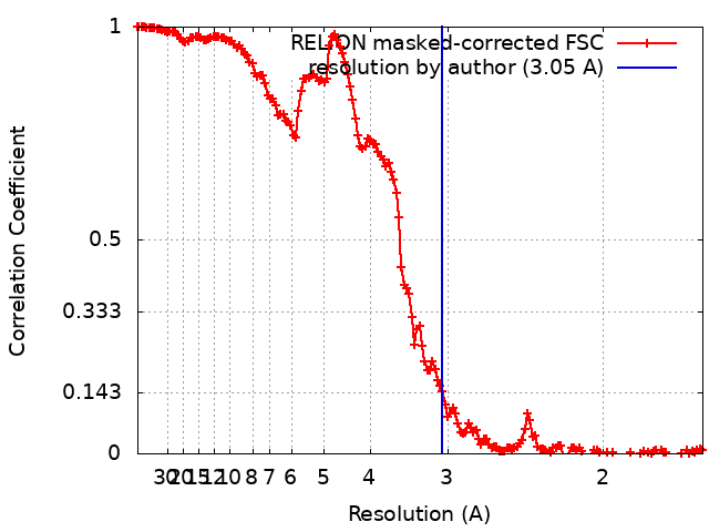
Journal: Acta Neuropathol Commun / Year: 2023 Title: Cryo-EM structures of tau filaments from the brains of mice transgenic for human mutant P301S Tau. Authors: Manuel Schweighauser / Alexey G Murzin / Jennifer Macdonald / Isabelle Lavenir / R Anthony Crowther / Sjors H W Scheres / Michel Goedert / Abstract: Mice transgenic for human mutant P301S tau are widely used as models for human tauopathies. They develop neurodegeneration and abundant filamentous inclusions made of human mutant four-repeat tau. ...Mice transgenic for human mutant P301S tau are widely used as models for human tauopathies. They develop neurodegeneration and abundant filamentous inclusions made of human mutant four-repeat tau. Here we used electron cryo-microscopy (cryo-EM) to determine the structures of tau filaments from the brains of Tg2541 and PS19 mice. Both lines express human P301S tau (0N4R for Tg2541 and 1N4R for PS19) on mixed genetic backgrounds and downstream of different promoters (murine Thy1 for Tg2541 and murine Prnp for PS19). The structures of tau filaments from Tg2541 and PS19 mice differ from each other and those of wild-type tau filaments from human brains. The structures of tau filaments from the brains of humans with mutations P301L, P301S or P301T in MAPT are not known. Filaments from the brains of Tg2541 and PS19 mice share a substructure at the junction of repeats 2 and 3, which comprises residues I297-V312 of tau and includes the P301S mutation. The filament core from the brainstem of Tg2541 mice consists of residues K274-H329 of tau and two disconnected protein densities. Two non-proteinaceous densities are also in evidence. The filament core from the cerebral cortex of line PS19 extends from residues G271-P364 of tau. One strong non-proteinaceous density is also present. Unlike the tau filaments from human brains, the sequences following repeat 4 are missing from the cores of tau filaments from the brains of Tg2541 and PS19 mice.
Protein or peptide: Microtubule-associated protein tau
-
Supramolecule #1: P301S Tau Protein Filament (PS19)
Supramolecule
Name: P301S Tau Protein Filament (PS19) / type: tissue / ID: 1 / Parent: 0 / Macromolecule list: all Details: Human P301S tau filaments extracted from the brains of transgenic mouse line PS19
In the structure databanks used in Yorodumi, some data are registered as the other names, "COVID-19 virus" and "2019-nCoV". Here are the details of the virus and the list of structure data.
Jan 31, 2019. EMDB accession codes are about to change! (news from PDBe EMDB page)
EMDB accession codes are about to change! (news from PDBe EMDB page)
The allocation of 4 digits for EMDB accession codes will soon come to an end. Whilst these codes will remain in use, new EMDB accession codes will include an additional digit and will expand incrementally as the available range of codes is exhausted. The current 4-digit format prefixed with “EMD-” (i.e. EMD-XXXX) will advance to a 5-digit format (i.e. EMD-XXXXX), and so on. It is currently estimated that the 4-digit codes will be depleted around Spring 2019, at which point the 5-digit format will come into force.
The EM Navigator/Yorodumi systems omit the EMD- prefix.
Related info.:Q: What is EMD? / ID/Accession-code notation in Yorodumi/EM Navigator
Yorodumi is a browser for structure data from EMDB, PDB, SASBDB, etc.
This page is also the successor to EM Navigator detail page, and also detail information page/front-end page for Omokage search.
The word "yorodu" (or yorozu) is an old Japanese word meaning "ten thousand". "mi" (miru) is to see.
Related info.:EMDB / PDB / SASBDB / Comparison of 3 databanks / Yorodumi Search / Aug 31, 2016. New EM Navigator & Yorodumi / Yorodumi Papers / Jmol/JSmol / Function and homology information / Changes in new EM Navigator and Yorodumi
 Movie
Movie Controller

 Yorodumi
 Open data
 Basic information
 Map data
 Sample
 Keywords
 Function and homology information
 Homo sapiens (human)
 Authors
 United Kingdom, 2 items
  Citation
 Structure visualization
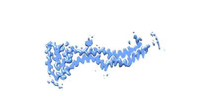
 Downloads & links
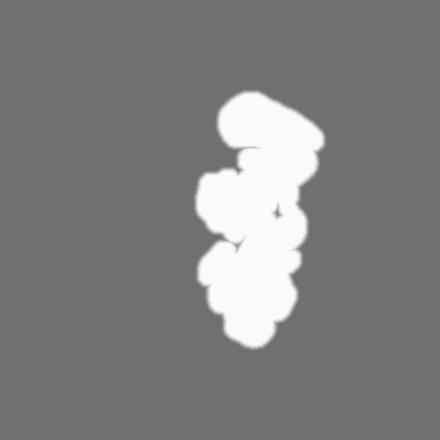
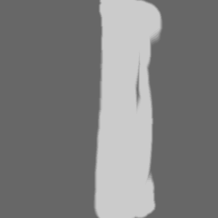
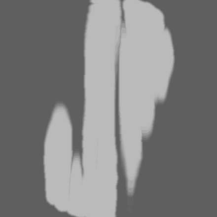
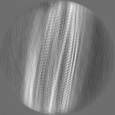
 emd_18268.png
 http://ftp.pdbj.org/pub/emdb/structures/EMD-18268
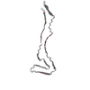


 Z (Sec.)
 Y (Row.)
 X (Col.)


 Sample components
 Processing
 Electron microscopy
 FIELD EMISSION GUN