- EMDB-17653: Mouse RPL39L integrated into the yeast 60S ribosomal subunit -
+
Open data
ID or keywords:
Loading...
-
Basic information
Entry
Database: EMDB / ID: EMD-17653
Title
Mouse RPL39L integrated into the yeast 60S ribosomal subunit
Map data
Sample
Complex: Mouse RPL39L in yeast 60S ribosomal subunit
RNA: x 3 types
Protein or peptide: x 40 types
Ligand: x 6 types
Keywords
RPL39 / RPL39L / ribosomal protein / protein exit tunnel / RIBOSOME
Function / homology
Function and homology information
Formation of a pool of free 40S subunits / SRP-dependent cotranslational protein targeting to membrane / Major pathway of rRNA processing in the nucleolus and cytosol / Nonsense Mediated Decay (NMD) independent of the Exon Junction Complex (EJC) / Nonsense Mediated Decay (NMD) enhanced by the Exon Junction Complex (EJC) / L13a-mediated translational silencing of Ceruloplasmin expression / GTP hydrolysis and joining of the 60S ribosomal subunit / pre-mRNA 5'-splice site binding / cytosolic large ribosomal subunit assembly / response to cycloheximide ...Formation of a pool of free 40S subunits / SRP-dependent cotranslational protein targeting to membrane / Major pathway of rRNA processing in the nucleolus and cytosol / Nonsense Mediated Decay (NMD) independent of the Exon Junction Complex (EJC) / Nonsense Mediated Decay (NMD) enhanced by the Exon Junction Complex (EJC) / L13a-mediated translational silencing of Ceruloplasmin expression / GTP hydrolysis and joining of the 60S ribosomal subunit / pre-mRNA 5'-splice site binding / cytosolic large ribosomal subunit assembly / response to cycloheximide / cleavage in ITS2 between 5.8S rRNA and LSU-rRNA of tricistronic rRNA transcript (SSU-rRNA, 5.8S rRNA, LSU-rRNA) / SRP-dependent cotranslational protein targeting to membrane / GTP hydrolysis and joining of the 60S ribosomal subunit / negative regulation of mRNA splicing, via spliceosome / preribosome, large subunit precursor / Formation of a pool of free 40S subunits / Nonsense Mediated Decay (NMD) independent of the Exon Junction Complex (EJC) / Nonsense Mediated Decay (NMD) enhanced by the Exon Junction Complex (EJC) / L13a-mediated translational silencing of Ceruloplasmin expression / translational elongation / ribosomal large subunit export from nucleus / translational termination / regulation of translational fidelity / protein-RNA complex assembly / maturation of LSU-rRNA / ribosomal large subunit biogenesis / maturation of LSU-rRNA from tricistronic rRNA transcript (SSU-rRNA, 5.8S rRNA, LSU-rRNA) / macroautophagy / translational initiation / maintenance of translational fidelity / modification-dependent protein catabolic process / protein tag activity / rRNA processing / ribosome biogenesis / 5S rRNA binding / ribosomal large subunit assembly / large ribosomal subunit rRNA binding / spermatogenesis / cytosolic large ribosomal subunit / cytoplasmic translation / negative regulation of translation / rRNA binding / structural constituent of ribosome / protein ubiquitination / ribosome / translation / response to antibiotic / mRNA binding / ubiquitin protein ligase binding / nucleolus / RNA binding / zinc ion binding / nucleus / cytoplasm / cytosol Similarity search - Function
: / Ribosomal protein L13e, conserved site / Ribosomal protein L13e signature. / Ribosomal protein L29e / Ribosomal L29e protein family / Ribosomal protein L27e, conserved site / Ribosomal protein L27e signature. / Ribosomal protein L22e / Ribosomal protein L22e superfamily / Ribosomal L22e protein family ...: / Ribosomal protein L13e, conserved site / Ribosomal protein L13e signature. / Ribosomal protein L29e / Ribosomal L29e protein family / Ribosomal protein L27e, conserved site / Ribosomal protein L27e signature. / Ribosomal protein L22e / Ribosomal protein L22e superfamily / Ribosomal L22e protein family / Ribosomal protein L13e / Ribosomal protein L13e / Ribosomal protein L38e / Ribosomal protein L38e superfamily / Ribosomal L38e protein family / Ribosomal protein L19, eukaryotic / : / Ribosomal protein L10e, conserved site / Ribosomal protein L6e signature. / Ribosomal protein L10e signature. / Ribosomal protein L19/L19e conserved site / Ribosomal protein L19e signature. / Ribosomal protein L44e signature. / 60S ribosomal protein L18a/ L20, eukaryotes / Ribosomal protein L10e / Ribosomal protein L24e, conserved site / Ribosomal protein L24e signature. / Ribosomal protein L18/L18-A/B/e, conserved site / Ribosomal protein L18e signature. / Ribosomal protein L34e, conserved site / Ribosomal protein L34e signature. / Ribosomal protein L5 eukaryotic, C-terminal / Ribosomal L18 C-terminal region / Ribosomal protein L23/L25, N-terminal / Ribosomal protein L23, N-terminal domain / : / Ribosomal L40e family / Ribosomal protein L36e signature. / Ribosomal protein L30e signature 1. / Ribosomal protein L44e / Ribosomal protein L44 / 50S ribosomal protein L18Ae/60S ribosomal protein L20 and L18a / Ribosomal protein L35Ae, conserved site / Ribosomal protein 50S-L18Ae/60S-L20/60S-L18A / Ribosomal proteins 50S-L18Ae/60S-L20/60S-L18A / Ribosomal protein 60S L18 and 50S L18e / Ribosomal protein L35Ae signature. / Eukaryotic Ribosomal Protein L27, KOW domain / Ribosomal_L40e / Ribosomal protein L40e / Ribosomal protein L40e superfamily / Ribosomal protein L27e / Ribosomal protein L27e superfamily / Ribosomal L27e protein family / : / Ribosomal Protein L6, KOW domain / Ribosomal protein L39e, conserved site / Ribosomal protein L39e signature. / 60S ribosomal protein L35 / Ribosomal protein L30e signature 2. / Ribosomal protein L7A/L8 / Ribosomal protein L30e, conserved site / : / Ribosomal protein L34Ae / Ribosomal protein L34e / 60S ribosomal protein L19 / Ribosomal protein L6e / Ribosomal protein L13, eukaryotic/archaeal / Ribosomal protein L7, eukaryotic / Ribosomal protein L30, N-terminal / Ribosomal L30 N-terminal domain / 60S ribosomal protein L6E / : / Ribosomal protein L30/YlxQ / 60S ribosomal protein L4, C-terminal domain / 60S ribosomal protein L4 C-terminal domain / Ribosomal protein L36e / Ribosomal protein L36e domain superfamily / Ribosomal protein L36e / Ribosomal protein L18e / Ribosomal protein L14e domain / Ribosomal protein L31e, conserved site / Ribosomal protein L14 / Ribosomal protein L31e signature. / : / : / Ribosomal protein L19e, C-terminal domain / Ribosomal_L19e / Ribosomal protein L19/L19e / Ribosomal protein L19/L19e, domain 1 / Ribosomal protein L19/L19e superfamily / Ribosomal protein L19e, N-terminal domain / Ribosomal protein L35A / Ribosomal protein L35Ae / Ribosomal protein L35A superfamily / Ribosomal protein L32e, conserved site / Ribosomal protein L32e signature. / Ribosomal protein L37ae / Ribosomal L37ae protein family / Ribosomal protein L6, conserved site-2 Similarity search - Domain/homology
Large ribosomal subunit protein uL15 / Large ribosomal subunit protein eL24A / Large ribosomal subunit protein uL23 / Large ribosomal subunit protein uL30A / Large ribosomal subunit protein uL6A / Large ribosomal subunit protein uL22A / Large ribosomal subunit protein uL24A / Large ribosomal subunit protein eL33A / Large ribosomal subunit protein eL36A / Large ribosomal subunit protein eL29 ...Large ribosomal subunit protein uL15 / Large ribosomal subunit protein eL24A / Large ribosomal subunit protein uL23 / Large ribosomal subunit protein uL30A / Large ribosomal subunit protein uL6A / Large ribosomal subunit protein uL22A / Large ribosomal subunit protein uL24A / Large ribosomal subunit protein eL33A / Large ribosomal subunit protein eL36A / Large ribosomal subunit protein eL29 / Large ribosomal subunit protein eL15A / Large ribosomal subunit protein eL22A / Large ribosomal subunit protein uL5A / Large ribosomal subunit protein eL27A / Large ribosomal subunit protein eL31A / Ubiquitin-ribosomal protein eL40A fusion protein / Large ribosomal subunit protein eL20A / Large ribosomal subunit protein eL43A / Large ribosomal subunit protein eL42A / Large ribosomal subunit protein uL14A / Large ribosomal subunit protein uL2A / Large ribosomal subunit protein eL18A / Large ribosomal subunit protein eL19A / Large ribosomal subunit protein uL29A / Large ribosomal subunit protein uL4A / Large ribosomal subunit protein eL30 / Large ribosomal subunit protein uL3 / Large ribosomal subunit protein eL8A / Large ribosomal subunit protein uL18 / Large ribosomal subunit protein uL13A / Large ribosomal subunit protein eL14A / Large ribosomal subunit protein eL32 / Large ribosomal subunit protein uL16 / Large ribosomal subunit protein eL37A / Large ribosomal subunit protein eL38 / Large ribosomal subunit protein eL34A / Large ribosomal subunit protein eL6A / Large ribosomal subunit protein eL21A / Large ribosomal subunit protein eL13A / Large ribosomal subunit protein eL39-like Similarity search - Component
Journal: Acta Crystallogr D Struct Biol / Year: 2018 Title: Real-space refinement in PHENIX for cryo-EM and crystallography. Authors: Pavel V Afonine / Billy K Poon / Randy J Read / Oleg V Sobolev / Thomas C Terwilliger / Alexandre Urzhumtsev / Paul D Adams / Abstract: This article describes the implementation of real-space refinement in the phenix.real_space_refine program from the PHENIX suite. The use of a simplified refinement target function enables very fast ...This article describes the implementation of real-space refinement in the phenix.real_space_refine program from the PHENIX suite. The use of a simplified refinement target function enables very fast calculation, which in turn makes it possible to identify optimal data-restraint weights as part of routine refinements with little runtime cost. Refinement of atomic models against low-resolution data benefits from the inclusion of as much additional information as is available. In addition to standard restraints on covalent geometry, phenix.real_space_refine makes use of extra information such as secondary-structure and rotamer-specific restraints, as well as restraints or constraints on internal molecular symmetry. The re-refinement of 385 cryo-EM-derived models available in the Protein Data Bank at resolutions of 6 Å or better shows significant improvement of the models and of the fit of these models to the target maps.
In the structure databanks used in Yorodumi, some data are registered as the other names, "COVID-19 virus" and "2019-nCoV". Here are the details of the virus and the list of structure data.
Jan 31, 2019. EMDB accession codes are about to change! (news from PDBe EMDB page)
EMDB accession codes are about to change! (news from PDBe EMDB page)
The allocation of 4 digits for EMDB accession codes will soon come to an end. Whilst these codes will remain in use, new EMDB accession codes will include an additional digit and will expand incrementally as the available range of codes is exhausted. The current 4-digit format prefixed with “EMD-” (i.e. EMD-XXXX) will advance to a 5-digit format (i.e. EMD-XXXXX), and so on. It is currently estimated that the 4-digit codes will be depleted around Spring 2019, at which point the 5-digit format will come into force.
The EM Navigator/Yorodumi systems omit the EMD- prefix.
Related info.:Q: What is EMD? / ID/Accession-code notation in Yorodumi/EM Navigator
Yorodumi is a browser for structure data from EMDB, PDB, SASBDB, etc.
This page is also the successor to EM Navigator detail page, and also detail information page/front-end page for Omokage search.
The word "yorodu" (or yorozu) is an old Japanese word meaning "ten thousand". "mi" (miru) is to see.
Related info.:EMDB / PDB / SASBDB / Comparison of 3 databanks / Yorodumi Search / Aug 31, 2016. New EM Navigator & Yorodumi / Yorodumi Papers / Jmol/JSmol / Function and homology information / Changes in new EM Navigator and Yorodumi
 Movie
Movie Controller


 Open data
 Basic information
 Map data
 Sample
 Keywords
 Function and homology information

 Authors
 Switzerland, 1 items
  Citation


 Structure visualization
 Downloads & links
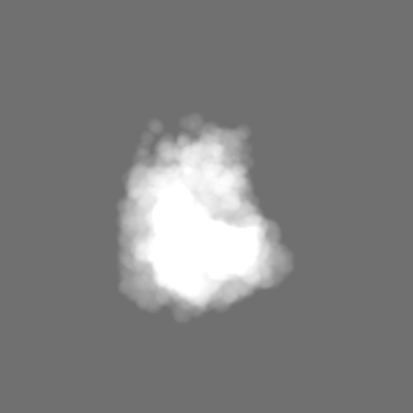
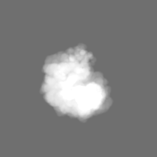
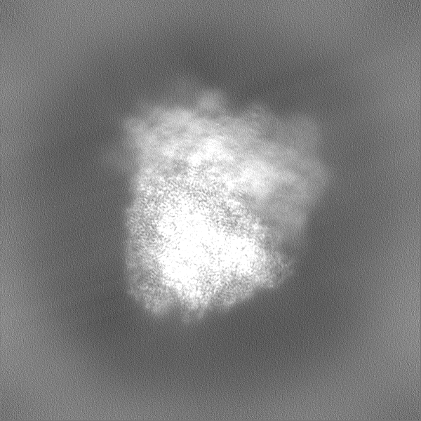
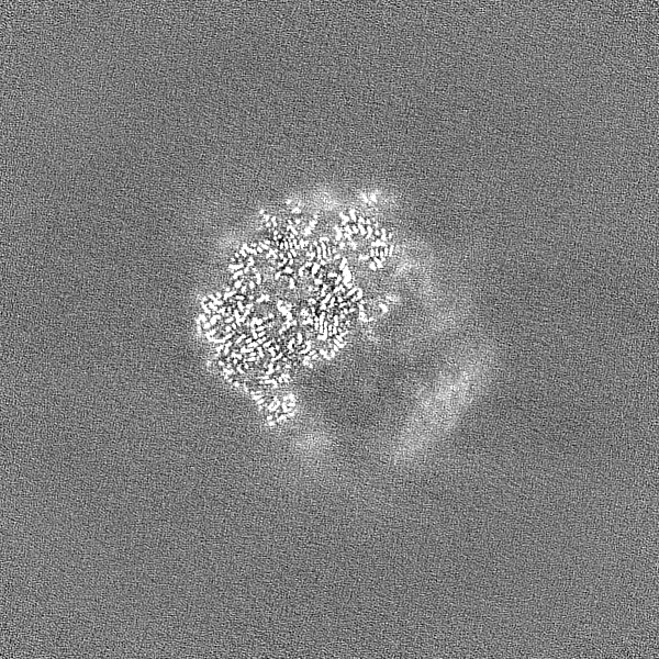
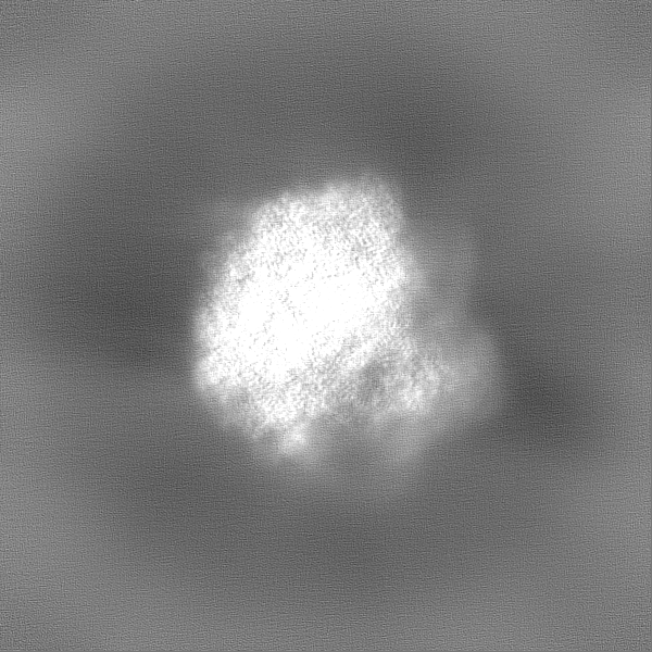
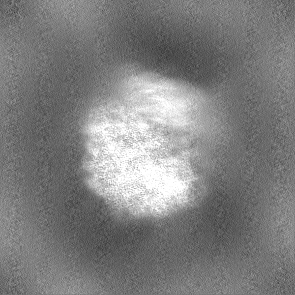
 emd_17653.png
 http://ftp.pdbj.org/pub/emdb/structures/EMD-17653


 Z (Sec.)
 Y (Row.)
 X (Col.)


 Sample components


 Processing
 Electron microscopy
 FIELD EMISSION GUN