 Movie
Movie Controller

[English] 日本語
 Yorodumi
- EMDB-17297: Structure of a human 48S translation initiation complex with eIF4... -
+ Open data

- Basic information
| Entry |  | ||||||||||||
|---|---|---|---|---|---|---|---|---|---|---|---|---|---|
| Title | Structure of a human 48S translation initiation complex with eIF4F and eIF4A | ||||||||||||
 Map data Map data | Human 48S complex | ||||||||||||
 Sample Sample |
| ||||||||||||
 Keywords Keywords | translation / eIF4A / eIF4F / initiation / ribosome / mRNA | ||||||||||||
| Function / homology |  Function and homology information Function and homology informationpositive regulation of eukaryotic translation initiation factor 4F complex assembly / male germ cell proliferation / eukaryotic initiation factor eIF2 binding / positive regulation of translation in response to endoplasmic reticulum stress / translation initiation ternary complex / regulation of translation in response to endoplasmic reticulum stress / glial limiting end-foot / HRI-mediated signaling / viral translational termination-reinitiation / response to manganese-induced endoplasmic reticulum stress ...positive regulation of eukaryotic translation initiation factor 4F complex assembly / male germ cell proliferation / eukaryotic initiation factor eIF2 binding / positive regulation of translation in response to endoplasmic reticulum stress / translation initiation ternary complex / regulation of translation in response to endoplasmic reticulum stress / glial limiting end-foot / HRI-mediated signaling / viral translational termination-reinitiation / response to manganese-induced endoplasmic reticulum stress / Cellular response to mitochondrial stress / positive regulation of type B pancreatic cell apoptotic process / Activation of the mRNA upon binding of the cap-binding complex and eIFs, and subsequent binding to 43S / Response of EIF2AK1 (HRI) to heme deficiency / eukaryotic translation initiation factor 3 complex, eIF3e / methionyl-initiator methionine tRNA binding / Recycling of eIF2:GDP / eukaryotic initiation factor 4E binding / negative regulation of translational initiation in response to stress / cap-dependent translational initiation / PERK-mediated unfolded protein response / eukaryotic translation initiation factor 3 complex, eIF3m / PERK regulates gene expression / IRES-dependent viral translational initiation / response to kainic acid / translation reinitiation / RNA cap binding / eukaryotic translation initiation factor 2 complex / eukaryotic translation initiation factor 4F complex / nuclear stress granule / Z-decay: degradation of maternal mRNAs by zygotically expressed factors / formation of cytoplasmic translation initiation complex / multi-eIF complex / regulation of cellular response to stress / regulation of translational initiation in response to stress / eukaryotic translation initiation factor 3 complex / eukaryotic 43S preinitiation complex / translation factor activity, RNA binding / Deadenylation of mRNA / formation of translation preinitiation complex / mRNA cap binding / miRNA-mediated gene silencing by inhibition of translation / eukaryotic 48S preinitiation complex / M-decay: degradation of maternal mRNAs by maternally stored factors / negative regulation of endoplasmic reticulum unfolded protein response / regulation of translational initiation / oxidized pyrimidine DNA binding / response to TNF agonist / positive regulation of base-excision repair / positive regulation of respiratory burst involved in inflammatory response / protein-synthesizing GTPase / positive regulation of gastrulation / nuclear-transcribed mRNA catabolic process, nonsense-mediated decay / positive regulation of ubiquitin-protein transferase activity / protein tyrosine kinase inhibitor activity / positive regulation of DNA-templated transcription initiation / positive regulation of intrinsic apoptotic signaling pathway in response to DNA damage / IRE1-RACK1-PP2A complex / positive regulation of Golgi to plasma membrane protein transport / nucleolus organization / TNFR1-mediated ceramide production / GDP-dissociation inhibitor activity / negative regulation of RNA splicing / positive regulation of protein localization to cell periphery / metal-dependent deubiquitinase activity / neural crest cell differentiation / supercoiled DNA binding / cytoplasmic translational initiation / NF-kappaB complex / negative regulation of DNA repair / oxidized purine DNA binding / cysteine-type endopeptidase activator activity involved in apoptotic process / rRNA modification in the nucleus and cytosol / negative regulation of intrinsic apoptotic signaling pathway in response to hydrogen peroxide / negative regulation of bicellular tight junction assembly / ubiquitin-like protein conjugating enzyme binding / regulation of establishment of cell polarity / negative regulation of phagocytosis / erythrocyte homeostasis / cytoplasmic side of rough endoplasmic reticulum membrane / Formation of the ternary complex, and subsequently, the 43S complex / ion channel inhibitor activity / protein kinase A binding / laminin receptor activity / pigmentation / Ribosomal scanning and start codon recognition / positive regulation of mitochondrial depolarization / Translation initiation complex formation / positive regulation of protein metabolic process / negative regulation of Wnt signaling pathway / fibroblast growth factor binding / Protein hydroxylation / monocyte chemotaxis / BH3 domain binding / negative regulation of translational frameshifting / regulation of adenylate cyclase-activating G protein-coupled receptor signaling pathway / positive regulation of GTPase activity / TOR signaling / mTORC1-mediated signalling / SARS-CoV-1 modulates host translation machinery Similarity search - Function | ||||||||||||
| Biological species |  Homo sapiens (human) Homo sapiens (human) | ||||||||||||
| Method | single particle reconstruction / cryo EM / Resolution: 3.5 Å | ||||||||||||
 Authors Authors | Brito Querido J / Sokabe M / Diaz-Lopez I / Gordiyenko Y / Fraser CS / Ramakrishnan V | ||||||||||||
| Funding support |  United Kingdom, United Kingdom,  United States, 3 items United States, 3 items
| ||||||||||||
 Citation Citation | Journal: Nat Struct Mol Biol / Year: 2024 Title: The structure of a human translation initiation complex reveals two independent roles for the helicase eIF4A. Authors: Jailson Brito Querido / Masaaki Sokabe / Irene Díaz-López / Yuliya Gordiyenko / Christopher S Fraser / V Ramakrishnan / Abstract: Eukaryotic translation initiation involves recruitment of the 43S pre-initiation complex to the 5' end of mRNA by the cap-binding complex eIF4F, forming the 48S translation initiation complex (48S), ...Eukaryotic translation initiation involves recruitment of the 43S pre-initiation complex to the 5' end of mRNA by the cap-binding complex eIF4F, forming the 48S translation initiation complex (48S), which then scans along the mRNA until the start codon is recognized. We have previously shown that eIF4F binds near the mRNA exit channel of the 43S, leaving open the question of how mRNA secondary structure is removed as it enters the mRNA channel on the other side of the 40S subunit. Here we report the structure of a human 48S that shows that, in addition to the eIF4A that is part of eIF4F, there is a second eIF4A helicase bound at the mRNA entry site, which could unwind RNA secondary structures as they enter the 48S. The structure also reveals conserved interactions between eIF4F and the 43S, probaby explaining how eIF4F can promote mRNA recruitment in all eukaryotes. | ||||||||||||
| History |
|
- Structure visualization
| Supplemental images |
|---|

- Downloads & links
-EMDB archive
| Map data | emd_17297.map.gz | 32.6 MB | EMDB map data format | |
|---|---|---|---|---|
| Header (meta data) | emd-17297-v30.xmlemd-17297.xml | 108.8 KB 108.8 KB | Display Display | EMDB header |
| FSC (resolution estimation) | emd_17297_fsc.xml | 14.2 KB | Display | FSC data file |
| Images |  emd_17297.png emd_17297.png | 85.4 KB | ||
| Masks | emd_17297_msk_1.map | 244.1 MB | Mask map | |
| Filedesc metadata | emd-17297.cif.gz | 20.8 KB | ||
| Others | emd_17297_additional_1.map.gzemd_17297_additional_10.map.gzemd_17297_additional_11.map.gzemd_17297_additional_2.map.gzemd_17297_additional_3.map.gzemd_17297_additional_4.map.gzemd_17297_additional_5.map.gzemd_17297_additional_6.map.gzemd_17297_additional_7.map.gzemd_17297_additional_8.map.gzemd_17297_additional_9.map.gzemd_17297_half_map_1.map.gzemd_17297_half_map_2.map.gz | 190.3 MB 13 MB 209.8 MB 152.6 MB 144.3 MB 209.3 MB 139.3 MB 5.1 MB 3.4 MB 192.5 MB 8.6 MB 194.6 MB 193.8 MB | ||
| Archive directory |  http://ftp.pdbj.org/pub/emdb/structures/EMD-17297ftp://ftp.pdbj.org/pub/emdb/structures/EMD-17297 http://ftp.pdbj.org/pub/emdb/structures/EMD-17297ftp://ftp.pdbj.org/pub/emdb/structures/EMD-17297 | HTTPS FTP |
-Related structure data
| Related structure data |  8oz0MC M: atomic model generated by this map C: citing same article ( |
|---|---|
| Similar structure data |
-Links
| EMDB pages | EMDB (EBI/PDBe) / EMDataResource |
|---|---|
| Related items in Molecule of the Month |


-Map
| File | Download / File: emd_17297.map.gz / Format: CCP4 / Size: 244.1 MB / Type: IMAGE STORED AS FLOATING POINT NUMBER (4 BYTES) | ||||||||||||||||||||||||||||||||||||
|---|---|---|---|---|---|---|---|---|---|---|---|---|---|---|---|---|---|---|---|---|---|---|---|---|---|---|---|---|---|---|---|---|---|---|---|---|---|
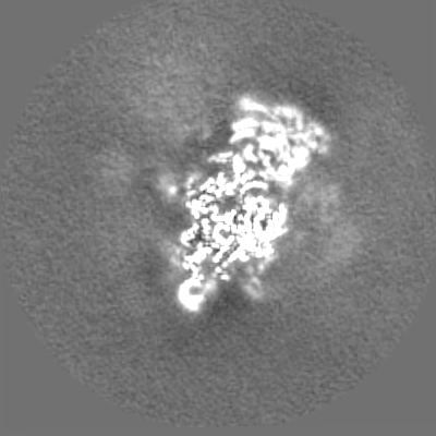
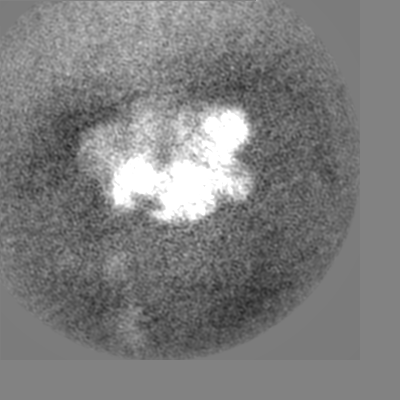
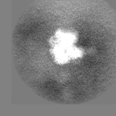
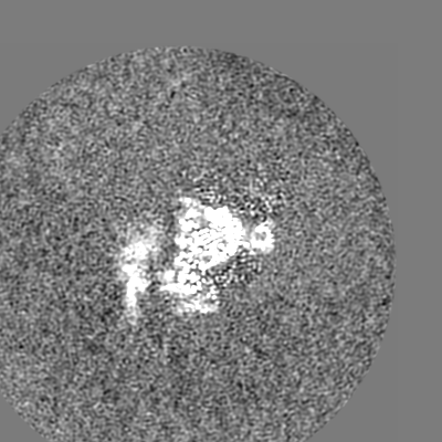
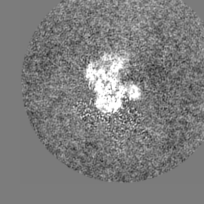
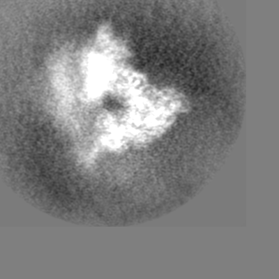
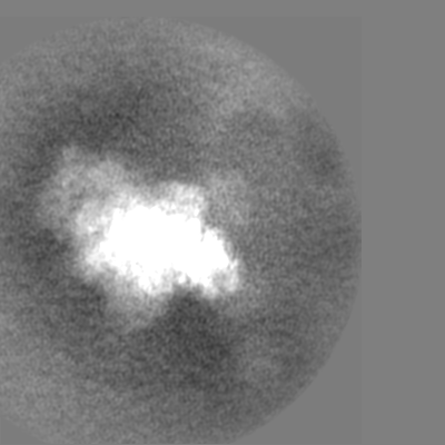
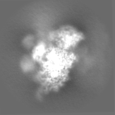
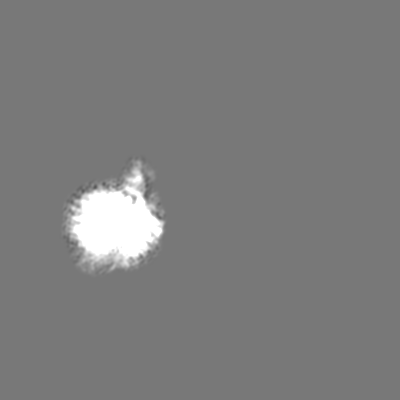
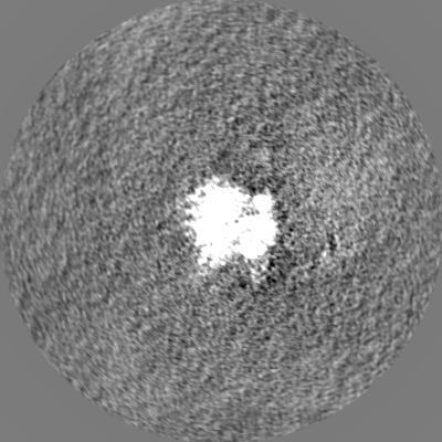
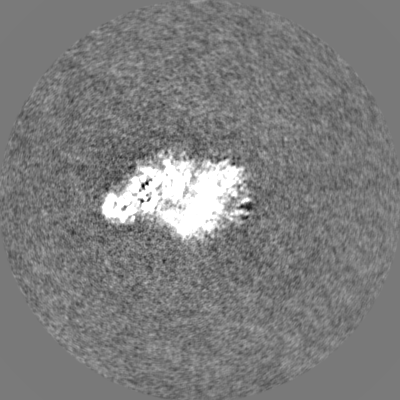
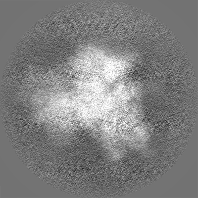
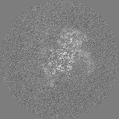
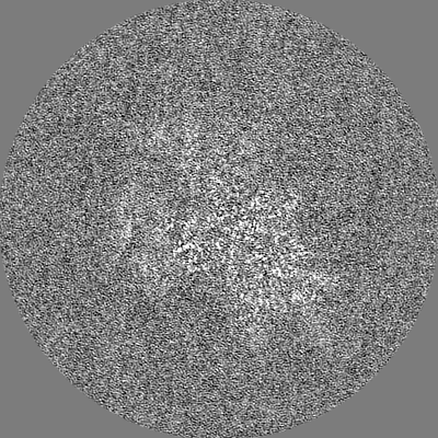
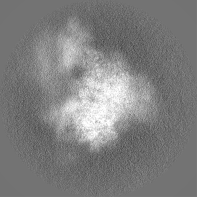
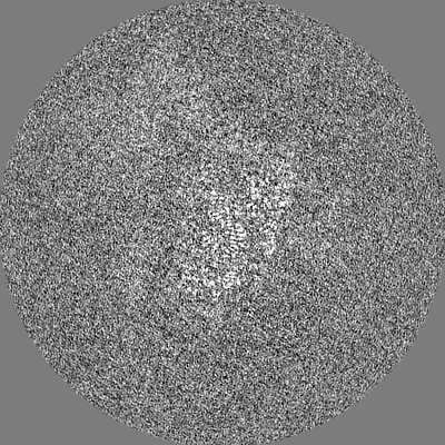
| Annotation | Human 48S complex | ||||||||||||||||||||||||||||||||||||
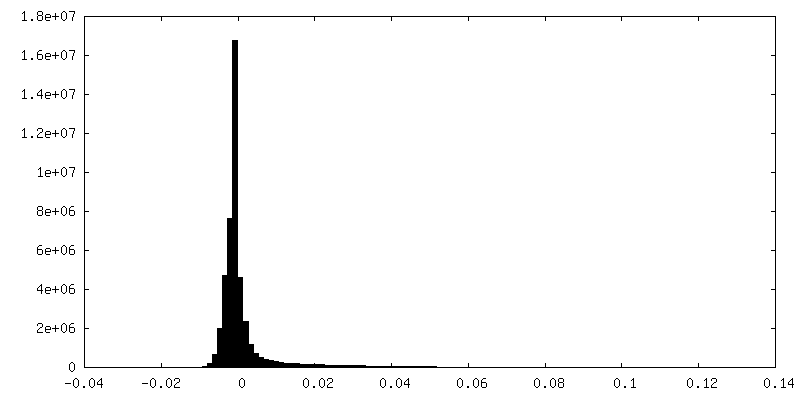
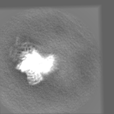
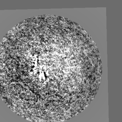
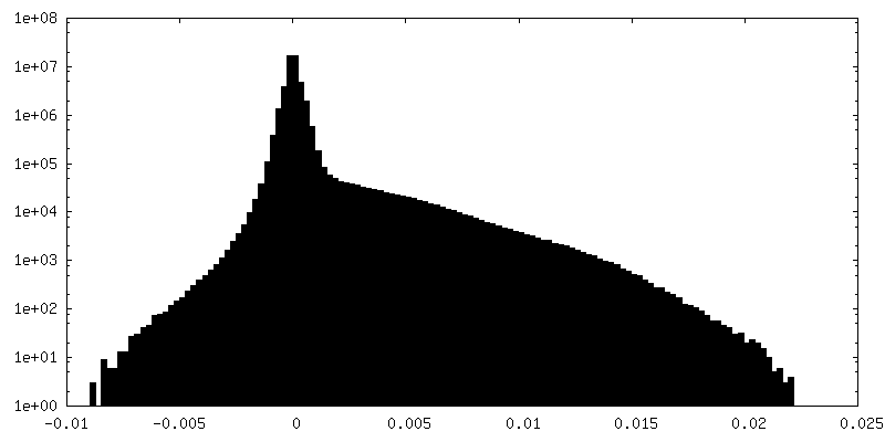
| Projections & slices | Image control
Images are generated by Spider. | ||||||||||||||||||||||||||||||||||||
| Voxel size | X=Y=Z: 1.06 Å | ||||||||||||||||||||||||||||||||||||
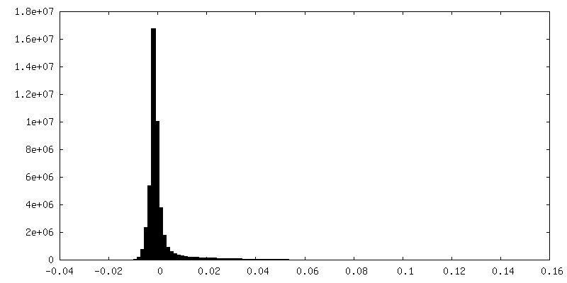
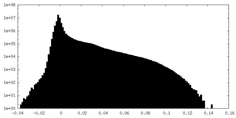
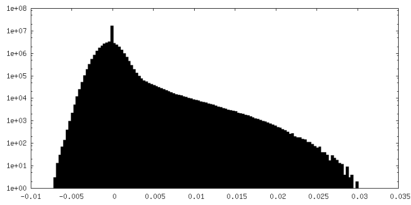
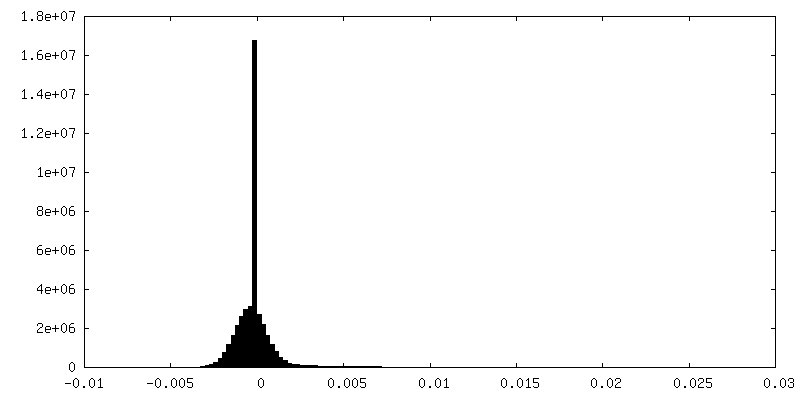
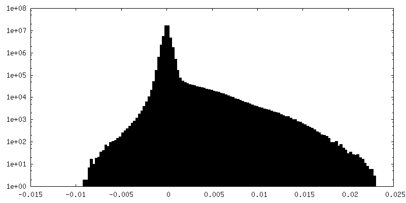
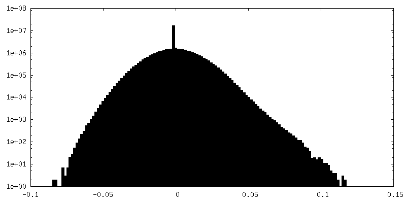
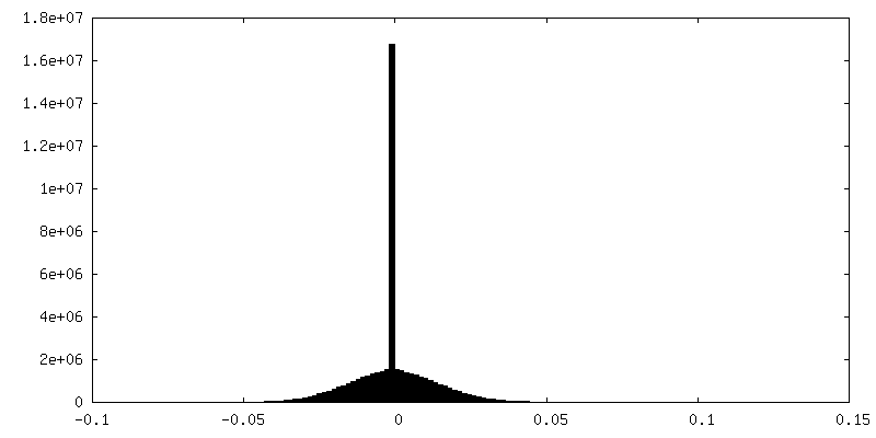
| Density |
| ||||||||||||||||||||||||||||||||||||
| Symmetry | Space group: 1 | ||||||||||||||||||||||||||||||||||||
| Details | EMDB XML:
|
 Z (Sec.)
 Y (Row.)
 X (Col.)


-Supplemental data
+Mask #1


+Additional map: Body1 - 3D multi-body refinement of 48S


+Additional map: eIF3 after focus 4F


+Additional map: PIC after focus EntrySite

+Additional map: Body2 - 3D multi-body refinement of 48S


+Additional map: Body3 - 3D multi-body refinement of 48S


+Additional map: PIC after focus EntrySite Resampled map


+Additional map: eIF4F-eIF3 after focus EntrySite Resampled map


+Additional map: eIF3 after focus 4F Resampled map


+Additional map: eIF4F after focus 4F Resampled map


+Additional map: eIF4F eIF3 after focus 4F


+Additional map: eIF4F after focus 4F


+Half map: Half map1 48S


+Half map: Half map1 48S


- Sample components
+Entire : Human 48S translation initiation complex
+Supramolecule #1: Human 48S translation initiation complex
+Macromolecule #1: Eukaryotic translation initiation factor 3 subunit E
+Macromolecule #2: Eukaryotic translation initiation factor 3 subunit F
+Macromolecule #3: Eukaryotic translation initiation factor 3 subunit G
+Macromolecule #4: Eukaryotic translation initiation factor 3 subunit H
+Macromolecule #5: 60S ribosomal protein L41
+Macromolecule #6: Eukaryotic translation initiation factor 3 subunit A
+Macromolecule #7: Eukaryotic translation initiation factor 3 subunit K
+Macromolecule #8: Eukaryotic translation initiation factor 3 subunit M
+Macromolecule #9: Eukaryotic translation initiation factor 2 subunit 1
+Macromolecule #10: Eukaryotic translation initiation factor 2 subunit 3
+Macromolecule #11: Eukaryotic translation initiation factor 3 subunit I
+Macromolecule #12: Eukaryotic translation initiation factor 1A, X-chromosomal

+Macromolecule #13: Eukaryotic translation initiation factor 5
+Macromolecule #14: Eukaryotic translation initiation factor 3 subunit B
+Macromolecule #15: Eukaryotic translation initiation factor 3 subunit C
+Macromolecule #16: Eukaryotic translation initiation factor 3 subunit L
+Macromolecule #17: 40S ribosomal protein S7
+Macromolecule #18: 40S ribosomal protein S27
+Macromolecule #19: 40S ribosomal protein S21
+Macromolecule #20: 40S ribosomal protein S2
+Macromolecule #21: 40S ribosomal protein S3a
+Macromolecule #22: 40S ribosomal protein SA
+Macromolecule #23: 40S ribosomal protein S26
+Macromolecule #24: 40S ribosomal protein S6
+Macromolecule #25: 40S ribosomal protein S14
+Macromolecule #26: Eukaryotic translation initiation factor 2 subunit 2
+Macromolecule #27: 40S ribosomal protein S13
+Macromolecule #29: 40S ribosomal protein S11
+Macromolecule #30: 40S ribosomal protein S4, X isoform
+Macromolecule #31: 40S ribosomal protein S9
+Macromolecule #32: 40S ribosomal protein S23
+Macromolecule #33: 40S ribosomal protein S30
+Macromolecule #34: 40S ribosomal protein S15a
+Macromolecule #35: 40S ribosomal protein S8
+Macromolecule #36: 40S ribosomal protein S24
+Macromolecule #37: 40S ribosomal protein S5
+Macromolecule #38: 40S ribosomal protein S16
+Macromolecule #39: 40S ribosomal protein S3
+Macromolecule #40: 40S ribosomal protein S10
+Macromolecule #41: 40S ribosomal protein S15
+Macromolecule #42: Receptor of activated protein C kinase 1
+Macromolecule #43: 40S ribosomal protein S19
+Macromolecule #44: 40S ribosomal protein S25
+Macromolecule #45: Small ribosomal subunit protein uS13
+Macromolecule #46: 40S ribosomal protein S29
+Macromolecule #47: Ubiquitin-40S ribosomal protein S27a
+Macromolecule #48: 40S ribosomal protein S12
+Macromolecule #49: 40S ribosomal protein S28
+Macromolecule #50: 40S ribosomal protein S17
+Macromolecule #51: 40S ribosomal protein S20
+Macromolecule #52: Eukaryotic translation initiation factor 3 subunit D
+Macromolecule #55: Eukaryotic initiation factor 4A-I
+Macromolecule #56: Eukaryotic translation initiation factor 4 gamma 1
 Baculovirus expression vector pFastBac1-HM
+Macromolecule #28: 18S rRNA
+Macromolecule #53: tRNAiMet
+Macromolecule #54: mRNA
+Macromolecule #57: ZINC ION
+Macromolecule #58: MAGNESIUM ION
-Experimental details
-Structure determination
| Method | cryo EM |
|---|---|
 Processing Processing | single particle reconstruction |
| Aggregation state | particle |
-Sample preparation
| Buffer | pH: 7.4 |
|---|---|
| Grid | Model: UltrAuFoil R1.2/1.3 / Support film - Material: GRAPHENE OXIDE / Pretreatment - Type: GLOW DISCHARGE |
| Vitrification | Cryogen name: ETHANE / Chamber humidity: 100 % / Chamber temperature: 277.15 K |
- Electron microscopy
| Microscope | FEI TITAN KRIOS |
|---|---|
| Software | Name: EPU |
| Image recording | Film or detector model: GATAN K3 (6k x 4k) / Average electron dose: 47.88 e/Å2 |
| Electron beam | Acceleration voltage: 300 kV / Electron source:  FIELD EMISSION GUN FIELD EMISSION GUN |
| Electron optics | Illumination mode: OTHER / Imaging mode: OTHER / Nominal defocus max: 3.0 µm / Nominal defocus min: 1.2 µm |
| Sample stage | Specimen holder model: FEI TITAN KRIOS AUTOGRID HOLDER / Cooling holder cryogen: NITROGEN |
| Experimental equipment |  Model: Titan Krios / Image courtesy: FEI Company |

