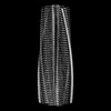
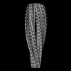
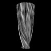
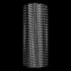
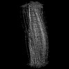
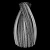
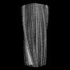
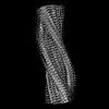
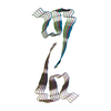
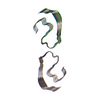
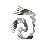
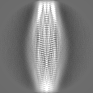
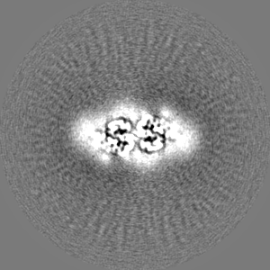
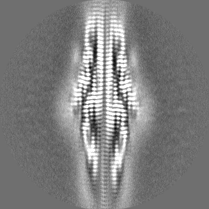
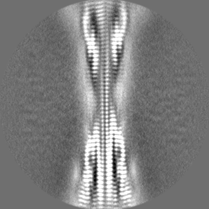
登録情報 データベース : EMDB / ID : EMD-16942タイトル Murine type II Abeta fibril from APP23 mouse Ex vivo Abeta42 fibril from APP23 mouse brain. 複合体 : Amyloid fibril of amyloid-betaタンパク質・ペプチド : Amyloid-beta protein 42 / 機能・相同性 分子機能 ドメイン・相同性 構成要素
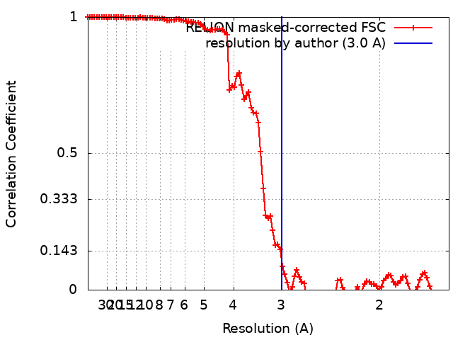
/ / / / / / / / / / / / / / / / / / / / / / / / / / / / / / / / / / / / / / / / / / / / / / / / / / / / / / / / / / / / / / / / / / / / / / / / / / / / / / / / / / / / / / / / / / / / / / / / / / / / / / / / / / / / / / / / / / / / / / / / / / / / / / / / / / / / / / / / / / / / / / / / / / / 生物種 Mus musculus (ハツカネズミ) / Homo sapiens (ヒト)手法 / / 解像度 : 3.0 Å Zielinski M / Peralta Reyes FS / Gremer L / Schemmert S / Frieg B / Willuweit A / Donner L / Elvers M / Nilsson LNG / Syvanen S ...Zielinski M / Peralta Reyes FS / Gremer L / Schemmert S / Frieg B / Willuweit A / Donner L / Elvers M / Nilsson LNG / Syvanen S / Sehlin D / Ingelsson M / Willbold D / Schroeder GF 資金援助 Organization Grant number 国 German Federal Ministry for Education and Research 16LW028 Swedish Research Council 2021-02793 Swedish Research Council 2021-01083 Swedish Research Council 2021-03524 Helmholtz Association
ジャーナル : Nat Neurosci / 年 : 2023タイトル : Cryo-EM of Aβ fibrils from mouse models find tg-APP fibrils resemble those found in patients with sporadic Alzheimer's disease.著者: Mara Zielinski / Fernanda S Peralta Reyes / Lothar Gremer / Sarah Schemmert / Benedikt Frieg / Luisa U Schäfer / Antje Willuweit / Lili Donner / Margitta Elvers / Lars N G Nilsson / Stina ... 著者 : Mara Zielinski / Fernanda S Peralta Reyes / Lothar Gremer / Sarah Schemmert / Benedikt Frieg / Luisa U Schäfer / Antje Willuweit / Lili Donner / Margitta Elvers / Lars N G Nilsson / Stina Syvänen / Dag Sehlin / Martin Ingelsson / Dieter Willbold / Gunnar F Schröder / 要旨 : The use of transgenic mice displaying amyloid-β (Aβ) brain pathology has been essential for the preclinical assessment of new treatment strategies for Alzheimer's disease. However, the properties ... The use of transgenic mice displaying amyloid-β (Aβ) brain pathology has been essential for the preclinical assessment of new treatment strategies for Alzheimer's disease. However, the properties of Aβ in such mice have not been systematically compared to Aβ in the brains of patients with Alzheimer's disease. Here, we determined the structures of nine ex vivo Aβ fibrils from six different mouse models by cryogenic-electron microscopy. We found novel Aβ fibril structures in the APP/PS1, ARTE10 and tg-SwDI models, whereas the human type II filament fold was found in the ARTE10, tg-APP and APP23 models. The tg-APP mice showed an Aβ fibril whose structure resembles the human type I filament found in patients with sporadic Alzheimer's disease. A detailed assessment of the Aβ fibril structure is key to the selection of adequate mouse models for the preclinical development of novel plaque-targeting therapeutics and positron emission tomography imaging tracers in Alzheimer's disease. 履歴 登録 2023年3月30日 - ヘッダ(付随情報) 公開 2023年11月29日 - マップ公開 2023年11月29日 - 更新 2023年12月13日 - 現状 2023年12月13日 処理サイト : PDBe / 状態 : 公開
すべて表示 表示を減らす
 ムービー
ムービー コントローラー


 データを開く
 基本情報
基本情報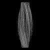
 マップデータ
 試料
試料 キーワード
キーワード 機能・相同性情報
 Homo sapiens (ヒト)
 データ登録者
 ドイツ,
ドイツ,  スウェーデン, 5件
  引用
引用

 構造の表示
構造の表示
 ダウンロードとリンク
 emd_16942.png
 http://ftp.pdbj.org/pub/emdb/structures/EMD-16942


 Z (Sec.)
 Y (Row.)
 X (Col.)


 試料の構成要素
 解析
解析 電子顕微鏡法
 FIELD EMISSION GUN