 Movie
Movie Controller


+ Open data

- Basic information
| Entry | 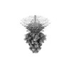 | |||||||||
|---|---|---|---|---|---|---|---|---|---|---|
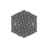
| Title | Carin 1 bacteriophage tail, connector and tail fibers assembly | |||||||||
 Map data Map data | ||||||||||
 Sample Sample | Bacteriophage sp. Cobetia Marina Virus Carin1 != Bacteriophage sp. Bacteriophage sp. Cobetia Marina Virus Carin1
| |||||||||
| Biological species |  Bacteriophage sp. (virus) Bacteriophage sp. (virus) | |||||||||
| Method | single particle reconstruction / cryo EM / Resolution: 3.9 Å | |||||||||
 Authors Authors | d'Acapito A / Neumann E / Schoehn G | |||||||||
| Funding support |  France, 1 items France, 1 items
| |||||||||
 Citation Citation | Journal: J Virol / Year: 2023 Title: Structural Study of the Cobetia marina Bacteriophage 1 (Carin-1) by Cryo-EM. Authors: Alessio d'Acapito / Thomas Roret / Eleftherios Zarkadas / Pierre-Yves Mocaër / Florian Lelchat / Anne-Claire Baudoux / Guy Schoehn / Emmanuelle Neumann / Abstract: Most of studied bacteriophages (phages) are terrestrial viruses. However, marine phages are shown to be highly involved in all levels of oceanic regulation. They are, however, still largely ...Most of studied bacteriophages (phages) are terrestrial viruses. However, marine phages are shown to be highly involved in all levels of oceanic regulation. They are, however, still largely overlooked by the scientific community. By inducing cell lysis on half of the bacterial population daily, their role and influence on the bacterial biomass and evolution, as well as their impact in the global biogeochemical cycles, is undeniable. Cobetia marina (Carin-1) is a member of the family infecting the γ. marina. Here, we present the almost complete, nearly-atomic resolution structure of Carin-1 comprising capsid, portal, and tail machineries at 3.5 Å, 3.8 Å and 3.9 Å, respectively, determined by cryo-electron microscopy (cryo-EM). Our experimental results, combined with AlphaFold2 (AF), allowed us to obtain the nearly-atomic structure of Carin-1 by fitting and refining the AF atomic models in the high resolution cryo-EM map, skipping the bottleneck of manual building and speeding up the structure determination process. Our structural results highlighted the T7-like nature of Carin1, as well as several novel structural features like the presence of short spikes on the capsid, reminiscent those described for Rhodobacter capsulatus gene transfer agent (RcGTA). This is, to our knowledge, the first time such assembly is described for a bacteriophage, shedding light into the common evolution and shared mechanisms between gene transfer agents and phages. This first full structure determined for a marine podophage allowed to propose an infection mechanism different than the one proposed for the archetypal podophage T7. Oceans play a central role in the carbon cycle on Earth and on the climate regulation (half of the planet's CO2 is absorbed by phytoplankton photosynthesis in the oceans and just as much O2 is liberated). The understanding of the biochemical equilibriums of marine biology represents a major goal for our future. By lysing half of the bacterial population every day, marine bacteriophages are key actors of these equilibriums. Despite their importance, these marine phages have, so far, only been studied a little and, in particular, structural insights are currently lacking, even though they are fundamental for the understanding of the molecular mechanisms of their mode of infection. The structures described in our manuscript allow us to propose an infection mechanism that differs from the one proposed for the terrestrial T7 virus, and might also allow us to, in the future, better understand the way bacteriophages shape the global ecosystem. | |||||||||
| History |
|
- Structure visualization
| Supplemental images |
|---|

- Downloads & links
-EMDB archive
| Map data | emd_16689.map.gz | 94.5 MB |  EMDB map data format EMDB map data format | |
|---|---|---|---|---|
| Header (meta data) | emd-16689-v30.xmlemd-16689.xml | 18.2 KB 18.2 KB | Display Display | EMDB header |
| FSC (resolution estimation) | emd_16689_fsc.xml | 10.7 KB | Display | FSC data file |
| Images |  emd_16689.png emd_16689.png | 52.5 KB | ||
| Others | emd_16689_half_map_1.map.gzemd_16689_half_map_2.map.gz | 79.1 MB 79.1 MB | ||
| Archive directory |  http://ftp.pdbj.org/pub/emdb/structures/EMD-16689ftp://ftp.pdbj.org/pub/emdb/structures/EMD-16689 http://ftp.pdbj.org/pub/emdb/structures/EMD-16689ftp://ftp.pdbj.org/pub/emdb/structures/EMD-16689 | HTTPS FTP |
-Validation report
| Summary document | emd_16689_validation.pdf.gz | 1.2 MB | Display | EMDB validaton report |
|---|---|---|---|---|
| Full document | emd_16689_full_validation.pdf.gz | 1.2 MB | Display | |
| Data in XML | emd_16689_validation.xml.gz | 17.6 KB | Display | |
| Data in CIF | emd_16689_validation.cif.gz | 23.2 KB | Display | |
| Arichive directory | https://ftp.pdbj.org/pub/emdb/validation_reports/EMD-16689ftp://ftp.pdbj.org/pub/emdb/validation_reports/EMD-16689 | HTTPS FTP |
-Related structure data
| Related structure data | 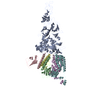 8ck1MC 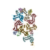 8cjzC  8ck0C M: atomic model generated by this map C: citing same article ( |
|---|
-Links
| EMDB pages | EMDB (EBI/PDBe) / EMDataResource |
|---|
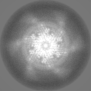
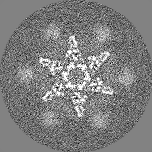
-Map
| File | Download / File: emd_16689.map.gz / Format: CCP4 / Size: 103 MB / Type: IMAGE STORED AS FLOATING POINT NUMBER (4 BYTES) | ||||||||||||||||||||
|---|---|---|---|---|---|---|---|---|---|---|---|---|---|---|---|---|---|---|---|---|---|
| Voxel size | X=Y=Z: 1.35 Å | ||||||||||||||||||||
| Density |
| ||||||||||||||||||||
| Symmetry | Space group: 1 | ||||||||||||||||||||
| Details | EMDB XML:
|
-Supplemental data
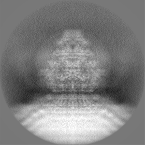
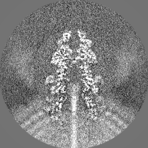
-Half map: #2
| File | emd_16689_half_map_1.map | ||||||||||||
|---|---|---|---|---|---|---|---|---|---|---|---|---|---|
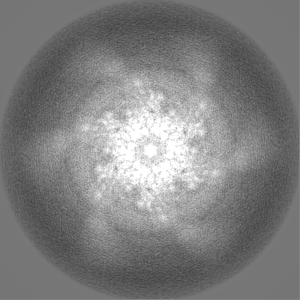
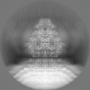
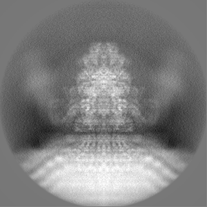
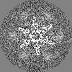
| Projections & Slices |
| ||||||||||||
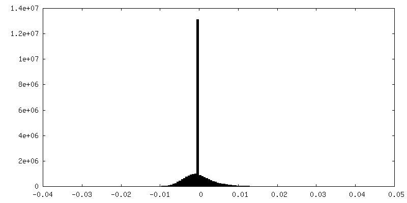
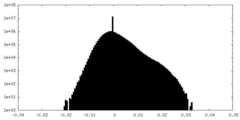
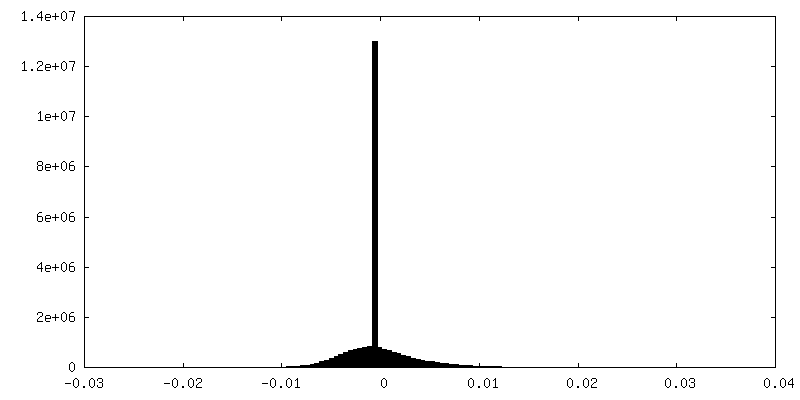
| Density Histograms |
 Z
Z Y
Y X
X
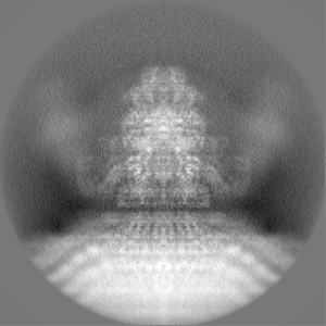

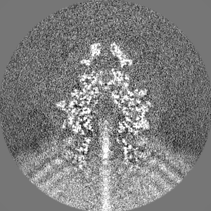
-Half map: #1
| File | emd_16689_half_map_2.map | ||||||||||||
|---|---|---|---|---|---|---|---|---|---|---|---|---|---|
| Projections & Slices |
| ||||||||||||
| Density Histograms |


- Sample components
-Entire : Bacteriophage sp. Cobetia Marina Virus Carin1
| Entire | Name: Bacteriophage sp. Cobetia Marina Virus Carin1 |
|---|---|
| Components |
|
-Supramolecule #1: Bacteriophage sp.
| Supramolecule | Name: Bacteriophage sp. / type: virus / ID: 1 / Parent: 0 / Macromolecule list: all / Details: Cobetia Marina Virus Carin1 / NCBI-ID: 3801 / Sci species name: Bacteriophage sp. / Virus type: VIRION / Virus isolate: OTHER / Virus enveloped: No / Virus empty: No |
|---|---|
| Host (natural) | Organism:  Cobetia marina (bacteria) / Strain: DSM 4741 Cobetia marina (bacteria) / Strain: DSM 4741 |
-Macromolecule #1: Tail Nozzle
| Macromolecule | Name: Tail Nozzle / type: protein_or_peptide / ID: 1 / Number of copies: 1 / Enantiomer: LEVO |
|---|---|
| Source (natural) | Organism: Bacteriophage sp. (virus) |
| Molecular weight | Theoretical: 91.324289 KDa |
| Sequence | String: MASNNYQPAS SYIQPSFAGG ELAPSLQGRV DLARYAISLK TCRNFVVQPY GGASNRPGFR FNTACKYKNY ATRLIPFSFN TEQTYVIEI GHQYMRFHRD GAPVLDGGEP VEVATSWHRD DIFEIKYVQS ADVLTLVHPD YKPRQLKRYS ETDWVLDFFD N EFGPLQDQ ...String: MASNNYQPAS SYIQPSFAGG ELAPSLQGRV DLARYAISLK TCRNFVVQPY GGASNRPGFR FNTACKYKNY ATRLIPFSFN TEQTYVIEI GHQYMRFHRD GAPVLDGGEP VEVATSWHRD DIFEIKYVQS ADVLTLVHPD YKPRQLKRYS ETDWVLDFFD N EFGPLQDQ NVDESITIIS NGVVDLVELT ASEAIFSEAM VGTTIKLQQV SSGEVAAWQN RSAVEQGDLA YVDERTYKAT SL SGGVDNT LTGDNTPAHT EGEQWDGPRT TIQGVTETLG VKWAYLHSGF GYVRITEHRD DTHIVGRVIG RLPEEIRTEG TYR WSFAAW DSDRGYPGTA SYYQQRLVFA NSRAEPQAFW MSETGIFNGF KVSFPIEADD AITFTLASRQ VNEIRHLIPL GSLL ALTSG AEWMISDNDQ GLAPDTVSAD VQGYRGASDV TPLLIGSSAL YVQARGTVIR DLAYSFELDG YTGDDLTIFS NHLLK DYTI KDWAYAQEPD SVVWLVRSDG ALLSMTYQRE QQVVAWARHD TVDGEFESVA VIAEGSRDVP YAIVKRQVGG ETVRYI EYL DSRRFSHVED FFCVDSGLTY DGRSSTGALL TIGGGTNWTT DEDLTLTASA SSFSPSDVGR RVRVYTGDKF ADVDVDA YV SATSVAVSAV RIVPEELRGV QGDRWGFMAK TLTGLDHLEG KTVSILADGN VHAPEVVTGG QVTLDYSAAV VHVGLPIE S DIETLPISSS GATVRDSHKA IVGVGIQLEK SRGVFAARSR RDFTSSDLIE LKQRDAEDWG EATGLETGLV ELGIPTSWD KDGSLFIRQS DPLPLTILSI IPRVVMGGKG |
-Macromolecule #2: Tail fibers Dpo36
| Macromolecule | Name: Tail fibers Dpo36 / type: protein_or_peptide / ID: 2 / Number of copies: 3 / Enantiomer: LEVO |
|---|---|
| Source (natural) | Organism: Bacteriophage sp. (virus) |
| Molecular weight | Theoretical: 86.858156 KDa |
| Sequence | String: MTVPTNDNRE QYAGNGATTV FPYAFRIFES SDLEVYLTDE DGDQALLIEG TDYTVSGAGD EEGGEITFPV SGDPLDDGET LTILRVIDI TQETDLKNQG AYYPEVVEDE FDRSRMIDQQ QQEQLDRALI KTETGDRWEG QGVPAKNFAM SDPVEDTDLP T VRWTKDYV ...String: MTVPTNDNRE QYAGNGATTV FPYAFRIFES SDLEVYLTDE DGDQALLIEG TDYTVSGAGD EEGGEITFPV SGDPLDDGET LTILRVIDI TQETDLKNQG AYYPEVVEDE FDRSRMIDQQ QQEQLDRALI KTETGDRWEG QGVPAKNFAM SDPVEDTDLP T VRWTKDYV TQMAEGITGD IGAYTVVAPT SGDEKRLDEW MDDIQRPDDS LVVADGGTEA RSLSERFADS ASYQDYGIAG DG TTNDTAA FAALESDRSS DAIELHGNTY LVDEIPNGNA YRDAVWSLDG EDLSISEYGG LVTGTPTTGA FEPAYTGGVN NTP TTSGRT NKHTRAILAS QNCRADFARS ACVASIYSWA YGNVSGNFAS RQSIAGAPQT VNIGSEEGQA LGFQSGNYTT QFCR AEGST TFNIGSDDCA ASGAHSGTIS SLESYAGRGH DFRGTPVFDD GVLVDITIDD AGAGYVPGSD VMYLQNRQFG NTTDA VITY TVDGTGGVSA ITITDGGSGY SGIVAARIDT FGDYSLVMAS ARSKIEDQFC AAIASDNARV RGRESAVIAS DGGVVN EDN SVVIGSVDST SNGARSGIYT GSGCETTGAG AVVIGGVNAK ASNDGAIVMG RGVDSEFARS LVFGDGGSGA AASTAGR KF QVTAAGNVTA AGTITGSTTY ADYAEYFENS ARGVIPLGVI VTLDGRKVRP ASAGDDIIGV VSGTAILAAG DSQFHWGG R YLAGEFGELL YHDVDVDGKI ERQPVENPEY DPSVPNVPRS QRPEEWSCIG LVGQLHVRVS SDVAAGDRVA AGDGGIGVP GDNGMICMEI KQAYDSGKGY AVALCLHK |
-Macromolecule #3: Connector Protein
| Macromolecule | Name: Connector Protein / type: protein_or_peptide / ID: 3 / Number of copies: 2 / Enantiomer: LEVO |
|---|---|
| Source (natural) | Organism: Bacteriophage sp. (virus) |
| Molecular weight | Theoretical: 24.697715 KDa |
| Sequence | String: MPSKVDICNR ALSNTGTDIT IASLTEKSKE ARLCQQWYDA TLASLLRTYQ WAFAQRRVTL ALIGVGPAGW RHKYRYPTDA ITIHDVFTA DTYPDGASEF TDGRYRQIFQ IASDGEGGRL VLANCEDAMC RYTSDIEDPN LMPPDFSTAL EMMLAKNIAM P MTGNPGLM ...String: MPSKVDICNR ALSNTGTDIT IASLTEKSKE ARLCQQWYDA TLASLLRTYQ WAFAQRRVTL ALIGVGPAGW RHKYRYPTDA ITIHDVFTA DTYPDGASEF TDGRYRQIFQ IASDGEGGRL VLANCEDAMC RYTSDIEDPN LMPPDFSTAL EMMLAKNIAM P MTGNPGLM TVLAQQAASL VSDAIARDQN EGYRNPLPYA SWTRANIGDS YPDDDHLPHR GGRR |
-Experimental details
-Structure determination
| Method | cryo EM |
|---|---|
 Processing Processing | single particle reconstruction |
| Aggregation state | particle |
-Sample preparation
| Buffer | pH: 7.5 / Component - Concentration: 1.0 x / Component - Name: PBS |
|---|---|
| Grid | Model: Quantifoil R1.2/1.3 / Material: COPPER/RHODIUM / Pretreatment - Type: GLOW DISCHARGE / Pretreatment - Time: 45 sec. / Details: 25mA |
| Vitrification | Cryogen name: ETHANE / Chamber humidity: 100 % / Chamber temperature: 293.15 K / Instrument: FEI VITROBOT MARK IV |
- Electron microscopy
| Microscope | TFS KRIOS |
|---|---|
| Image recording | Film or detector model: GATAN K3 (6k x 4k) / Average electron dose: 30.0 e/Å2 |
| Electron beam | Acceleration voltage: 300 kV / Electron source:  FIELD EMISSION GUN FIELD EMISSION GUN |
| Electron optics | Illumination mode: OTHER / Imaging mode: BRIGHT FIELD / Nominal defocus max: 4.0 µm / Nominal defocus min: 0.7000000000000001 µm |
| Sample stage | Cooling holder cryogen: NITROGEN |
| Experimental equipment |  Model: Titan Krios / Image courtesy: FEI Company |
-Image processing
| Final reconstruction | Applied symmetry - Point group: C6 (6 fold cyclic) / Resolution.type: BY AUTHOR / Resolution: 3.9 Å / Resolution method: FSC 0.143 CUT-OFF / Software - Name: RELION (ver. 3.1) / Number images used: 11395 |
|---|---|
| Initial angle assignment | Type: MAXIMUM LIKELIHOOD / Software - Name: RELION |
| Final angle assignment | Type: MAXIMUM LIKELIHOOD / Software - Name: RELION |
| FSC plot (resolution estimation) | 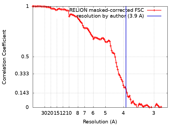 |
