[English] 日本語
Yorodumi
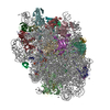
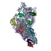
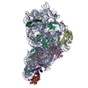
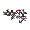
- EMDB-16615: Tetracycline bound to the 30S head -

+
Open data


ID or keywords:

Loading...

-
Basic information

Entry
Database: EMDB / ID: EMD-16615
TitleTetracycline bound to the 30S head
Map data
Sample
  • Complex: 70S ribosomes with antibiotic cocktail
    • RNA: x 1 types
    • Protein or peptide: x 9 types
  • Ligand: x 4 types
KeywordsAntibiotic / RIBOSOME
Function / homology
Function and homology information


transcription antitermination factor activity, RNA binding / transcription elongation factor complex / regulation of DNA-templated transcription elongation / transcription antitermination / maintenance of translational fidelity / ribosome biogenesis / ribosomal small subunit assembly / small ribosomal subunit / cytosolic small ribosomal subunit / cytoplasmic translation ...transcription antitermination factor activity, RNA binding / transcription elongation factor complex / regulation of DNA-templated transcription elongation / transcription antitermination / maintenance of translational fidelity / ribosome biogenesis / ribosomal small subunit assembly / small ribosomal subunit / cytosolic small ribosomal subunit / cytoplasmic translation / tRNA binding / negative regulation of translation / rRNA binding / ribosome / structural constituent of ribosome / translation / response to antibiotic / mRNA binding / RNA binding / zinc ion binding / membrane / cytosol / cytoplasm
Similarity search - Function
Ribosomal protein S14, bacterial/plastid / Ribosomal protein S3, bacterial-type / Ribosomal protein S19, bacterial-type / Ribosomal protein S13, bacterial-type / Ribosomal protein S7, bacterial/organellar-type / Ribosomal protein S9, bacterial/plastid / Ribosomal protein S5, bacterial-type / Ribosomal protein S2, bacteria/mitochondria/plastid / Ribosomal protein S2 signature 2. / K Homology domain ...Ribosomal protein S14, bacterial/plastid / Ribosomal protein S3, bacterial-type / Ribosomal protein S19, bacterial-type / Ribosomal protein S13, bacterial-type / Ribosomal protein S7, bacterial/organellar-type / Ribosomal protein S9, bacterial/plastid / Ribosomal protein S5, bacterial-type / Ribosomal protein S2, bacteria/mitochondria/plastid / Ribosomal protein S2 signature 2. / K Homology domain / K homology RNA-binding domain / Ribosomal protein S10, conserved site / Ribosomal protein S10 signature. / Ribosomal protein S3, conserved site / Ribosomal protein S3 signature. / Ribosomal protein S14, conserved site / Ribosomal protein S14 signature. / Ribosomal protein S2 signature 1. / KH domain / Type-2 KH domain profile. / K Homology domain, type 2 / Ribosomal protein S3, C-terminal / Ribosomal protein S3, C-terminal domain / Ribosomal protein S3, C-terminal domain superfamily / Ribosomal protein S15/S19, conserved site / Ribosomal protein S19 signature. / Ribosomal protein S10 / Ribosomal protein S19/S15 / Ribosomal protein S19/S15, superfamily / Ribosomal protein S19 / Ribosomal protein S5, N-terminal, conserved site / Ribosomal protein S5 signature. / Ribosomal protein S2, conserved site / Ribosomal protein S7, conserved site / Ribosomal protein S7 signature. / Ribosomal protein S2 / Ribosomal protein S2, flavodoxin-like domain superfamily / Ribosomal protein S2 / Ribosomal protein S5 / S5 double stranded RNA-binding domain profile. / K homology domain superfamily, prokaryotic type / Ribosomal protein S5, N-terminal / Ribosomal protein S5, N-terminal domain / Ribosomal protein S5, C-terminal / Ribosomal protein S5, C-terminal domain / Ribosomal protein S13, conserved site / Ribosomal protein S13 signature. / Ribosomal protein S13 / 30s ribosomal protein S13, C-terminal / Ribosomal protein S13/S18 / Ribosomal protein S13 family profile. / Ribosomal protein S14 / Ribosomal protein S14p/S29e / K homology domain-like, alpha/beta / Ribosomal protein S10p/S20e / Ribosomal protein S10 domain / Ribosomal protein S10 domain superfamily / Ribosomal protein S10p/S20e / Ribosomal protein S9, conserved site / Ribosomal protein S9 signature. / Ribosomal protein S13-like, H2TH / Ribosomal protein S5/S7 / Ribosomal protein S7 domain / Ribosomal protein S7 domain superfamily / Ribosomal protein S7p/S5e / Ribosomal protein S9 / Ribosomal protein S9/S16 / Ribosomal protein S5 domain 2-type fold, subgroup / Ribosomal protein S5 domain 2-type fold
Similarity search - Domain/homology
Small ribosomal subunit protein uS7 / Small ribosomal subunit protein uS10 / Small ribosomal subunit protein uS13 / Small ribosomal subunit protein uS19 / Small ribosomal subunit protein uS2 / Small ribosomal subunit protein uS3 / Small ribosomal subunit protein uS5 / Small ribosomal subunit protein uS9 / Small ribosomal subunit protein uS14
Similarity search - Component
Biological speciesEscherichia coli BW25113 (bacteria)
Methodsingle particle reconstruction / cryo EM / Resolution: 1.82 Å
AuthorsPaternoga H / Crowe-McAuliffe C / Beckert B / Wilson DN
Funding supportEuropean Union, 3 items
OrganizationGrant numberCountry
Other governmentDLR01Kl1820
iNEXT-Discovery871037European Union
Other governmentLM2018127
CitationJournal: Nat Struct Mol Biol / Year: 2023
Title: Structural conservation of antibiotic interaction with ribosomes.
Authors: Helge Paternoga / Caillan Crowe-McAuliffe / Lars V Bock / Timm O Koller / Martino Morici / Bertrand Beckert / Alexander G Myasnikov / Helmut Grubmüller / Jiří Nováček / Daniel N Wilson /
Abstract: The ribosome is a major target for clinically used antibiotics, but multidrug resistant pathogenic bacteria are making our current arsenal of antimicrobials obsolete. Here we present cryo-electron- ...The ribosome is a major target for clinically used antibiotics, but multidrug resistant pathogenic bacteria are making our current arsenal of antimicrobials obsolete. Here we present cryo-electron-microscopy structures of 17 distinct compounds from six different antibiotic classes bound to the bacterial ribosome at resolutions ranging from 1.6 to 2.2 Å. The improved resolution enables a precise description of antibiotic-ribosome interactions, encompassing solvent networks that mediate multiple additional interactions between the drugs and their target. Our results reveal a high structural conservation in the binding mode between antibiotics with the same scaffold, including ordered water molecules. Water molecules are visualized within the antibiotic binding sites that are preordered, become ordered in the presence of the drug and that are physically displaced on drug binding. Insight into RNA-ligand interactions will facilitate development of new antimicrobial agents, as well as other RNA-targeting therapies.
History
DepositionFeb 2, 2023-
Header (metadata) releaseJul 26, 2023-
Map releaseJul 26, 2023-
UpdateApr 24, 2024-
Current statusApr 24, 2024Processing site: PDBe / Status: Released

-
Structure visualization

Supplemental images

Downloads & links

-
Map

FileDownload / File: emd_16615.map.gz / Format: CCP4 / Size: 1.9 GB / Type: IMAGE STORED AS FLOATING POINT NUMBER (4 BYTES)
Projections & slices

Image control

Size
Brightness
Contrast
Others
AxesZ (Sec.)Y (Row.)X (Col.)
0.68 Å/pix.
x 800 pix.
= 544.8 Å
0.68 Å/pix.
x 800 pix.
= 544.8 Å
0.68 Å/pix.
x 800 pix.
= 544.8 Å

Surface

Projections

Slices (1/3)

Slices (1/2)

Slices (2/3)

Images are generated by Spider.

Voxel sizeX=Y=Z: 0.681 Å
Density
Contour LevelBy AUTHOR: 0.009
Minimum - Maximum-0.032056328 - 0.09248401
Average (Standard dev.)0.0000030593576 (±0.00041963652)
SymmetrySpace group: 1
Details

EMDB XML:

Map geometry
Axis orderXYZ
Origin000
Dimensions800800800
Spacing800800800
CellA=B=C: 544.8 Å
α=β=γ: 90.0 °

-
Supplemental data

-
Additional map: #1

Fileemd_16615_additional_1.map
Projections & Slices
AxesZYX

Projections

Slices (1/2)
Density Histograms

-
Half map: #2

Fileemd_16615_half_map_1.map
Projections & Slices
AxesZYX

Projections

Slices (1/2)
Density Histograms

-
Half map: #1

Fileemd_16615_half_map_2.map
Projections & Slices
AxesZYX

Projections

Slices (1/2)
Density Histograms

-
Sample components

+
Entire : 70S ribosomes with antibiotic cocktail

EntireName: 70S ribosomes with antibiotic cocktail
Components
  • Complex: 70S ribosomes with antibiotic cocktail
    • RNA: 16S rRNA
    • Protein or peptide: 30S ribosomal protein S2
    • Protein or peptide: Small ribosomal subunit protein uS3
    • Protein or peptide: Small ribosomal subunit protein uS5
    • Protein or peptide: 30S ribosomal protein S7
    • Protein or peptide: Small ribosomal subunit protein uS9
    • Protein or peptide: Small ribosomal subunit protein uS10
    • Protein or peptide: Small ribosomal subunit protein uS13
    • Protein or peptide: Small ribosomal subunit protein uS14
    • Protein or peptide: Small ribosomal subunit protein uS19
  • Ligand: TETRACYCLINE
  • Ligand: MAGNESIUM ION
  • Ligand: POTASSIUM ION
  • Ligand: water

+
Supramolecule #1: 70S ribosomes with antibiotic cocktail

SupramoleculeName: 70S ribosomes with antibiotic cocktail / type: complex / ID: 1 / Parent: 0 / Macromolecule list: #1-#10
Details: 70S + Tetracycline, Lincomycin, Avilamycin, Streptomycin
Source (natural)Organism: Escherichia coli BW25113 (bacteria)

+
Macromolecule #1: 16S rRNA

MacromoleculeName: 16S rRNA / type: rna / ID: 1 / Number of copies: 1
Source (natural)Organism: Escherichia coli BW25113 (bacteria)
Molecular weightTheoretical: 499.197938 KDa
SequenceString: AAAUUGAAGA GUUUGAUCAU GGCUCAGAUU GAACGCUGGC GGCAGGCCUA ACACAUGCAA GUCGAACGGU AACAGGAAGA AGCUUGCUU CUUUGCUGAC GAGUGGCGGA CGGGUGAGUA AUGUCUGGGA AACUGCCUGA UGGAGGGGGA UAACUACUGG A AACGGUAG ...String:
AAAUUGAAGA GUUUGAUCAU GGCUCAGAUU GAACGCUGGC GGCAGGCCUA ACACAUGCAA GUCGAACGGU AACAGGAAGA AGCUUGCUU CUUUGCUGAC GAGUGGCGGA CGGGUGAGUA AUGUCUGGGA AACUGCCUGA UGGAGGGGGA UAACUACUGG A AACGGUAG CUAAUACCGC AUAACGUCGC AAGACCAAAG AGGGGGACCU UCGGGCCUCU UGCCAUCGGA UGUGCCCAGA UG GGAUUAG CUAGUAGGUG GGGUAACGGC UCACCUAGGC GACGAUCCCU AGCUGGUCUG AGAGGAUGAC CAGCCACACU GGA ACUGAG ACACGGUCCA GACUCCUACG GGAGGCAGCA GUGGGGAAUA UUGCACAAUG GGCGCAAGCC UGAUGCAGCC AUGC CGCGU GUAUGAAGAA GCCCUUCGGG UUGUAAAGUA CUUUCAGCGG GGAGGAAGGG AGUAAAGUUA AUACCUUUGC UCAUU GACG UUACCCGCAG AAGAAGCACC GGCUAACUCC G(PSU)GCCAGCAG CC(G7M)CGGUAAU ACGGAGGGUG CAAGCGUU A AUCGGAAUUA CUGGGCGUAA AGCGCACGCA GGCGGUUUGU UAAGUCAGAU GUGAAAUCCC CGGGCUCAAC CUGGGAACU GCAUCUGAUA CUGGCAAGCU UGAGUCUCGU AGAGGGGGGU AGAAUUCCAG GUGUAGCGGU GAAAUGCGUA GAGAUCUGGA GGAAUACCG GUGGCGAAGG CGGCCCCCUG GACGAAGACU GACGCUCAGG UGCGAAAGCG UGGGGAGCAA ACAGGAUUAG A UACCCUGG UAGUCCACGC CGUAAACGAU GUCGACUUGG AGGUUGUGCC CUUGAGGCGU GGCUUCCGGA GCUAACGCGU UA AGUCGAC CGCCUGGGGA GUACGGCCGC AAGGUUAAAA CUCAAAUGAA UUGACGGGGG CCCGCACAAG CGGUGGAGCA UGU GGUUUA AUUCGAU(2MG)(5MC)A ACGCGAAGAA CCUUACCUGG UCUUGACAUC CACGGAAGUU UUCAGAGAUG AGAAUG UGC CUUCGGGAAC CGUGAGACAG GUGCUGCAUG GCUGUCGUCA GCUCGUGUUG UGAAAUGUUG GGUUAAGUCC CGCAACG AG CGCAACCCUU AUCCUUUGUU GCCAGCGGUC CGGCCGGGAA CUCAAAGGAG ACUGCCAGUG AUAAACUGGA GGAAGGUG G GGAUGACGUC AAGUCAUCAU G(2MG)CCCUUACG ACCAGGGCUA CACACGUGCU ACAAUGGCGC AUACAAAGAG AAGCG ACCU CGCGAGAGCA AGCGGACCUC AUAAAGUGCG UCGUAGUCCG GAUUGGAGUC UGCAACUCGA CUCCAUGAAG UCGGAA UCG CUAGUAAUCG UGGAUCAGAA UGCCACGGUG AAUACGUUCC CGGGCCUUGU ACACACCG(4OC)C CGU(5MC)ACACC AUGGGAGUGG GUUGCAAAAG AAGUAGGUAG CUUAACCUUC GGGAGGGCGC UUACCACUUU GUGAUUCAUG ACUGGGGUGA AGUCG(UR3)AAC AAGGUAACCG UAGG(2MG)G(MA6)(MA6)CC UGCGGUUGGA UCACCUCCU

+
Macromolecule #2: 30S ribosomal protein S2

MacromoleculeName: 30S ribosomal protein S2 / type: protein_or_peptide / ID: 2 / Number of copies: 1 / Enantiomer: LEVO
Source (natural)Organism: Escherichia coli BW25113 (bacteria)
Molecular weightTheoretical: 26.78167 KDa
SequenceString: MATVSMRDML KAGVHFGHQT RYWNPKMKPF IFGARNKVHI INLEKTVPMF NEALAELNKI ASRKGKILFV GTKRAASEAV KDAALSCDQ FFVNHRWLGG MLTNWKTVRQ SIKRLKDLET QSQDGTFDKL TKKEALMRTR ELEKLENSLG GIKDMGGLPD A LFVIDADH ...String:
MATVSMRDML KAGVHFGHQT RYWNPKMKPF IFGARNKVHI INLEKTVPMF NEALAELNKI ASRKGKILFV GTKRAASEAV KDAALSCDQ FFVNHRWLGG MLTNWKTVRQ SIKRLKDLET QSQDGTFDKL TKKEALMRTR ELEKLENSLG GIKDMGGLPD A LFVIDADH EHIAIKEANN LGIPVFAIVD TNSDPDGVDF VIPGNDDAIR AVTLYLGAVA ATVREGRSQD LASQAEESFV EA E

UniProtKB: Small ribosomal subunit protein uS2

+
Macromolecule #3: Small ribosomal subunit protein uS3

MacromoleculeName: Small ribosomal subunit protein uS3 / type: protein_or_peptide / ID: 3 / Number of copies: 1 / Enantiomer: LEVO
Source (natural)Organism: Escherichia coli BW25113 (bacteria)
Molecular weightTheoretical: 26.031316 KDa
SequenceString: MGQKVHPNGI RLGIVKPWNS TWFANTKEFA DNLDSDFKVR QYLTKELAKA SVSRIVIERP AKSIRVTIHT ARPGIVIGKK GEDVEKLRK VVADIAGVPA QINIAEVRKP ELDAKLVADS ITSQLERRVM FRRAMKRAVQ NAMRLGAKGI KVEVSGRLGG A EIARTEWY ...String:
MGQKVHPNGI RLGIVKPWNS TWFANTKEFA DNLDSDFKVR QYLTKELAKA SVSRIVIERP AKSIRVTIHT ARPGIVIGKK GEDVEKLRK VVADIAGVPA QINIAEVRKP ELDAKLVADS ITSQLERRVM FRRAMKRAVQ NAMRLGAKGI KVEVSGRLGG A EIARTEWY REGRVPLHTL RADIDYNTSE AHTTYGVIGV KVWIFKGEIL GGMAAVEQPE KPAAQPKKQQ RKGRK

UniProtKB: Small ribosomal subunit protein uS3

+
Macromolecule #4: Small ribosomal subunit protein uS5

MacromoleculeName: Small ribosomal subunit protein uS5 / type: protein_or_peptide / ID: 4 / Number of copies: 1 / Enantiomer: LEVO
Source (natural)Organism: Escherichia coli BW25113 (bacteria)
Molecular weightTheoretical: 17.629398 KDa
SequenceString:
MAHIEKQAGE LQEKLIAVNR VSKTVKGGRI FSFTALTVVG DGNGRVGFGY GKAREVPAAI QKAMEKARRN MINVALNNGT LQHPVKGVH TGSRVFMQPA SEGTGIIAGG AMRAVLEVAG VHNVLAKAYG STNPINVVRA TIDGLENMNS PEMVAAKRGK S VEEILGK

UniProtKB: Small ribosomal subunit protein uS5

+
Macromolecule #5: 30S ribosomal protein S7

MacromoleculeName: 30S ribosomal protein S7 / type: protein_or_peptide / ID: 5 / Number of copies: 1 / Enantiomer: LEVO
Source (natural)Organism: Escherichia coli BW25113 (bacteria)
Molecular weightTheoretical: 20.055156 KDa
SequenceString:
MPRRRVIGQR KILPDPKFGS ELLAKFVNIL MVDGKKSTAE SIVYSALETL AQRSGKSELE AFEVALENVR PTVEVKSRRV GGSTYQVPV EVRPVRRNAL AMRWIVEAAR KRGDKSMALR LANELSDAAE NKGTAVKKRE DVHRMAEANK AFAHYRWLSL R SFSHQAGA SSKQPALGYL N

UniProtKB: Small ribosomal subunit protein uS7

+
Macromolecule #6: Small ribosomal subunit protein uS9

MacromoleculeName: Small ribosomal subunit protein uS9 / type: protein_or_peptide / ID: 6 / Number of copies: 1 / Enantiomer: LEVO
Source (natural)Organism: Escherichia coli BW25113 (bacteria)
Molecular weightTheoretical: 14.88627 KDa
SequenceString:
MAENQYYGTG RRKSSAARVF IKPGNGKIVI NQRSLEQYFG RETARMVVRQ PLELVDMVEK LDLYITVKGG GISGQAGAIR HGITRALME YDESLRSELR KAGFVTRDAR QVERKKVGLR KARRRPQFSK R

UniProtKB: Small ribosomal subunit protein uS9

+
Macromolecule #7: Small ribosomal subunit protein uS10

MacromoleculeName: Small ribosomal subunit protein uS10 / type: protein_or_peptide / ID: 7 / Number of copies: 1 / Enantiomer: LEVO
Source (natural)Organism: Escherichia coli BW25113 (bacteria)
Molecular weightTheoretical: 11.755597 KDa
SequenceString:
MQNQRIRIRL KAFDHRLIDQ ATAEIVETAK RTGAQVRGPI PLPTRKERFT VLISPHVNKD ARDQYEIRTH LRLVDIVEPT EKTVDALMR LDLAAGVDVQ ISLG

UniProtKB: Small ribosomal subunit protein uS10

+
Macromolecule #8: Small ribosomal subunit protein uS13

MacromoleculeName: Small ribosomal subunit protein uS13 / type: protein_or_peptide / ID: 8 / Number of copies: 1 / Enantiomer: LEVO
Source (natural)Organism: Escherichia coli BW25113 (bacteria)
Molecular weightTheoretical: 13.128467 KDa
SequenceString:
MARIAGINIP DHKHAVIALT SIYGVGKTRS KAILAAAGIA EDVKISELSE GQIDTLRDEV AKFVVEGDLR REISMSIKRL MDLGCYRGL RHRRGLPVRG QRTKTNARTR KGPRKPIKK

UniProtKB: Small ribosomal subunit protein uS13

+
Macromolecule #9: Small ribosomal subunit protein uS14

MacromoleculeName: Small ribosomal subunit protein uS14 / type: protein_or_peptide / ID: 9 / Number of copies: 1 / Enantiomer: LEVO
Source (natural)Organism: Escherichia coli BW25113 (bacteria)
Molecular weightTheoretical: 11.60656 KDa
SequenceString:
MAKQSMKARE VKRVALADKY FAKRAELKAI ISDVNASDED RWNAVLKLQT LPRDSSPSRQ RNRCRQTGRP HGFLRKFGLS RIKVREAAM RGEIPGLKKA SW

UniProtKB: Small ribosomal subunit protein uS14

+
Macromolecule #10: Small ribosomal subunit protein uS19

MacromoleculeName: Small ribosomal subunit protein uS19 / type: protein_or_peptide / ID: 10 / Number of copies: 1 / Enantiomer: LEVO
Source (natural)Organism: Escherichia coli BW25113 (bacteria)
Molecular weightTheoretical: 10.455355 KDa
SequenceString:
MPRSLKKGPF IDLHLLKKVE KAVESGDKKP LRTWSRRSTI FPNMIGLTIA VHNGRQHVPV FVTDEMVGHK LGEFAPTRTY RGHAADKKA KKK

UniProtKB: Small ribosomal subunit protein uS19

+
Macromolecule #11: TETRACYCLINE

MacromoleculeName: TETRACYCLINE / type: ligand / ID: 11 / Number of copies: 1 / Formula: TAC
Molecular weightTheoretical: 444.435 Da
Chemical component information

ChemComp-TAC:
TETRACYCLINE / medication, antibiotic*YM

+
Macromolecule #12: MAGNESIUM ION

MacromoleculeName: MAGNESIUM ION / type: ligand / ID: 12 / Number of copies: 31 / Formula: MG
Molecular weightTheoretical: 24.305 Da

+
Macromolecule #13: POTASSIUM ION

MacromoleculeName: POTASSIUM ION / type: ligand / ID: 13 / Number of copies: 12 / Formula: K
Molecular weightTheoretical: 39.098 Da

+
Macromolecule #14: water

MacromoleculeName: water / type: ligand / ID: 14 / Number of copies: 677 / Formula: HOH
Molecular weightTheoretical: 18.015 Da
Chemical component information

ChemComp-HOH:
WATER

-
Experimental details

-
Structure determination

Methodcryo EM
Processingsingle particle reconstruction
Aggregation stateparticle

-
Sample preparation

BufferpH: 7.5
Component:
ConcentrationFormulaName
50.0 mMC8H18N2O4SHEPES
25.0 mMMg(OAc)2Magnesium acetate
80.0 mMNH4ClAmmonium chloride
100.0 mMKOAcPotassium acetate
1.0 mMC4H10O2S2DTT
0.05 % (w/v)C24H46O11DDM
GridModel: Quantifoil R1.2/1.3 / Material: GOLD / Mesh: 300 / Support film - Material: GRAPHENE
VitrificationCryogen name: ETHANE / Chamber humidity: 100 % / Chamber temperature: 277.15 K

-
Electron microscopy

MicroscopeFEI TITAN KRIOS
Image recordingFilm or detector model: FEI FALCON IV (4k x 4k) / Detector mode: COUNTING / Average exposure time: 4.5 sec. / Average electron dose: 60.0 e/Å2
Electron beamAcceleration voltage: 300 kV / Electron source: FIELD EMISSION GUN
Electron opticsIllumination mode: FLOOD BEAM / Imaging mode: BRIGHT FIELD / Nominal defocus max: 1.0 µm / Nominal defocus min: 0.4 µm
Experimental equipment
Model: Titan Krios / Image courtesy: FEI Company

-
Image processing

Startup modelType of model: PDB ENTRY
PDB model - PDB ID:
Final reconstructionResolution.type: BY AUTHOR / Resolution: 1.82 Å / Resolution method: FSC 0.143 CUT-OFF / Number images used: 419159
Initial angle assignmentType: MAXIMUM LIKELIHOOD
Final angle assignmentType: MAXIMUM LIKELIHOOD

+
About Yorodumi

-
News

-
Feb 9, 2022. New format data for meta-information of EMDB entries

New format data for meta-information of EMDB entries

  • Version 3 of the EMDB header file is now the official format.
  • The previous official version 1.9 will be removed from the archive.

Related info.:EMDB header

External links:wwPDB to switch to version 3 of the EMDB data model

-
Aug 12, 2020. Covid-19 info

Covid-19 info

URL: https://pdbj.org/emnavi/covid19.php

New page: Covid-19 featured information page in EM Navigator.

Related info.:Covid-19 info / Mar 5, 2020. Novel coronavirus structure data

+
Mar 5, 2020. Novel coronavirus structure data

Novel coronavirus structure data

Related info.:Yorodumi Speices / Aug 12, 2020. Covid-19 info

External links:COVID-19 featured content - PDBj / Molecule of the Month (242):Coronavirus Proteases

+
Jan 31, 2019. EMDB accession codes are about to change! (news from PDBe EMDB page)

EMDB accession codes are about to change! (news from PDBe EMDB page)

  • The allocation of 4 digits for EMDB accession codes will soon come to an end. Whilst these codes will remain in use, new EMDB accession codes will include an additional digit and will expand incrementally as the available range of codes is exhausted. The current 4-digit format prefixed with “EMD-” (i.e. EMD-XXXX) will advance to a 5-digit format (i.e. EMD-XXXXX), and so on. It is currently estimated that the 4-digit codes will be depleted around Spring 2019, at which point the 5-digit format will come into force.
  • The EM Navigator/Yorodumi systems omit the EMD- prefix.

Related info.:Q: What is EMD? / ID/Accession-code notation in Yorodumi/EM Navigator

External links:EMDB Accession Codes are Changing Soon! / Contact to PDBj

+
Jul 12, 2017. Major update of PDB

Major update of PDB

  • wwPDB released updated PDB data conforming to the new PDBx/mmCIF dictionary.
  • This is a major update changing the version number from 4 to 5, and with Remediation, in which all the entries are updated.
  • In this update, many items about electron microscopy experimental information are reorganized (e.g. em_software).
  • Now, EM Navigator and Yorodumi are based on the updated data.

External links:wwPDB Remediation / Enriched Model Files Conforming to OneDep Data Standards Now Available in the PDB FTP Archive

-
Yorodumi

Thousand views of thousand structures

  • Yorodumi is a browser for structure data from EMDB, PDB, SASBDB, etc.
  • This page is also the successor to EM Navigator detail page, and also detail information page/front-end page for Omokage search.
  • The word "yorodu" (or yorozu) is an old Japanese word meaning "ten thousand". "mi" (miru) is to see.

Related info.:EMDB / PDB / SASBDB / Comparison of 3 databanks / Yorodumi Search / Aug 31, 2016. New EM Navigator & Yorodumi / Yorodumi Papers / Jmol/JSmol / Function and homology information / Changes in new EM Navigator and Yorodumi

Read more