 Movie
Movie Controller

[English] 日本語
 Yorodumi
- EMDB-15574: Symmetric hexamer of vaccinia virus DNA helicase D5 residues 323-785 -
+ Open data

- Basic information
| Entry |  | ||||||||||||||||||
|---|---|---|---|---|---|---|---|---|---|---|---|---|---|---|---|---|---|---|---|
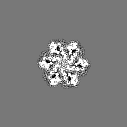
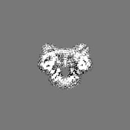
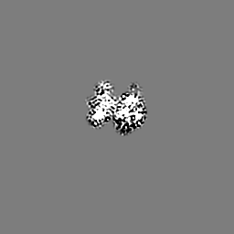
| Title | Symmetric hexamer of vaccinia virus DNA helicase D5 residues 323-785 | ||||||||||||||||||
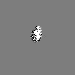
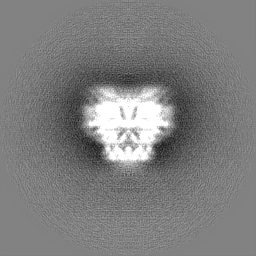
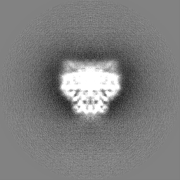
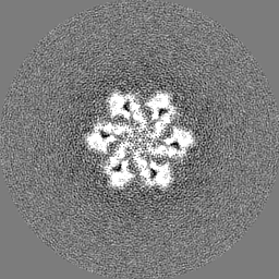
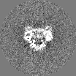
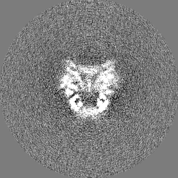
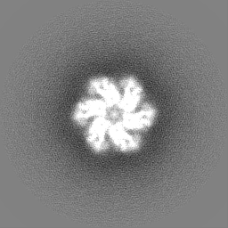
 Map data Map data | Flipped to correct hand, masked map from Relion PostRefine | ||||||||||||||||||
 Sample Sample |
| ||||||||||||||||||
 Keywords Keywords | DNA helicase / D5_N domain / DUF5906 domain / Pox_D5 domain / SF3 helicase / viral protein | ||||||||||||||||||
| Function / homology |  Function and homology information Function and homology informationhelicase activity / Hydrolases; Acting on acid anhydrides; Acting on acid anhydrides to facilitate cellular and subcellular movement / host cell cytoplasm / hydrolase activity / ATP binding Similarity search - Function | ||||||||||||||||||
| Biological species |  Vaccinia virus Copenhagen Vaccinia virus Copenhagen | ||||||||||||||||||
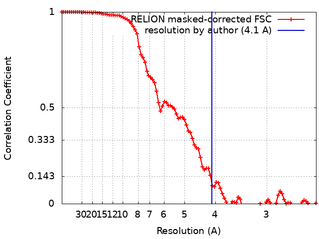
| Method | single particle reconstruction / cryo EM / Resolution: 4.1 Å | ||||||||||||||||||
 Authors Authors | Burmeister WP / Hutin S / Ling WL / Grimm C / Schoehn G | ||||||||||||||||||
| Funding support |  France, 5 items France, 5 items
| ||||||||||||||||||
 Citation Citation | Journal: Viruses / Year: 2022 Title: The Vaccinia Virus DNA Helicase Structure from Combined Single-Particle Cryo-Electron Microscopy and AlphaFold2 Prediction. Authors: Stephanie Hutin / Wai Li Ling / Nicolas Tarbouriech / Guy Schoehn / Clemens Grimm / Utz Fischer / Wim P Burmeister /  Abstract: Poxviruses are large DNA viruses with a linear double-stranded DNA genome circularized at the extremities. The helicase-primase D5, composed of six identical 90 kDa subunits, is required for DNA ...Poxviruses are large DNA viruses with a linear double-stranded DNA genome circularized at the extremities. The helicase-primase D5, composed of six identical 90 kDa subunits, is required for DNA replication. D5 consists of a primase fragment flexibly attached to the hexameric C-terminal polypeptide (res. 323-785) with confirmed nucleotide hydrolase and DNA-binding activity but an elusive helicase activity. We determined its structure by single-particle cryo-electron microscopy. It displays an AAA+ helicase core flanked by N- and C-terminal domains. Model building was greatly helped by the predicted structure of D5 using AlphaFold2. The 3.9 Å structure of the N-terminal domain forms a well-defined tight ring while the resolution decreases towards the C-terminus, still allowing the fit of the predicted structure. The N-terminal domain is partially present in papillomavirus E1 and polyomavirus LTA helicases, as well as in a bacteriophage NrS-1 helicase domain, which is also closely related to the AAA+ helicase domain of D5. Using the Pfam domain database, a D5_N domain followed by DUF5906 and Pox_D5 domains could be assigned to the cryo-EM structure, providing the first 3D structures for D5_N and Pox_D5 domains. The same domain organization has been identified in a family of putative helicases from large DNA viruses, bacteriophages, and selfish DNA elements. | ||||||||||||||||||
| History |
|
- Structure visualization
| Supplemental images |
|---|

- Downloads & links
-EMDB archive
| Map data | emd_15574.map.gz | 5 MB | EMDB map data format | |
|---|---|---|---|---|
| Header (meta data) | emd-15574-v30.xmlemd-15574.xml | 19.8 KB 19.8 KB | Display Display | EMDB header |
| FSC (resolution estimation) | emd_15574_fsc.xml | 9.1 KB | Display | FSC data file |
| Images | 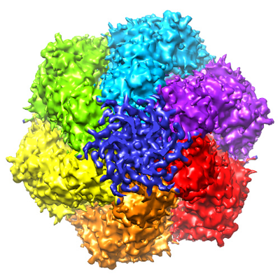 emd_15574.png emd_15574.png | 238.8 KB | ||
| Filedesc metadata | emd-15574.cif.gz | 6.3 KB | ||
| Others | emd_15574_half_map_1.map.gzemd_15574_half_map_2.map.gz | 48.7 MB 48.6 MB | ||
| Archive directory |  http://ftp.pdbj.org/pub/emdb/structures/EMD-15574ftp://ftp.pdbj.org/pub/emdb/structures/EMD-15574 http://ftp.pdbj.org/pub/emdb/structures/EMD-15574ftp://ftp.pdbj.org/pub/emdb/structures/EMD-15574 | HTTPS FTP |
-Related structure data
| Related structure data |  8aplMC  8apmC M: atomic model generated by this map C: citing same article ( |
|---|---|
| Similar structure data |

-Links
| EMDB pages | EMDB (EBI/PDBe) / EMDataResource |
|---|---|
| Related items in Molecule of the Month |


-Map
| File | Download / File: emd_15574.map.gz / Format: CCP4 / Size: 64 MB / Type: IMAGE STORED AS FLOATING POINT NUMBER (4 BYTES) | ||||||||||||||||||||||||||||||||||||
|---|---|---|---|---|---|---|---|---|---|---|---|---|---|---|---|---|---|---|---|---|---|---|---|---|---|---|---|---|---|---|---|---|---|---|---|---|---|
| Annotation | Flipped to correct hand, masked map from Relion PostRefine | ||||||||||||||||||||||||||||||||||||
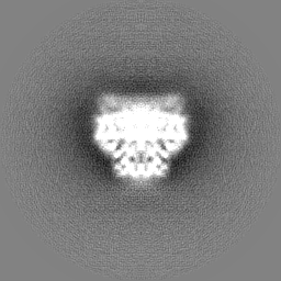
| Projections & slices | Image control
Images are generated by Spider. | ||||||||||||||||||||||||||||||||||||
| Voxel size | X=Y=Z: 1.21 Å | ||||||||||||||||||||||||||||||||||||
| Density |
| ||||||||||||||||||||||||||||||||||||
| Symmetry | Space group: 1 | ||||||||||||||||||||||||||||||||||||
| Details | EMDB XML:
|
 Z (Sec.)
 Y (Row.)
 X (Col.)


-Supplemental data
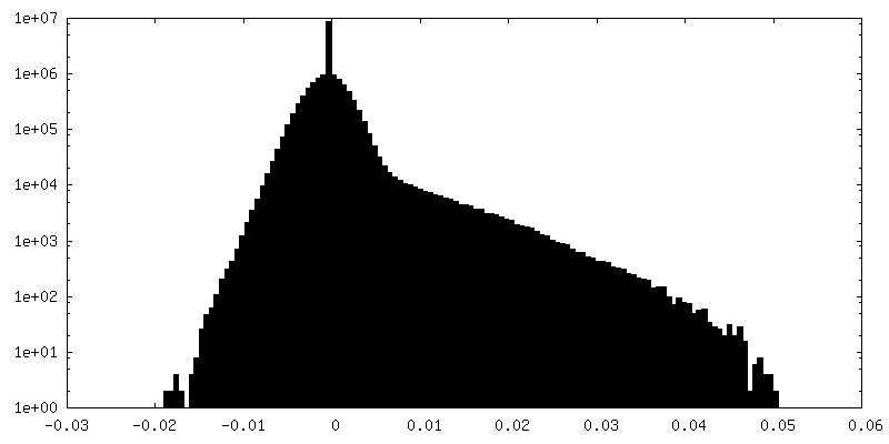
-Half map: Flipped to correct hand, half map from Relion...
| File | emd_15574_half_map_1.map | ||||||||||||
|---|---|---|---|---|---|---|---|---|---|---|---|---|---|
| Annotation | Flipped to correct hand, half map from Relion Refine3D after polishing. | ||||||||||||
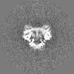
| Projections & Slices |
| ||||||||||||
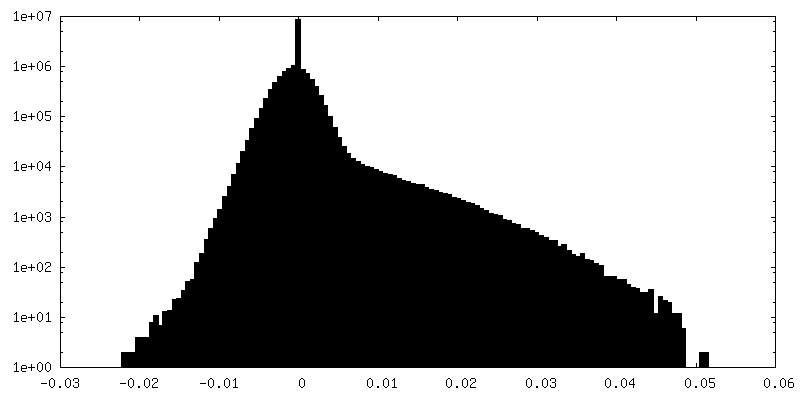
| Density Histograms |


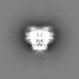
-Half map: Flipped to correct hand, half map from Relion Refine3D after polishin
| File | emd_15574_half_map_2.map | ||||||||||||
|---|---|---|---|---|---|---|---|---|---|---|---|---|---|
| Annotation | Flipped to correct hand, half map from Relion Refine3D after polishin | ||||||||||||
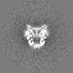
| Projections & Slices |
| ||||||||||||
| Density Histograms |


- Sample components
-Entire : D5 C-terminal fragment res. 323 - res. 785
| Entire | Name: D5 C-terminal fragment res. 323 - res. 785 |
|---|---|
| Components |
|
-Supramolecule #1: D5 C-terminal fragment res. 323 - res. 785
| Supramolecule | Name: D5 C-terminal fragment res. 323 - res. 785 / type: complex / ID: 1 / Parent: 0 / Macromolecule list: all / Details: construct |
|---|---|
| Source (natural) | Organism: Vaccinia virus Copenhagen |
| Molecular weight | Theoretical: 321 KDa |
-Macromolecule #1: Primase D5
| Macromolecule | Name: Primase D5 / type: protein_or_peptide / ID: 1 / Number of copies: 6 / Enantiomer: LEVO EC number: Hydrolases; Acting on acid anhydrides; Acting on acid anhydrides to facilitate cellular and subcellular movement |
|---|---|
| Source (natural) | Organism: Vaccinia virus Copenhagen / Strain: Copenhagen |
| Molecular weight | Theoretical: 53.495285 KDa |
| Recombinant expression | Organism:   Spodoptera frugiperda (fall armyworm) Spodoptera frugiperda (fall armyworm) |
| Sequence | String: AMGNKLFNIA QRILDTNSVL LTERGDYIVW INNSWKFNSE EPLITKLILS IRHQLPKEYS SELLCPRKRK TVEANIRDML VDSVETDTY PDKLPFKNGV LDLVDGMFYS GDDAKKYTCT VSTGFKFDDT KFVEDSPEME ELMNIINDIQ PLTDENKKNR E LYEKTLSS ...String: AMGNKLFNIA QRILDTNSVL LTERGDYIVW INNSWKFNSE EPLITKLILS IRHQLPKEYS SELLCPRKRK TVEANIRDML VDSVETDTY PDKLPFKNGV LDLVDGMFYS GDDAKKYTCT VSTGFKFDDT KFVEDSPEME ELMNIINDIQ PLTDENKKNR E LYEKTLSS CLCGATKGCL TFFFGETATG KSTTKRLLKS AIGDLFVETG QTILTDVLDK GPNPFIANMH LKRSVFCSEL PD FACSGSK KIRSDNIKKL TEPCVIGRPC FSNKINNRNH ATIIIDTNYK PVFDRIDNAL MRRIAVVRFR THFSQPSGRE AAE NNDAYD KVKLLDEGLD GKIQNNRYRF AFLYLLVKWY KKYHVPIMKL YPTPEEIPDF AFYLKIGTLL VSSSVKHIPL MTDL SKKGY ILYDNVVTLP LTTFQQKISK YFNSRLFGHD IESFINRHKK FANVSDEYLQ YIFIEDISSP UniProtKB: Uncoating factor OPG117 |
-Experimental details
-Structure determination
| Method | cryo EM |
|---|---|
 Processing Processing | single particle reconstruction |
| Aggregation state | particle |
-Sample preparation
| Concentration | 0.24 mg/mL | |||||||||||||||||||||
|---|---|---|---|---|---|---|---|---|---|---|---|---|---|---|---|---|---|---|---|---|---|---|
| Buffer | pH: 7 Component:
| |||||||||||||||||||||
| Grid | Model: Quantifoil R1.2/1.3 / Material: COPPER / Mesh: 300 / Support film - Material: CARBON / Support film - topology: HOLEY / Pretreatment - Type: GLOW DISCHARGE / Pretreatment - Atmosphere: AIR | |||||||||||||||||||||
| Vitrification | Cryogen name: ETHANE / Chamber humidity: 100 % / Chamber temperature: 293 K / Instrument: FEI VITROBOT MARK IV | |||||||||||||||||||||
| Details | The sample contained also a dsDNA oligomer in a close to stoechiometric concentration not visible in this structure. |
- Electron microscopy
| Microscope | FEI POLARA 300 |
|---|---|
| Image recording | Film or detector model: GATAN K2 SUMMIT (4k x 4k) / Detector mode: COUNTING / Digitization - Frames/image: 1-40 / Number grids imaged: 1 / Number real images: 830 / Average electron dose: 1.0 e/Å2 |
| Electron beam | Acceleration voltage: 300 kV / Electron source:  FIELD EMISSION GUN FIELD EMISSION GUN |
| Electron optics | Calibrated defocus max: 3.0 µm / Calibrated defocus min: 1.5 µm / Illumination mode: FLOOD BEAM / Imaging mode: BRIGHT FIELD / Nominal defocus max: 3.0 µm / Nominal defocus min: 1.5 µm |
| Sample stage | Cooling holder cryogen: NITROGEN |
| Experimental equipment |  Model: Tecnai Polara / Image courtesy: FEI Company |
+Image processing
-Atomic model buiding 1
| Refinement | Space: REAL / Protocol: OTHER / Overall B value: 152 |
|---|---|
| Output model | PDB-8apl: |
