 Movie
Movie Controller

[English] 日本語
 Yorodumi
- EMDB-15051: Localized reconstruction of flavobacterium infecting lipid-contai... -
+ Open data

- Basic information
| Entry |  | |||||||||
|---|---|---|---|---|---|---|---|---|---|---|
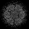
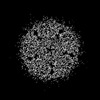
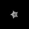
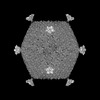
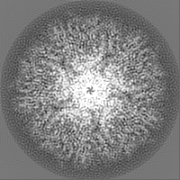
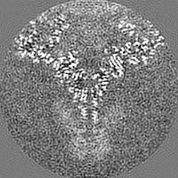
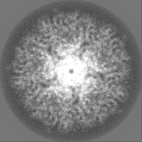
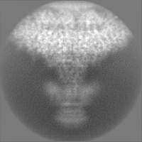
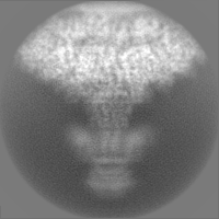
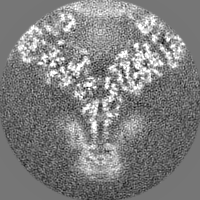
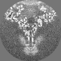
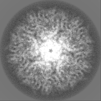
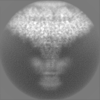
| Title | Localized reconstruction of flavobacterium infecting lipid-containing phage FLiP vertex | |||||||||
 Map data Map data | ||||||||||
 Sample Sample |
| |||||||||
 Keywords Keywords | bacteriophage / Flavobacterium / lipid-containing / phage / FLiP / penton protein / VIRUS | |||||||||
| Function / homology | DUF3168 domain-containing protein Function and homology information Function and homology information | |||||||||
| Biological species |  Flavobacterium phage FLiP (virus) Flavobacterium phage FLiP (virus) | |||||||||
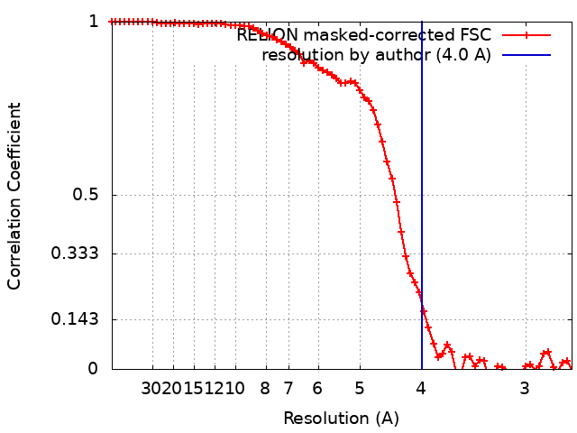
| Method | single particle reconstruction / cryo EM / Resolution: 4.0 Å | |||||||||
 Authors Authors | Kejzar N / Abrishami V / Selvaraj M / Huiskonen JT | |||||||||
| Funding support | 1 items
| |||||||||
 Citation Citation | Journal: Nat Commun / Year: 2022 Title: Cryo-EM structure of ssDNA bacteriophage ΦCjT23 provides insight into early virus evolution. Authors: Nejc Kejzar / Elina Laanto / Ilona Rissanen / Vahid Abrishami / Muniyandi Selvaraj / Sylvain Moineau / Janne Ravantti / Lotta-Riina Sundberg / Juha T Huiskonen /    Abstract: The origin of viruses remains an open question. While lack of detectable sequence similarity hampers the analysis of distantly related viruses, structural biology investigations of conserved capsid ...The origin of viruses remains an open question. While lack of detectable sequence similarity hampers the analysis of distantly related viruses, structural biology investigations of conserved capsid protein structures facilitate the study of distant evolutionary relationships. Here we characterize the lipid-containing ssDNA temperate bacteriophage ΦCjT23, which infects Flavobacterium sp. (Bacteroidetes). We report ΦCjT23-like sequences in the genome of strains belonging to several Flavobacterium species. The virion structure determined by cryogenic electron microscopy reveals similarities to members of the viral kingdom Bamfordvirae that currently consists solely of dsDNA viruses with a major capsid protein composed of two upright β-sandwiches. The minimalistic structure of ΦCjT23 suggests that this phage serves as a model for the last common ancestor between ssDNA and dsDNA viruses in the Bamfordvirae. Both ΦCjT23 and the related phage FLiP infect Flavobacterium species found in several environments, suggesting that these types of viruses have a global distribution and a shared evolutionary origin. Detailed comparisons to related, more complex viruses not only expand our knowledge about this group of viruses but also provide a rare glimpse into early virus evolution. #1: Journal: Proc Natl Acad Sci U S A / Year: 2017Title: Virus found in a boreal lake links ssDNA and dsDNA viruses. Authors: Laanto E / Mantynen S / De Colibus L / Marjakangas J / Gillum A / Stuart DI / Ravantti JJ / Huiskonen JT / Sundberg LR | |||||||||
| History |
|
- Structure visualization
| Supplemental images |
|---|

- Downloads & links
-EMDB archive
| Map data | emd_15051.map.gz | 28.5 MB | EMDB map data format | |
|---|---|---|---|---|
| Header (meta data) | emd-15051-v30.xmlemd-15051.xml | 15.3 KB 15.3 KB | Display Display | EMDB header |
| FSC (resolution estimation) | emd_15051_fsc.xml | 7.1 KB | Display | FSC data file |
| Images | 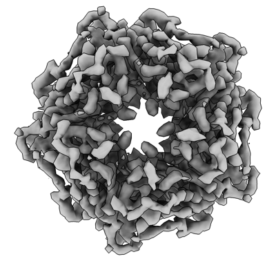 emd_15051.png emd_15051.png | 124 KB | ||
| Masks | emd_15051_msk_1.map | 30.5 MB | Mask map | |
| Filedesc metadata | emd-15051.cif.gz | 5.2 KB | ||
| Others | emd_15051_half_map_1.map.gzemd_15051_half_map_2.map.gz | 23.3 MB 23.3 MB | ||
| Archive directory |  http://ftp.pdbj.org/pub/emdb/structures/EMD-15051ftp://ftp.pdbj.org/pub/emdb/structures/EMD-15051 http://ftp.pdbj.org/pub/emdb/structures/EMD-15051ftp://ftp.pdbj.org/pub/emdb/structures/EMD-15051 | HTTPS FTP |
-Validation report
| Summary document | emd_15051_validation.pdf.gz | 1.2 MB | Display | EMDB validaton report |
|---|---|---|---|---|
| Full document | emd_15051_full_validation.pdf.gz | 1.2 MB | Display | |
| Data in XML | emd_15051_validation.xml.gz | 12.6 KB | Display | |
| Data in CIF | emd_15051_validation.cif.gz | 17.2 KB | Display | |
| Arichive directory | https://ftp.pdbj.org/pub/emdb/validation_reports/EMD-15051ftp://ftp.pdbj.org/pub/emdb/validation_reports/EMD-15051 | HTTPS FTP |
-Related structure data
| Related structure data | 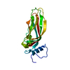 8a06MC 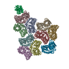 7zzzC 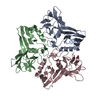 8a01C 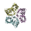 8a02C 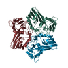 8a03C 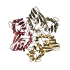 8a04C 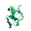 8a05C M: atomic model generated by this map C: citing same article ( |
|---|---|
| Similar structure data |

-Links
| EMDB pages | EMDB (EBI/PDBe) / EMDataResource |
|---|
-Map
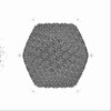
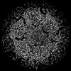
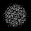
| File | Download / File: emd_15051.map.gz / Format: CCP4 / Size: 30.5 MB / Type: IMAGE STORED AS FLOATING POINT NUMBER (4 BYTES) | ||||||||||||||||||||||||||||||||||||
|---|---|---|---|---|---|---|---|---|---|---|---|---|---|---|---|---|---|---|---|---|---|---|---|---|---|---|---|---|---|---|---|---|---|---|---|---|---|
| Projections & slices | Image control
Images are generated by Spider. | ||||||||||||||||||||||||||||||||||||
| Voxel size | X=Y=Z: 1.35 Å | ||||||||||||||||||||||||||||||||||||
| Density |
| ||||||||||||||||||||||||||||||||||||
| Symmetry | Space group: 1 | ||||||||||||||||||||||||||||||||||||
| Details | EMDB XML:
|
 Z (Sec.)
 Y (Row.)
 X (Col.)


-Supplemental data
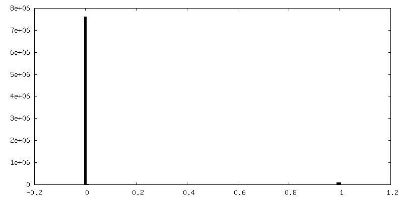
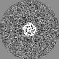
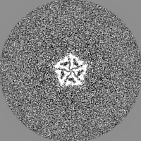
-Mask #1
| File | emd_15051_msk_1.map | ||||||||||||
|---|---|---|---|---|---|---|---|---|---|---|---|---|---|
| Projections & Slices |
| ||||||||||||
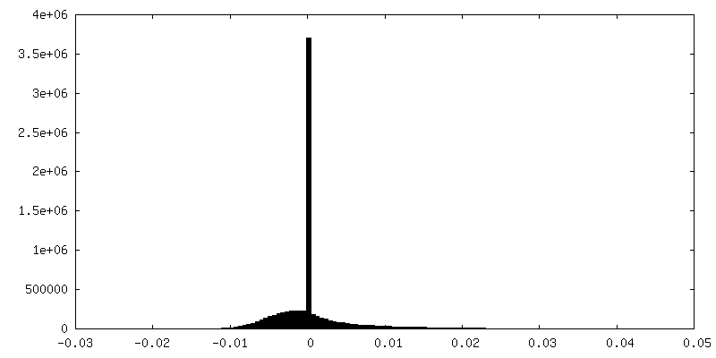
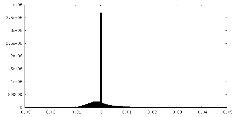
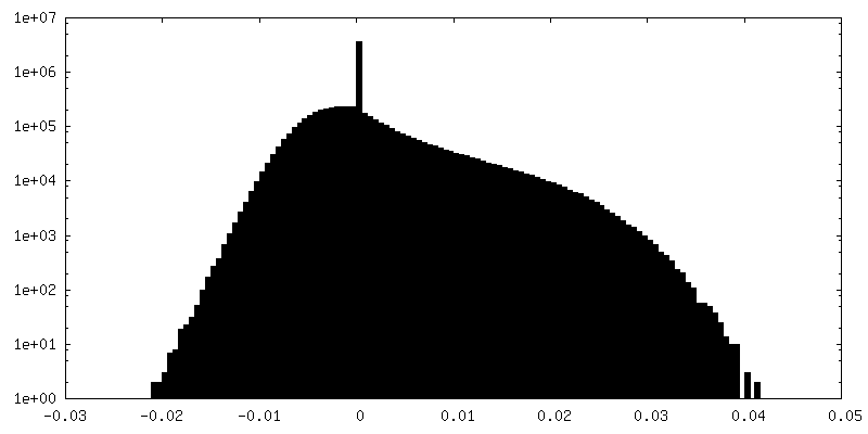
| Density Histograms |


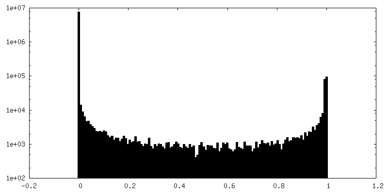
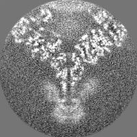
-Half map: #1
| File | emd_15051_half_map_1.map | ||||||||||||
|---|---|---|---|---|---|---|---|---|---|---|---|---|---|
| Projections & Slices |
| ||||||||||||
| Density Histograms |

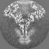
-Half map: #2
| File | emd_15051_half_map_2.map | ||||||||||||
|---|---|---|---|---|---|---|---|---|---|---|---|---|---|
| Projections & Slices |
| ||||||||||||
| Density Histograms |

- Sample components
-Entire : Flavobacterium phage FLiP
| Entire | Name: Flavobacterium phage FLiP (virus) |
|---|---|
| Components |
|
-Supramolecule #1: Flavobacterium phage FLiP
| Supramolecule | Name: Flavobacterium phage FLiP / type: virus / ID: 1 / Parent: 0 / Macromolecule list: all / NCBI-ID: 2023716 / Sci species name: Flavobacterium phage FLiP / Virus type: VIRION / Virus isolate: SPECIES / Virus enveloped: Yes / Virus empty: No |
|---|---|
| Host (natural) | Organism:  Flavobacterium (bacteria) Flavobacterium (bacteria) |
-Macromolecule #1: Penton protein P12
| Macromolecule | Name: Penton protein P12 / type: protein_or_peptide / ID: 1 / Number of copies: 1 / Enantiomer: LEVO |
|---|---|
| Source (natural) | Organism: Flavobacterium phage FLiP (virus) |
| Molecular weight | Theoretical: 16.851057 KDa |
| Sequence | String: DFSTIPIDYV KAKDPNTIDF CLSYLELYHT TKAVKACTPF SFILGSDAGM QRATETTESL YWGKVILDIN PNLSPLVNTT IVLEIESML SSNSINRSEN KRITRYIEKE NFVNESSERF EFFKSMELSH LSTAYDVYVT FIGFKIDL UniProtKB: DUF3168 domain-containing protein |
-Experimental details
-Structure determination
| Method | cryo EM |
|---|---|
 Processing Processing | single particle reconstruction |
| Aggregation state | particle |
-Sample preparation
| Buffer | pH: 7.2 |
|---|---|
| Vitrification | Cryogen name: ETHANE |
- Electron microscopy
| Microscope | FEI POLARA 300 |
|---|---|
| Image recording | Film or detector model: GATAN K2 SUMMIT (4k x 4k) / Average electron dose: 22.0 e/Å2 |
| Electron beam | Acceleration voltage: 300 kV / Electron source:  FIELD EMISSION GUN FIELD EMISSION GUN |
| Electron optics | Illumination mode: FLOOD BEAM / Imaging mode: BRIGHT FIELD / Nominal defocus max: 2.5 µm / Nominal defocus min: 0.7000000000000001 µm |
| Experimental equipment |  Model: Tecnai Polara / Image courtesy: FEI Company |
+Image processing
-Atomic model buiding 1
| Refinement | Protocol: AB INITIO MODEL |
|---|---|
| Output model | PDB-8a06: |
