 Movie
Movie Controller


+ Open data

- Basic information
| Entry |  | |||||||||
|---|---|---|---|---|---|---|---|---|---|---|
| Title | RNA origami 5-helix tile | |||||||||
 Map data Map data | ||||||||||
 Sample Sample |
| |||||||||
 Keywords Keywords | RNA / origami / nanostructure | |||||||||
| Biological species | synthetic construct (others) | |||||||||
| Method | single particle reconstruction / cryo EM / Resolution: 4.08 Å | |||||||||
 Authors Authors | McRae EKS / Bogglid A / Boesen T / Andersen ES | |||||||||
| Funding support |  Denmark, 1 items Denmark, 1 items
| |||||||||
 Citation Citation | Journal: Nat Nanotechnol / Year: 2023 Title: Structure, folding and flexibility of co-transcriptional RNA origami. Authors: Ewan K S McRae / Helena Østergaard Rasmussen / Jianfang Liu / Andreas Bøggild / Michael T A Nguyen / Nestor Sampedro Vallina / Thomas Boesen / Jan Skov Pedersen / Gang Ren / Cody Geary / ...Authors: Ewan K S McRae / Helena Østergaard Rasmussen / Jianfang Liu / Andreas Bøggild / Michael T A Nguyen / Nestor Sampedro Vallina / Thomas Boesen / Jan Skov Pedersen / Gang Ren / Cody Geary / Ebbe Sloth Andersen /  Abstract: RNA origami is a method for designing RNA nanostructures that can self-assemble through co-transcriptional folding with applications in nanomedicine and synthetic biology. However, to advance the ...RNA origami is a method for designing RNA nanostructures that can self-assemble through co-transcriptional folding with applications in nanomedicine and synthetic biology. However, to advance the method further, an improved understanding of RNA structural properties and folding principles is required. Here we use cryogenic electron microscopy to study RNA origami sheets and bundles at sub-nanometre resolution revealing structural parameters of kissing-loop and crossover motifs, which are used to improve designs. In RNA bundle designs, we discover a kinetic folding trap that forms during folding and is only released after 10 h. Exploration of the conformational landscape of several RNA designs reveal the flexibility of helices and structural motifs. Finally, sheets and bundles are combined to construct a multidomain satellite shape, which is characterized by individual-particle cryo-electron tomography to reveal the domain flexibility. Together, the study provides a structural basis for future improvements to the design cycle of genetically encoded RNA nanodevices. | |||||||||
| History |
|
- Structure visualization
| Supplemental images |
|---|

- Downloads & links
-EMDB archive
| Map data | emd_13633.map.gz | 32.1 MB |  EMDB map data format EMDB map data format | |
|---|---|---|---|---|
| Header (meta data) | emd-13633-v30.xmlemd-13633.xml | 23.3 KB 23.3 KB | Display Display | EMDB header |
| FSC (resolution estimation) | emd_13633_fsc.xml | 8.9 KB | Display | FSC data file |
| Images | 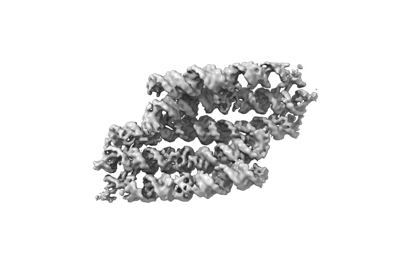 emd_13633.png emd_13633.png | 39.6 KB | ||
| Masks | emd_13633_msk_1.map | 64 MB | Mask map | |
| Filedesc metadata | emd-13633.cif.gz | 6.1 KB | ||
| Others | emd_13633_additional_1.map.gzemd_13633_additional_2.map.gzemd_13633_half_map_1.map.gzemd_13633_half_map_2.map.gz | 59.3 MB 904.4 KB 59.2 MB 59.2 MB | ||
| Archive directory |  http://ftp.pdbj.org/pub/emdb/structures/EMD-13633ftp://ftp.pdbj.org/pub/emdb/structures/EMD-13633 http://ftp.pdbj.org/pub/emdb/structures/EMD-13633ftp://ftp.pdbj.org/pub/emdb/structures/EMD-13633 | HTTPS FTP |
-Validation report
| Summary document | emd_13633_validation.pdf.gz | 829.6 KB | Display | EMDB validaton report |
|---|---|---|---|---|
| Full document | emd_13633_full_validation.pdf.gz | 829.1 KB | Display | |
| Data in XML | emd_13633_validation.xml.gz | 16.1 KB | Display | |
| Data in CIF | emd_13633_validation.cif.gz | 20.5 KB | Display | |
| Arichive directory | https://ftp.pdbj.org/pub/emdb/validation_reports/EMD-13633ftp://ftp.pdbj.org/pub/emdb/validation_reports/EMD-13633 | HTTPS FTP |
-Related structure data
| Related structure data |  7ptqMC  7ptkC  7ptlC 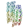 7ptsC 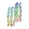 7qduC M: atomic model generated by this map C: citing same article ( |
|---|


-Links
| EMDB pages | EMDB (EBI/PDBe) / EMDataResource |
|---|
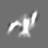
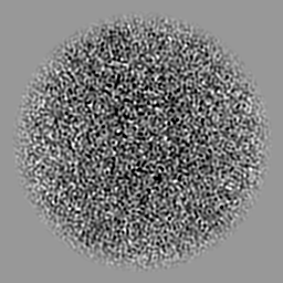
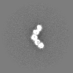
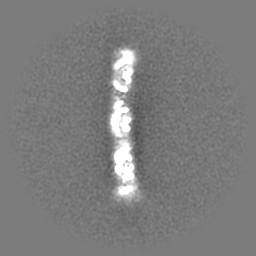
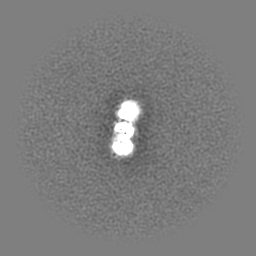
-Map
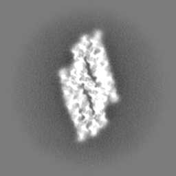
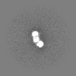
| File | Download / File: emd_13633.map.gz / Format: CCP4 / Size: 64 MB / Type: IMAGE STORED AS FLOATING POINT NUMBER (4 BYTES) | ||||||||||||||||||||||||||||||||||||
|---|---|---|---|---|---|---|---|---|---|---|---|---|---|---|---|---|---|---|---|---|---|---|---|---|---|---|---|---|---|---|---|---|---|---|---|---|---|
| Projections & slices | Image control
Images are generated by Spider. | ||||||||||||||||||||||||||||||||||||
| Voxel size | X=Y=Z: 1.29 Å | ||||||||||||||||||||||||||||||||||||
| Density |
| ||||||||||||||||||||||||||||||||||||
| Symmetry | Space group: 1 | ||||||||||||||||||||||||||||||||||||
| Details | EMDB XML:
|
 Z (Sec.)
 Y (Row.)
 X (Col.)


-Supplemental data
-Mask #1
| File | emd_13633_msk_1.map | ||||||||||||
|---|---|---|---|---|---|---|---|---|---|---|---|---|---|
| Projections & Slices |
| ||||||||||||
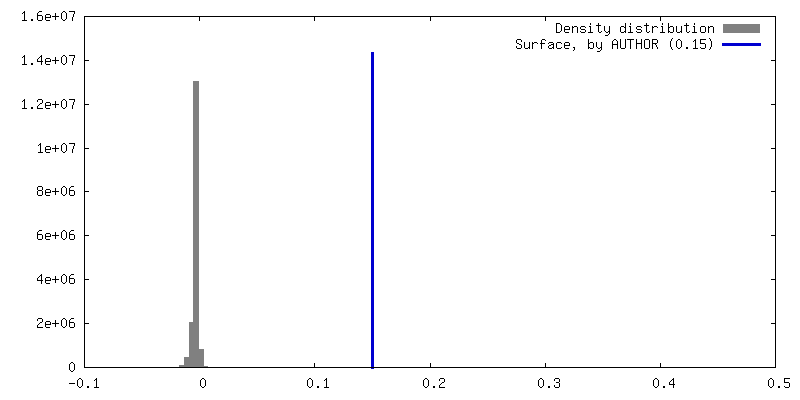
| Density Histograms |


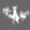
-Additional map: Sharpened map from cryoSPARC with bfactor of 163
| File | emd_13633_additional_1.map | ||||||||||||
|---|---|---|---|---|---|---|---|---|---|---|---|---|---|
| Annotation | Sharpened map from cryoSPARC with bfactor of 163 | ||||||||||||
| Projections & Slices |
| ||||||||||||
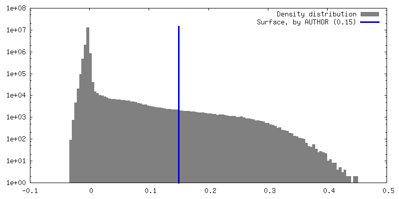
| Density Histograms |


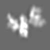
-Additional map: Local refinement from cryoSPARC using mask covering the...
| File | emd_13633_additional_2.map | ||||||||||||
|---|---|---|---|---|---|---|---|---|---|---|---|---|---|
| Annotation | Local refinement from cryoSPARC using mask covering the central 3 helices. | ||||||||||||
| Projections & Slices |
| ||||||||||||
| Density Histograms |


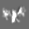
-Half map: #2
| File | emd_13633_half_map_1.map | ||||||||||||
|---|---|---|---|---|---|---|---|---|---|---|---|---|---|
| Projections & Slices |
| ||||||||||||
| Density Histograms |


-Half map: #1
| File | emd_13633_half_map_2.map | ||||||||||||
|---|---|---|---|---|---|---|---|---|---|---|---|---|---|
| Projections & Slices |
| ||||||||||||
| Density Histograms |


- Sample components
-Entire : 5 helix tile A
| Entire | Name: 5 helix tile A |
|---|---|
| Components |
|
-Supramolecule #1: 5 helix tile A
| Supramolecule | Name: 5 helix tile A / type: complex / ID: 1 / Parent: 0 / Macromolecule list: all Details: In vitro transcribed RNA purified by SEC. This is the young conformer. |
|---|---|
| Source (natural) | Organism: synthetic construct (others) |
| Molecular weight | Theoretical: 175 KDa |
-Macromolecule #1: Chains: C
| Macromolecule | Name: Chains: C / type: rna / ID: 1 / Details: In vitro transcribed RNA / Number of copies: 1 |
|---|---|
| Source (natural) | Organism: synthetic construct (others) |
| Molecular weight | Theoretical: 175.236297 KDa |
| Sequence | String: GGAAUUAGAG UGUGUUCCUG AACUGCUUCG GCGGUUCGCU ACGUUCUUCG GAAUGUAUAU AGUGUUCGCA UUAUACCGUA GUCCAAGCC GUGUGGUUCG CCGCACGUCG UUCGUUCGCG GACGAGCAGG UGCCAUAACC UCCAAAUGGU ACCUGCGCGU G UUGUCAGC ...String: GGAAUUAGAG UGUGUUCCUG AACUGCUUCG GCGGUUCGCU ACGUUCUUCG GAAUGUAUAU AGUGUUCGCA UUAUACCGUA GUCCAAGCC GUGUGGUUCG CCGCACGUCG UUCGUUCGCG GACGAGCAGG UGCCAUAACC UCCAAAUGGU ACCUGCGCGU G UUGUCAGC AAGGUCUAAG CUGAUAACAC UAUGCUAACG ACUGAAGCAU AUUGGAUUAC GGGCCAAGGG CAGCUAAGAU CG GAAGCUG UCCUUGAGGA ACGCACUCUG AUUCCCCUCC GGAAGGAGCC CCACAGGUAA UAAACCGAUC AUAUUACUUG UGC ACUCGC AACAGUCGAG CGGGUGGUAA UGAUUGCGCC CGUUGGCUAG AAUAGACCAC UAGCUAACGG CGGGUCUUGG AUCA AUGGA GGAGAUCCAG GACCCGACCC GGACUUCGGU UCGGGUGCGG CCUUAGUUCG CUGAGGCCCG UCGCAUUCGU GUGAC GGUG GUUAUUGCGG UCAAGGUUUC GACUUUGAGU AUUCCUUCGG GGAUACGCUU CUUCUGGAGG |
-Experimental details
-Structure determination
| Method | cryo EM |
|---|---|
 Processing Processing | single particle reconstruction |
| Aggregation state | particle |
-Sample preparation
| Concentration | 3.3 mg/mL | ||||||||||||
|---|---|---|---|---|---|---|---|---|---|---|---|---|---|
| Buffer | pH: 8 Component:
Details: Freshly prepared and filtered through 0.22 micron filter prior to use. | ||||||||||||
| Vitrification | Cryogen name: ETHANE / Chamber humidity: 99 % / Chamber temperature: 294 K / Instrument: LEICA EM GP Details: 3 microlitre droplet, 4 second delay before blotting, 6 second blot, 0 second delay before plunging.. | ||||||||||||
| Details | Sample was purified by size exclusion chromatography and concentrated in an Amicon spin concentrator. |
- Electron microscopy
| Microscope | FEI TITAN KRIOS |
|---|---|
| Specialist optics | Energy filter - Name: GIF Bioquantum / Energy filter - Slit width: 20 eV |
| Image recording | Film or detector model: GATAN K3 BIOQUANTUM (6k x 4k) / Number grids imaged: 1 / Number real images: 13524 / Average exposure time: 1.5 sec. / Average electron dose: 60.0 e/Å2 / Details: Images were collected as 56 frame movies. |
| Electron beam | Acceleration voltage: 300 kV / Electron source:  FIELD EMISSION GUN FIELD EMISSION GUN |
| Electron optics | C2 aperture diameter: 50.0 µm / Illumination mode: FLOOD BEAM / Imaging mode: BRIGHT FIELD / Cs: 2.7 mm / Nominal defocus max: 2.5 µm / Nominal defocus min: 0.75 µm / Nominal magnification: 130000 |
| Sample stage | Specimen holder model: FEI TITAN KRIOS AUTOGRID HOLDER / Cooling holder cryogen: NITROGEN |
| Experimental equipment |  Model: Titan Krios / Image courtesy: FEI Company |
+Image processing

-Atomic model buiding 1
| Initial model | PDB ID: Chain - Source name: PDB / Chain - Initial model type: experimental model |
|---|---|
| Details | Helical templates and tetraloops were generated in RNAbuild. Kissing loops were modelling based on the PDB:2D1B structure. A combination of PHENIX RSR and ISOLDE were used to arrive at the final structure. |
| Refinement | Space: REAL / Protocol: FLEXIBLE FIT / Overall B value: 303 / Target criteria: Correlation Coefficient |
| Output model | PDB-7ptq: |

