+データを開く
-基本情報
登録情報 | データベース: EMDB / ID: EMD-1344 | |||||||||
---|---|---|---|---|---|---|---|---|---|---|
タイトル | Structures of modified eEF2 80S ribosome complexes reveal the role of GTP hydrolysis in translocation. | |||||||||
マップデータ | Cryo-EM map of the 80S ribosome:eEF2:GDPNP complex | |||||||||
試料 |
| |||||||||
機能・相同性 | 機能・相同性情報 Peptide chain elongation / Synthesis of diphthamide-EEF2 / positive regulation of translational elongation / Protein methylation / translational elongation / translation elongation factor activity / Neutrophil degranulation / 加水分解酵素; 酸無水物に作用; GTPに作用・細胞または細胞小器官の運動に関与 / maintenance of translational fidelity / ribosome binding ...Peptide chain elongation / Synthesis of diphthamide-EEF2 / positive regulation of translational elongation / Protein methylation / translational elongation / translation elongation factor activity / Neutrophil degranulation / 加水分解酵素; 酸無水物に作用; GTPに作用・細胞または細胞小器官の運動に関与 / maintenance of translational fidelity / ribosome binding / protein-folding chaperone binding / rRNA binding / ribonucleoprotein complex / GTPase activity / GTP binding / identical protein binding / cytosol 類似検索 - 分子機能 | |||||||||
生物種 | Saccharomyces cerevisiae (パン酵母) / Thermomyces lanuginosus (菌類) | |||||||||
手法 | 単粒子再構成法 / クライオ電子顕微鏡法 / ネガティブ染色法 / 解像度: 11.7 Å | |||||||||
データ登録者 | Taylor DJ / Nilsson J / Merrill AR / Andersen GR / Nissen P / Frank J | |||||||||
引用 | 
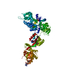
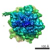
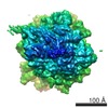
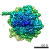
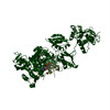
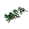
ジャーナル: EMBO J / 年: 2007 タイトル: Structures of modified eEF2 80S ribosome complexes reveal the role of GTP hydrolysis in translocation. 著者: Derek J Taylor / Jakob Nilsson / A Rod Merrill / Gregers Rom Andersen / Poul Nissen / Joachim Frank / 要旨: On the basis of kinetic data on ribosome protein synthesis, the mechanical energy for translocation of the mRNA-tRNA complex is thought to be provided by GTP hydrolysis of an elongation factor (eEF2 ...On the basis of kinetic data on ribosome protein synthesis, the mechanical energy for translocation of the mRNA-tRNA complex is thought to be provided by GTP hydrolysis of an elongation factor (eEF2 in eukaryotes, EF-G in bacteria). We have obtained cryo-EM reconstructions of eukaryotic ribosomes complexed with ADP-ribosylated eEF2 (ADPR-eEF2), before and after GTP hydrolysis, providing a structural basis for analyzing the GTPase-coupled mechanism of translocation. Using the ADP-ribosyl group as a distinct marker, we observe conformational changes of ADPR-eEF2 that are due strictly to GTP hydrolysis. These movements are likely representative of native eEF2 motions in a physiological context and are sufficient to uncouple the mRNA-tRNA complex from two universally conserved bases in the ribosomal decoding center (A1492 and A1493 in Escherichia coli) during translocation. Interpretation of these data provides a detailed two-step model of translocation that begins with the eEF2/EF-G binding-induced ratcheting motion of the small ribosomal subunit. GTP hydrolysis then uncouples the mRNA-tRNA complex from the decoding center so translocation of the mRNA-tRNA moiety may be completed by a head rotation of the small subunit. | |||||||||
履歴 |
|
-構造の表示
ムービー |
ムービービューア |
---|---|
構造ビューア | EMマップ: SurfViewMolmilJmol/JSmol |
添付画像 |
-ダウンロードとリンク
-EMDBアーカイブ
マップデータ | emd_1344.map.gz | 30.9 MB | EMDBマップデータ形式 | |
---|---|---|---|---|
ヘッダ (付随情報) | emd-1344-v30.xml emd-1344.xml | 10.7 KB 10.7 KB | 表示 表示 | EMDBヘッダ |
画像 | 1344.gif | 74.2 KB | ||
アーカイブディレクトリ | http://ftp.pdbj.org/pub/emdb/structures/EMD-1344
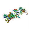
 ftp://ftp.pdbj.org/pub/emdb/structures/EMD-1344 | HTTPS FTP |
-検証レポート
文書・要旨 | emd_1344_validation.pdf.gz | 328.2 KB | 表示 | EMDB検証レポート |
---|---|---|---|---|
文書・詳細版 | emd_1344_full_validation.pdf.gz | 327.7 KB | 表示 | |
XML形式データ | emd_1344_validation.xml.gz | 6.4 KB | 表示 | |
アーカイブディレクトリ | https://ftp.pdbj.org/pub/emdb/validation_reports/EMD-1344 ftp://ftp.pdbj.org/pub/emdb/validation_reports/EMD-1344 | HTTPS FTP |
-関連構造データ
-リンク
EMDBのページ | EMDB (EBI/PDBe) / EMDataResource |
---|---|
「今月の分子」の関連する項目 |
-マップ
ファイル | ダウンロード / ファイル: emd_1344.map.gz / 形式: CCP4 / 大きさ: 32.1 MB / タイプ: IMAGE STORED AS FLOATING POINT NUMBER (4 BYTES) | ||||||||||||||||||||||||||||||||||||||||||||||||||||||||||||||||||||
---|---|---|---|---|---|---|---|---|---|---|---|---|---|---|---|---|---|---|---|---|---|---|---|---|---|---|---|---|---|---|---|---|---|---|---|---|---|---|---|---|---|---|---|---|---|---|---|---|---|---|---|---|---|---|---|---|---|---|---|---|---|---|---|---|---|---|---|---|---|
注釈 | Cryo-EM map of the 80S ribosome:eEF2:GDPNP complex | ||||||||||||||||||||||||||||||||||||||||||||||||||||||||||||||||||||
ボクセルのサイズ | X=Y=Z: 1.86 Å | ||||||||||||||||||||||||||||||||||||||||||||||||||||||||||||||||||||
密度 |
| ||||||||||||||||||||||||||||||||||||||||||||||||||||||||||||||||||||
対称性 | 空間群: 1 | ||||||||||||||||||||||||||||||||||||||||||||||||||||||||||||||||||||
詳細 | EMDB XML:
CCP4マップ ヘッダ情報:
|
-添付データ
-試料の構成要素
-全体 : Thermomyces lanuginosus 80S ribosome
全体 | 名称: Thermomyces lanuginosus 80S ribosome |
---|---|
要素 |
|
-超分子 #1000: Thermomyces lanuginosus 80S ribosome
超分子 | 名称: Thermomyces lanuginosus 80S ribosome / タイプ: sample / ID: 1000 / Number unique components: 3 |
---|---|
分子量 | 理論値: 3.0 MDa |
-超分子 #1: Thermomyces lanuginosus
超分子 | 名称: Thermomyces lanuginosus / タイプ: complex / ID: 1 / Ribosome-details: ribosome-eukaryote: ALL |
---|---|
分子量 | 理論値: 3.0 MDa |
-分子 #1: ADPR-eEF2
分子 | 名称: ADPR-eEF2 / タイプ: protein_or_peptide / ID: 1 / 組換発現: No |
---|---|
由来(天然) | 生物種: Saccharomyces cerevisiae (パン酵母) |
分子量 | 理論値: 93 KDa |
-分子 #2: tRNA
分子 | 名称: tRNA / タイプ: rna / ID: 2 / 分類: TRANSFER / Structure: SINGLE STRANDED / Synthetic?: No |
---|---|
由来(天然) | 生物種: Thermomyces lanuginosus (菌類) |
-実験情報
-構造解析
手法 | ネガティブ染色法, クライオ電子顕微鏡法 |
---|---|
解析 | 単粒子再構成法 |
試料の集合状態 | particle |
-試料調製
緩衝液 | pH: 7.5 / 詳細: see Methods |
---|---|
染色 | タイプ: NEGATIVE / 詳細: No staining (Cryo-EM) |
グリッド | 詳細: Quanti-foil grids coated with a thin carbon layer |
凍結 | 凍結剤: ETHANE / チャンバー内湿度: 90 % / チャンバー内温度: 279 K / 装置: OTHER / 詳細: Vitrification instrument: Vitrobot 手法: Apply sample, wait 30s, blot for 6s, plunge in liquid ethane |
-電子顕微鏡法
顕微鏡 | FEI TECNAI F30 |
---|---|
温度 | 平均: 84 K |
日付 | 2005年7月7日 |
撮影 | カテゴリ: FILM / フィルム・検出器のモデル: KODAK SO-163 FILM / デジタル化 - スキャナー: ZEISS SCAI / デジタル化 - サンプリング間隔: 7 µm / 実像数: 79 / 平均電子線量: 25 e/Å2 / ビット/ピクセル: 12 |
Tilt angle min | 0 |
Tilt angle max | 0 |
電子線 | 加速電圧: 300 kV / 電子線源: FIELD EMISSION GUN |
電子光学系 | 倍率(補正後): 37642 / 照射モード: FLOOD BEAM / 撮影モード: BRIGHT FIELD / Cs: 2.26 mm / 最大 デフォーカス(公称値): 3.7 µm / 最小 デフォーカス(公称値): 0.8 µm / 倍率(公称値): 39000 |
試料ステージ | 試料ホルダー: cartridge / 試料ホルダーモデル: OTHER |
実験機器 | モデル: Tecnai F30 / 画像提供: FEI Company |
-画像解析
CTF補正 | 詳細: CTF correction of 3D map |
---|---|
最終 再構成 | 想定した対称性 - 点群: C1 (非対称) / アルゴリズム: OTHER / 解像度のタイプ: BY AUTHOR / 解像度: 11.7 Å / 解像度の算出法: FSC 0.5 CUT-OFF / ソフトウェア - 名称: SPIDER package / 使用した粒子像数: 70510 |