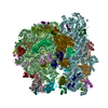
- EMDB-13058: Cryo-EM structure of an Escherichia coli 70S ribosome in complex ... -
+
データを開く
IDまたはキーワード:
読み込み中...
-
基本情報
登録情報
データベース: EMDB / ID: EMD-13058
タイトル
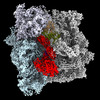
Cryo-EM structure of an Escherichia coli 70S ribosome in complex with elongation factor G and the antibiotic Argyrin B
マップデータ
Locally filtered and merged multibody refined map of an E. coli 70S - EF-G - Argyrin B complex
試料
複合体: Cryo-EM structure of an Escherichia coli 70S ribosome in complex with elongation factor G and the antibiotic ArgyrinB
RNA: x 3種
タンパク質・ペプチド: x 49種
リガンド: x 7種
キーワード
antibiotic / ribosome / translation
機能・相同性
機能・相同性情報
translation elongation factor activity / large ribosomal subunit / small ribosomal subunit / transferase activity / 5S rRNA binding / large ribosomal subunit rRNA binding / cytoplasmic translation / cytosolic large ribosomal subunit / tRNA binding / rRNA binding ...translation elongation factor activity / large ribosomal subunit / small ribosomal subunit / transferase activity / 5S rRNA binding / large ribosomal subunit rRNA binding / cytoplasmic translation / cytosolic large ribosomal subunit / tRNA binding / rRNA binding / negative regulation of translation / ribosome / structural constituent of ribosome / ribonucleoprotein complex / translation / GTPase activity / mRNA binding / GTP binding / metal ion binding / cytoplasm / cytosol 類似検索 - 分子機能
Translation elongation factor EFG/EF2 / Elongation factor G, domain III / EFG, domain V / Elongation Factor G, domain II / Elongation Factor G, domain III / Translation elongation factor EFG/EF2, domain IV / Elongation factor G, domain IV / Elongation factor G, domain IV / Elongation factor G C-terminus / Elongation factor EFG, domain V-like ...Translation elongation factor EFG/EF2 / Elongation factor G, domain III / EFG, domain V / Elongation Factor G, domain II / Elongation Factor G, domain III / Translation elongation factor EFG/EF2, domain IV / Elongation factor G, domain IV / Elongation factor G, domain IV / Elongation factor G C-terminus / Elongation factor EFG, domain V-like / Elongation factor G C-terminus / EF-G domain III/V-like / Tr-type G domain, conserved site / Translational (tr)-type guanine nucleotide-binding (G) domain signature. / Ribosomal protein S21, conserved site / Ribosomal protein S21 signature. / Translation elongation factor EFTu-like, domain 2 / Ribosomal protein L25, short-form / Ribosomal protein S14, bacterial/plastid / Elongation factor Tu domain 2 / Ribosomal protein L31 type A / Ribosomal protein S21 superfamily / Ribosomal protein S21 / Ribosomal protein S16, conserved site / Ribosomal protein S16 signature. / Translational (tr)-type GTP-binding domain / Elongation factor Tu GTP binding domain / Translational (tr)-type guanine nucleotide-binding (G) domain profile. / Ribosomal protein S21 / Ribosomal protein L31 signature. / Ribosomal protein L31 / Ribosomal protein L31 superfamily / Ribosomal protein L31 / : / Ribosomal protein L21, conserved site / Ribosomal protein L21 signature. / Ribosomal protein L16 signature 1. / : / Ribosomal protein L6, conserved site / Ribosomal protein L6 signature 1. / Ribosomal protein L16, conserved site / Ribosomal protein L16 signature 2. / Ribosomal protein L17 signature. / Ribosomal L25p family / Ribosomal protein L25 / Ribosomal protein L36 signature. / Ribosomal protein L28/L24 superfamily / Ribosomal protein L25/Gln-tRNA synthetase, N-terminal / Ribosomal protein L32p, bacterial type / Ribosomal protein L25/Gln-tRNA synthetase, anti-codon-binding domain superfamily / Ribosomal protein L28 / Ribosomal protein L35, conserved site / Ribosomal protein L35 signature. / Ribosomal protein L33, conserved site / Ribosomal protein L33 signature. / Ribosomal protein L35, non-mitochondrial / Ribosomal protein L5, bacterial-type / Ribosomal protein L18, bacterial-type / Ribosomal protein L6, bacterial-type / Ribosomal protein L19, conserved site / Ribosomal protein L19 signature. / Ribosomal protein S3, bacterial-type / Ribosomal protein S6, conserved site / Ribosomal protein S6 signature. / Ribosomal protein S19, bacterial-type / Ribosomal protein L36 / Ribosomal protein L36 superfamily / Ribosomal protein L36 / Ribosomal protein L20 signature. / Ribosomal protein S7, bacterial/organellar-type / Ribosomal protein S11, bacterial-type / Ribosomal protein S13, bacterial-type / Ribosomal protein S20 / Ribosomal protein S20 superfamily / Ribosomal protein S20 / Ribosomal protein S9, bacterial/plastid / Ribosomal protein L27, conserved site / Ribosomal protein L27 signature. / Ribosomal protein S4, bacterial-type / 30S ribosomal protein S17 / Ribosomal protein S5, bacterial-type / Ribosomal protein L14P, bacterial-type / Ribosomal protein L34, conserved site / Ribosomal protein L34 signature. / Ribosomal protein S6, plastid/chloroplast / Ribosomal protein L22, bacterial/chloroplast-type / Ribosomal protein L2, bacterial/organellar-type / Ribosomal protein S2, bacteria/mitochondria/plastid / Ribosomal protein L35 / Ribosomal protein L35 superfamily / Ribosomal protein L35 / Ribosomal L28 family / Ribosomal protein L33 / Ribosomal protein L33 / Ribosomal protein L28/L24 / Ribosomal protein L18 / Ribosomal L18 of archaea, bacteria, mitoch. and chloroplast / Ribosomal protein L33 superfamily / Ribosomal protein L30, bacterial-type / : 類似検索 - ドメイン・相同性
Large ribosomal subunit protein uL22 / Large ribosomal subunit protein uL6 / 30S ribosomal protein S9 / Large ribosomal subunit protein bL27 / Large ribosomal subunit protein uL3 / Large ribosomal subunit protein uL14 / Large ribosomal subunit protein bL28 / 30S ribosomal protein S12 / 30S ribosomal protein S3 / Large ribosomal subunit protein uL18 ...Large ribosomal subunit protein uL22 / Large ribosomal subunit protein uL6 / 30S ribosomal protein S9 / Large ribosomal subunit protein bL27 / Large ribosomal subunit protein uL3 / Large ribosomal subunit protein uL14 / Large ribosomal subunit protein bL28 / 30S ribosomal protein S12 / 30S ribosomal protein S3 / Large ribosomal subunit protein uL18 / Large ribosomal subunit protein bL17 / 30S ribosomal protein S19 / 30S ribosomal protein S8 / 30S ribosomal protein S4 / Large ribosomal subunit protein uL13 / Large ribosomal subunit protein uL5 / 30S ribosomal protein S13 / Large ribosomal subunit protein bL21 / Large ribosomal subunit protein uL4 / Large ribosomal subunit protein uL24 / Large ribosomal subunit protein bL36 / Large ribosomal subunit protein bL33 / Large ribosomal subunit protein bL19 / 30S ribosomal protein S7 / 30S ribosomal protein S10 / Large ribosomal subunit protein uL16 / 30S ribosomal protein S17 / 30S ribosomal protein S5 / Large ribosomal subunit protein uL15 / Elongation factor G / 30S ribosomal protein S16 / Large ribosomal subunit protein uL29 / Large ribosomal subunit protein uL30 / Large ribosomal subunit protein uL2 / 30S ribosomal protein S14 / 30S ribosomal protein S11 / 30S ribosomal protein S15 / Large ribosomal subunit protein bL25 / 30S ribosomal protein S21 / Large ribosomal subunit protein bL35 / Large ribosomal subunit protein bL32 / Large ribosomal subunit protein bL20 / 30S ribosomal protein S2 / 30S ribosomal protein S20 / 30S ribosomal protein S6 / 30S ribosomal protein S18 / Large ribosomal subunit protein bL34 / 50S ribosomal protein L31 / Large ribosomal subunit protein uL23 類似検索 - 構成要素
ジャーナル: Proc Natl Acad Sci U S A / 年: 2022 タイトル: The cyclic octapeptide antibiotic argyrin B inhibits translation by trapping EF-G on the ribosome during translocation. 著者: Maximiliane Wieland / Mikael Holm / Emily J Rundlet / Martino Morici / Timm O Koller / Tinashe P Maviza / Domen Pogorevc / Ilya A Osterman / Rolf Müller / Scott C Blanchard / Daniel N Wilson / 要旨: Argyrins are a family of naturally produced octapeptides that display promising antimicrobial activity against Pseudomonas aeruginosa. Argyrin B (ArgB) has been shown to interact with an elongated ...Argyrins are a family of naturally produced octapeptides that display promising antimicrobial activity against Pseudomonas aeruginosa. Argyrin B (ArgB) has been shown to interact with an elongated form of the translation elongation factor G (EF-G), leading to the suggestion that argyrins inhibit protein synthesis by interfering with EF-G binding to the ribosome. Here, using a combination of cryo-electron microscopy (cryo-EM) and single-molecule fluorescence resonance energy transfer (smFRET), we demonstrate that rather than interfering with ribosome binding, ArgB rapidly and specifically binds EF-G on the ribosome to inhibit intermediate steps of the translocation mechanism. Our data support that ArgB inhibits conformational changes within EF-G after GTP hydrolysis required for translocation and factor dissociation, analogous to the mechanism of fusidic acid, a chemically distinct antibiotic that binds a different region of EF-G. These findings shed light on the mechanism of action of the argyrin-class antibiotics on protein synthesis as well as the nature and importance of rate-limiting, intramolecular conformational events within the EF-G-bound ribosome during late-steps of translocation.
全体 : Cryo-EM structure of an Escherichia coli 70S ribosome in complex ...
全体
名称: Cryo-EM structure of an Escherichia coli 70S ribosome in complex with elongation factor G and the antibiotic ArgyrinB
要素
複合体: Cryo-EM structure of an Escherichia coli 70S ribosome in complex with elongation factor G and the antibiotic ArgyrinB
RNA: 16S ribosomal RNA
タンパク質・ペプチド: 30S ribosomal protein S2
タンパク質・ペプチド: 30S ribosomal protein S3
タンパク質・ペプチド: 30S ribosomal protein S4
タンパク質・ペプチド: 30S ribosomal protein S5
タンパク質・ペプチド: 30S ribosomal protein S6
タンパク質・ペプチド: 30S ribosomal protein S7
タンパク質・ペプチド: 30S ribosomal protein S8
タンパク質・ペプチド: 30S ribosomal protein S9
タンパク質・ペプチド: 30S ribosomal protein S10
タンパク質・ペプチド: 30S ribosomal protein S11
タンパク質・ペプチド: 30S ribosomal protein S12
タンパク質・ペプチド: 30S ribosomal protein S13
タンパク質・ペプチド: 30S ribosomal protein S14
タンパク質・ペプチド: 30S ribosomal protein S15
タンパク質・ペプチド: 30S ribosomal protein S16
タンパク質・ペプチド: 30S ribosomal protein S17
タンパク質・ペプチド: 30S ribosomal protein S18
タンパク質・ペプチド: 30S ribosomal protein S19
タンパク質・ペプチド: 30S ribosomal protein S20
タンパク質・ペプチド: 30S ribosomal protein S21
RNA: 23S ribosomal RNA
RNA: 5S ribosomal RNA
タンパク質・ペプチド: 50S ribosomal protein L2
タンパク質・ペプチド: 50S ribosomal protein L3
タンパク質・ペプチド: 50S ribosomal protein L4
タンパク質・ペプチド: 50S ribosomal protein L5
タンパク質・ペプチド: 50S ribosomal protein L6
タンパク質・ペプチド: 50S ribosomal protein L13
タンパク質・ペプチド: 50S ribosomal protein L14
タンパク質・ペプチド: 50S ribosomal protein L15
タンパク質・ペプチド: 50S ribosomal protein L16
タンパク質・ペプチド: 50S ribosomal protein L17
タンパク質・ペプチド: 50S ribosomal protein L18
タンパク質・ペプチド: 50S ribosomal protein L19
タンパク質・ペプチド: 50S ribosomal protein L20
タンパク質・ペプチド: 50S ribosomal protein L21
タンパク質・ペプチド: 50S ribosomal protein L22
タンパク質・ペプチド: 50S ribosomal protein L23
タンパク質・ペプチド: 50S ribosomal protein L24
タンパク質・ペプチド: 50S ribosomal protein L25
タンパク質・ペプチド: 50S ribosomal protein L27
タンパク質・ペプチド: 50S ribosomal protein L28
タンパク質・ペプチド: 50S ribosomal protein L29
タンパク質・ペプチド: 50S ribosomal protein L30
タンパク質・ペプチド: 50S ribosomal protein L31
タンパク質・ペプチド: 50S ribosomal protein L32
タンパク質・ペプチド: 50S ribosomal protein L33
タンパク質・ペプチド: 50S ribosomal protein L34
タンパク質・ペプチド: 50S ribosomal protein L35
タンパク質・ペプチド: 50S ribosomal protein L36
タンパク質・ペプチド: Elongation factor G
リガンド: MAGNESIUM ION
リガンド: ADENOSINE-5'-TRIPHOSPHATE
リガンド: 1,4-DIAMINOBUTANE
リガンド: SPERMIDINE
リガンド: ZINC ION
リガンド: GUANOSINE-5'-DIPHOSPHATE
リガンド: Argyrin B
+
超分子 #1: Cryo-EM structure of an Escherichia coli 70S ribosome in complex ...
超分子
名称: Cryo-EM structure of an Escherichia coli 70S ribosome in complex with elongation factor G and the antibiotic ArgyrinB タイプ: complex / ID: 1 / 親要素: 0 / 含まれる分子: #1-#52
 ムービー
ムービー コントローラー


 データを開く
 基本情報
基本情報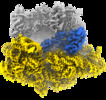
 マップデータ
 試料
試料 キーワード
キーワード 機能・相同性情報
 データ登録者
 ドイツ, 1件
  引用
引用

 構造の表示
構造の表示
 ダウンロードとリンク
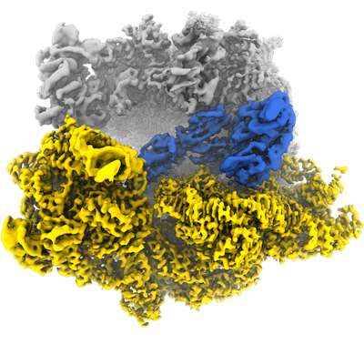 emd_13058.png
 http://ftp.pdbj.org/pub/emdb/structures/EMD-13058
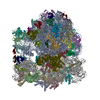


 Z
Z Y
Y X
X


 試料の構成要素


 解析
解析 電子顕微鏡法
 FIELD EMISSION GUN