transcriptional attenuation / endoribonuclease inhibitor activity / RNA-binding transcription regulator activity / negative regulation of cytoplasmic translation / DnaA-L2 complex / translation repressor activity / negative regulation of DNA-templated DNA replication initiation / ribosome assembly / assembly of large subunit precursor of preribosome / cytosolic ribosome assembly ...transcriptional attenuation / endoribonuclease inhibitor activity / RNA-binding transcription regulator activity / negative regulation of cytoplasmic translation / DnaA-L2 complex / translation repressor activity / negative regulation of DNA-templated DNA replication initiation / ribosome assembly / assembly of large subunit precursor of preribosome / cytosolic ribosome assembly / ribosomal large subunit assembly / DNA-templated transcription termination / response to radiation / mRNA 5'-UTR binding / large ribosomal subunit / ribosomal small subunit assembly / small ribosomal subunit / transferase activity / 5S rRNA binding / large ribosomal subunit rRNA binding / cytosolic small ribosomal subunit / cytosolic large ribosomal subunit / cytoplasmic translation / tRNA binding / rRNA binding / negative regulation of translation / ribosome / structural constituent of ribosome / translation / ribonucleoprotein complex / response to antibiotic / negative regulation of DNA-templated transcription / mRNA binding / DNA binding / RNA binding / zinc ion binding / membrane / metal ion binding / cytoplasm / cytosol 類似検索 - 分子機能
Cold shock, CspA / Cold-shock (CSD) domain / Cold-shock (CSD) domain signature. / Cold-shock (CSD) domain profile. / Cold-shock protein, DNA-binding / 'Cold-shock' DNA-binding domain / Cold shock domain / Cold shock protein domain / Ribosomal protein S21, conserved site / Ribosomal protein S21 signature. ...Cold shock, CspA / Cold-shock (CSD) domain / Cold-shock (CSD) domain signature. / Cold-shock (CSD) domain profile. / Cold-shock protein, DNA-binding / 'Cold-shock' DNA-binding domain / Cold shock domain / Cold shock protein domain / Ribosomal protein S21, conserved site / Ribosomal protein S21 signature. / Ribosomal protein L25, short-form / Ribosomal protein S14, bacterial/plastid / Ribosomal protein L31 type A / Ribosomal protein S21 superfamily / Ribosomal protein S21 / Ribosomal protein S16, conserved site / Ribosomal protein S16 signature. / Ribosomal protein S21 / Ribosomal protein L31 signature. / Ribosomal protein L31 / Ribosomal protein L31 superfamily / Ribosomal protein L31 / : / Ribosomal protein L21, conserved site / Ribosomal protein L21 signature. / Ribosomal protein L16 signature 1. / Ribosomal protein L6, conserved site / Ribosomal protein L6 signature 1. / Ribosomal protein L16, conserved site / Ribosomal protein L16 signature 2. / Ribosomal protein L9 signature. / Ribosomal protein L9, bacteria/chloroplast / Ribosomal protein L9, C-terminal / Ribosomal protein L9, C-terminal domain / Ribosomal protein L9, C-terminal domain superfamily / Ribosomal L25p family / Ribosomal protein L25 / Ribosomal protein L36 signature. / Ribosomal protein L28/L24 superfamily / Ribosomal protein L25/Gln-tRNA synthetase, N-terminal / Ribosomal protein L25/Gln-tRNA synthetase, anti-codon-binding domain superfamily / Ribosomal protein L9, N-terminal domain superfamily / Ribosomal protein L9 / Ribosomal protein L9, N-terminal / Ribosomal protein L9, N-terminal domain / Ribosomal protein L28 / Ribosomal protein L33, conserved site / Ribosomal protein L33 signature. / Ribosomal protein L5, bacterial-type / Ribosomal protein L18, bacterial-type / Ribosomal protein L6, bacterial-type / Ribosomal protein L19, conserved site / Ribosomal protein L19 signature. / Ribosomal protein S3, bacterial-type / Ribosomal protein L9/RNase H1, N-terminal / Ribosomal protein S6, conserved site / Ribosomal protein S6 signature. / Ribosomal protein S19, bacterial-type / Ribosomal protein L36 / Ribosomal protein L36 superfamily / Ribosomal protein L36 / Ribosomal protein S7, bacterial/organellar-type / Ribosomal protein L20 signature. / Ribosomal protein S11, bacterial-type / Ribosomal protein S13, bacterial-type / Ribosomal protein S20 / Ribosomal protein S20 superfamily / Ribosomal protein S20 / Ribosomal protein S9, bacterial/plastid / Ribosomal protein L27, conserved site / Ribosomal protein L27 signature. / Ribosomal protein S4, bacterial-type / 30S ribosomal protein S17 / Ribosomal protein L14P, bacterial-type / Ribosomal protein L34, conserved site / Ribosomal protein L34 signature. / Ribosomal protein S6, plastid/chloroplast / Ribosomal protein L22, bacterial/chloroplast-type / Ribosomal protein L2, bacterial/organellar-type / Ribosomal protein S2, bacteria/mitochondria/plastid / Ribosomal L28 family / Ribosomal protein L33 / Ribosomal protein L33 / Ribosomal protein L28/L24 / Ribosomal protein L18 / Ribosomal L18 of archaea, bacteria, mitoch. and chloroplast / Ribosomal protein L33 superfamily / Ribosomal protein L30, bacterial-type / Ribosomal protein S18, conserved site / Ribosomal protein S18 signature. / Ribosomal protein L16 / : / Ribosomal protein S16 / Ribosomal protein S16 / Ribosomal protein S16 domain superfamily / L28p-like / Ribosomal protein L20 / Ribosomal protein S15, bacterial-type / Ribosomal protein L20 / Ribosomal protein L20, C-terminal 類似検索 - ドメイン・相同性
50S ribosomal protein L32 / : / 50S ribosomal protein L20 / : / 50S ribosomal protein L31 / 50S ribosomal protein L30 / 30S ribosomal protein S14 / 30S ribosomal protein S3 / 50S ribosomal protein L28 / Small ribosomal subunit protein uS11 ...50S ribosomal protein L32 / : / 50S ribosomal protein L20 / : / 50S ribosomal protein L31 / 50S ribosomal protein L30 / 30S ribosomal protein S14 / 30S ribosomal protein S3 / 50S ribosomal protein L28 / Small ribosomal subunit protein uS11 / 30S ribosomal protein S2 / 30S ribosomal protein S9 / 30S ribosomal protein S6 / : / Cold shock protein CspB / 30S ribosomal protein S19 / 50S ribosomal protein L24 / Small ribosomal subunit protein uS13 / Small ribosomal subunit protein bS16 / Small ribosomal subunit protein bS21 / 30S ribosomal protein S15 / 50S ribosomal protein L18 / 50S ribosomal protein L33 / Large ribosomal subunit protein uL23 / 50S ribosomal protein L5 / 30S ribosomal protein S8 / 50S ribosomal protein L29 / 30S ribosomal protein S18 / 50S ribosomal protein L6 / 50S ribosomal protein L19 / 30S ribosomal protein S20 / 30S ribosomal protein S10 / 50S ribosomal protein L27 / Small ribosomal subunit protein uS7 / Large ribosomal subunit protein uL15 / Large ribosomal subunit protein bL34 / Large ribosomal subunit protein bL36A / Large ribosomal subunit protein bL9 / Large ribosomal subunit protein uL13 / Large ribosomal subunit protein uL14 / Large ribosomal subunit protein uL16 / Large ribosomal subunit protein bL21 / Large ribosomal subunit protein uL2 / Large ribosomal subunit protein uL3 / Large ribosomal subunit protein uL4 / Large ribosomal subunit protein uL22 / Large ribosomal subunit protein bL25 / 30S ribosomal protein S17 / Small ribosomal subunit protein uS4 / : 類似検索 - 構成要素
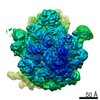
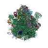
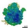
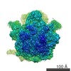
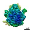
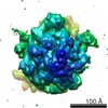
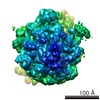
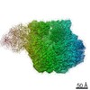
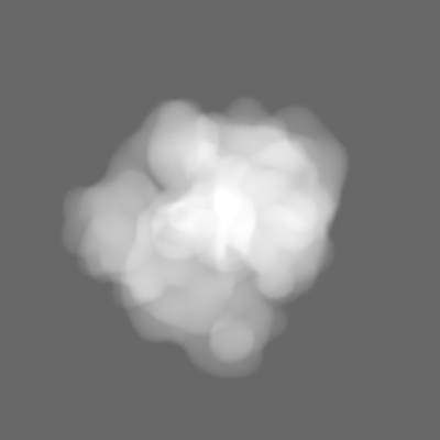
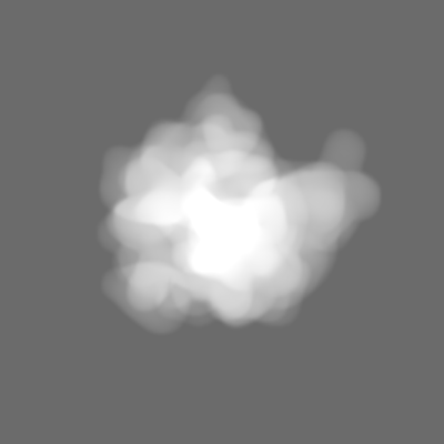
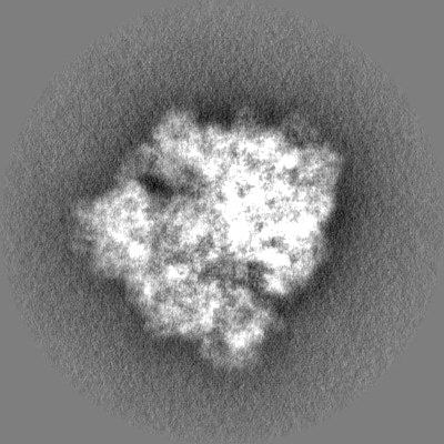
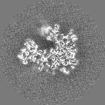
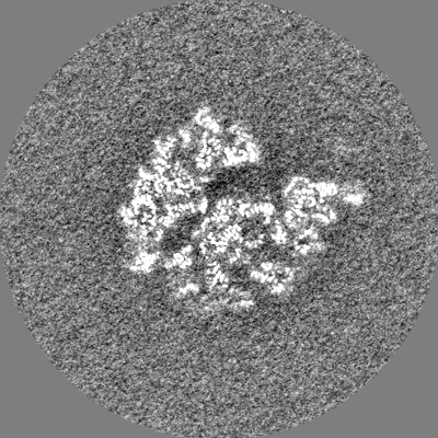
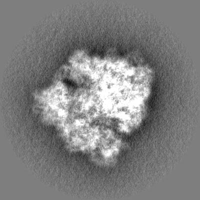
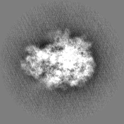
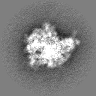
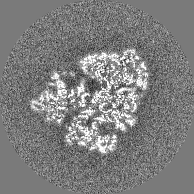
ジャーナル: EMBO J / 年: 2022 タイトル: A switch from α-helical to β-strand conformation during co-translational protein folding. 著者: Xabier Agirrezabala / Ekaterina Samatova / Meline Macher / Marija Liutkute / Manisankar Maiti / David Gil-Carton / Jiri Novacek / Mikel Valle / Marina V Rodnina / 要旨: Cellular proteins begin to fold as they emerge from the ribosome. The folding landscape of nascent chains is not only shaped by their amino acid sequence but also by the interactions with the ...Cellular proteins begin to fold as they emerge from the ribosome. The folding landscape of nascent chains is not only shaped by their amino acid sequence but also by the interactions with the ribosome. Here, we combine biophysical methods with cryo-EM structure determination to show that folding of a β-barrel protein begins with formation of a dynamic α-helix inside the ribosome. As the growing peptide reaches the end of the tunnel, the N-terminal part of the nascent chain refolds to a β-hairpin structure that remains dynamic until its release from the ribosome. Contacts with the ribosome and structure of the peptidyl transferase center depend on nascent chain conformation. These results indicate that proteins may start out as α-helices inside the tunnel and switch into their native folds only as they emerge from the ribosome. Moreover, the correlation of nascent chain conformations with reorientation of key residues of the ribosomal peptidyl-transferase center suggest that protein folding could modulate ribosome activity.
 ムービー
ムービー コントローラー


 データを開く
 基本情報
基本情報 マップデータ
 試料
試料 キーワード
キーワード 機能・相同性情報
 データ登録者
 スペイン, European Union, 3件
  引用
引用

 構造の表示
構造の表示
 ダウンロードとリンク
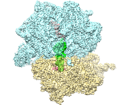 emd_13055.png
 http://ftp.pdbj.org/pub/emdb/structures/EMD-13055


 Z
Z Y
Y X
X


 試料の構成要素
 解析
解析 電子顕微鏡法
 FIELD EMISSION GUN