Ministry of Education, Youth and Sports of the Czech Republic
LM20115043
Czech Republic
Ministry of Education, Youth and Sports of the Czech Republic
LM20115070
Czech Republic
Ministry of Education, Youth and Sports of the Czech Republic
LM20115042
Czech Republic
European Research Council (ERC)
649030
Czech Republic
Ministry of Education, Youth and Sports of the Czech Republic
LQ1601
Czech Republic
Czech Science Foundation
GA18-11397S
Czech Republic
Citation
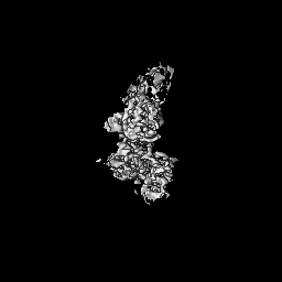
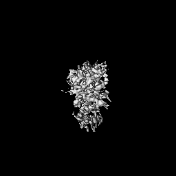
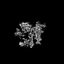
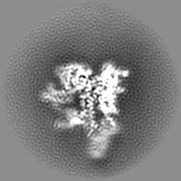
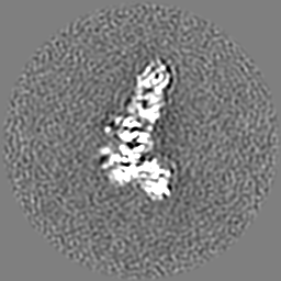
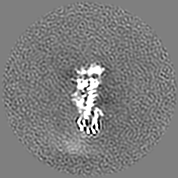
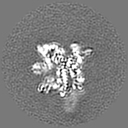
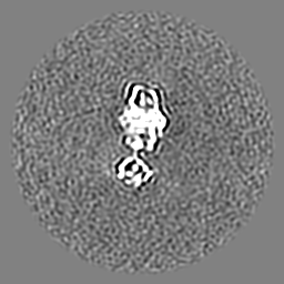
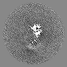
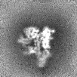
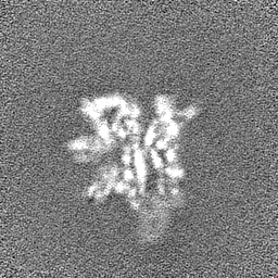
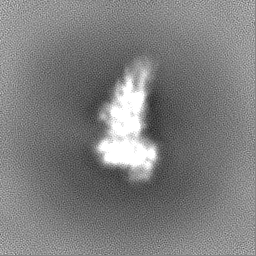
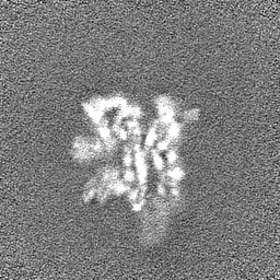
Journal: Nucleic Acids Res / Year: 2022 Title: Cooperation between intrinsically disordered and ordered regions of Spt6 regulates nucleosome and Pol II CTD binding, and nucleosome assembly. Authors: Aiste Kasiliauskaite / Karel Kubicek / Tomas Klumpler / Martina Zanova / David Zapletal / Eliska Koutna / Jiri Novacek / Richard Stefl / Abstract: Transcription elongation factor Spt6 associates with RNA polymerase II (Pol II) and acts as a histone chaperone, which promotes the reassembly of nucleosomes following the passage of Pol II. The ...Transcription elongation factor Spt6 associates with RNA polymerase II (Pol II) and acts as a histone chaperone, which promotes the reassembly of nucleosomes following the passage of Pol II. The precise mechanism of nucleosome reassembly mediated by Spt6 remains unclear. In this study, we used a hybrid approach combining cryo-electron microscopy and small-angle X-ray scattering to visualize the architecture of Spt6 from Saccharomyces cerevisiae. The reconstructed overall architecture of Spt6 reveals not only the core of Spt6, but also its flexible N- and C-termini, which are critical for Spt6's function. We found that the acidic N-terminal region of Spt6 prevents the binding of Spt6 not only to the Pol II CTD and Pol II CTD-linker, but also to pre-formed intact nucleosomes and nucleosomal DNA. The N-terminal region of Spt6 self-associates with the tSH2 domain and the core of Spt6 and thus controls binding to Pol II and nucleosomes. Furthermore, we found that Spt6 promotes the assembly of nucleosomes in vitro. These data indicate that the cooperation between the intrinsically disordered and structured regions of Spt6 regulates nucleosome and Pol II CTD binding, and also nucleosome assembly.
Name: Transcription elongation factor SPT6 / type: protein_or_peptide / ID: 1 Details: the sequence stretches of the sample that do not make part of the coordinates are flexible regions that were not observed in the cryoEM map Number of copies: 1 / Enantiomer: LEVO
Details: Initial PDB coordinates of the Spt6 structure comprising residues 298-1248 were produced from PDBs of a crystal structure of Spt6, namely 3PSI and 3PSF. The PDB and the cryoSPARC sharpened ...Details: Initial PDB coordinates of the Spt6 structure comprising residues 298-1248 were produced from PDBs of a crystal structure of Spt6, namely 3PSI and 3PSF. The PDB and the cryoSPARC sharpened electron density map were imported into the program Coot and the tool 'Real Space Refine Zone' was used to achiev e optimal fit of the PDB coordinates within the map. Low-resolution regions of the map (regions corresponding to residues: 1226-1243 and the S1-domain) were also excluded from the PDB and were docked with a rigid body strategy using Phenix once the fit of the well-resolved regions exhibited no clashes and deviations.
Final reconstruction
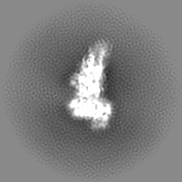
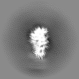
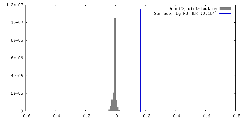
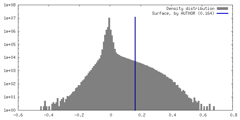
Number classes used: 313 / Applied symmetry - Point group: C1 (asymmetric) / Resolution.type: BY AUTHOR / Resolution: 3.71 Å / Resolution method: FSC 0.143 CUT-OFF / Software - Name: cryoSPARC (ver. 3.1) / Number images used: 718639
Initial angle assignment
Type: MAXIMUM LIKELIHOOD / Software - Name: cryoSPARC (ver. 3.1)
Final angle assignment
Type: MAXIMUM LIKELIHOOD / Software - Name: cryoSPARC (ver. 3.1)
Space: REAL / Protocol: FLEXIBLE FIT / Target criteria: Correlation coefficients
Output model
PDB-7o3d: Cooperation between the intrinsically disordered and ordered regions of Spt6 regulates nucleosome and Pol II CTD binding, and nucleosome assembly
+
About Yorodumi
-
News
-
Feb 9, 2022. New format data for meta-information of EMDB entries
New format data for meta-information of EMDB entries
Version 3 of the EMDB header file is now the official format.
The previous official version 1.9 will be removed from the archive.
In the structure databanks used in Yorodumi, some data are registered as the other names, "COVID-19 virus" and "2019-nCoV". Here are the details of the virus and the list of structure data.
Jan 31, 2019. EMDB accession codes are about to change! (news from PDBe EMDB page)
EMDB accession codes are about to change! (news from PDBe EMDB page)
The allocation of 4 digits for EMDB accession codes will soon come to an end. Whilst these codes will remain in use, new EMDB accession codes will include an additional digit and will expand incrementally as the available range of codes is exhausted. The current 4-digit format prefixed with “EMD-” (i.e. EMD-XXXX) will advance to a 5-digit format (i.e. EMD-XXXXX), and so on. It is currently estimated that the 4-digit codes will be depleted around Spring 2019, at which point the 5-digit format will come into force.
The EM Navigator/Yorodumi systems omit the EMD- prefix.
Related info.:Q: What is EMD? / ID/Accession-code notation in Yorodumi/EM Navigator
Yorodumi is a browser for structure data from EMDB, PDB, SASBDB, etc.
This page is also the successor to EM Navigator detail page, and also detail information page/front-end page for Omokage search.
The word "yorodu" (or yorozu) is an old Japanese word meaning "ten thousand". "mi" (miru) is to see.
Related info.:EMDB / PDB / SASBDB / Comparison of 3 databanks / Yorodumi Search / Aug 31, 2016. New EM Navigator & Yorodumi / Yorodumi Papers / Jmol/JSmol / Function and homology information / Changes in new EM Navigator and Yorodumi
 Movie
Movie Controller

 Yorodumi
 Open data
 Basic information
 Map data
 Sample
 Function and homology information
 Authors
 Czech Republic, 6 items
  Citation
 Structure visualization
 Downloads & links
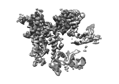 emd_12704.png
 http://ftp.pdbj.org/pub/emdb/structures/EMD-12704


 Z (Sec.)
 Y (Row.)
 X (Col.)


 Sample components
 Processing
 Electron microscopy
 FIELD EMISSION GUN


