Movie
Movie Controller


+ Open data

- Basic information
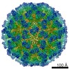
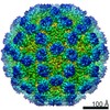
| Entry | Database: EMDB / ID: EMD-11115 | |||||||||
|---|---|---|---|---|---|---|---|---|---|---|
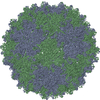
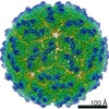
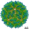
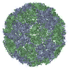
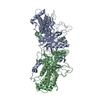
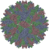
| Title | Human Picobirnavirus CP VLP | |||||||||
 Map data Map data | HPBV CP VLP | |||||||||
 Sample Sample |
| |||||||||
 Keywords Keywords | Human Picobirnavirus / CryoEM / assembly / capsid protein / VIRUS LIKE PARTICLE | |||||||||
| Function / homology | : / : / Picobirnavirus, Capsid protein / T=3 icosahedral viral capsid / Capsid protein precursor Function and homology information Function and homology information | |||||||||
| Biological species |  Human picobirnavirus Human picobirnavirus | |||||||||
| Method | single particle reconstruction / cryo EM / Resolution: 2.63 Å | |||||||||
 Authors Authors | Ortega-Esteban A / Mata CP | |||||||||
| Funding support |  Spain, 1 items Spain, 1 items
| |||||||||
 Citation Citation | Journal: J Virol / Year: 2020 Title: Cryo-electron Microscopy Structure, Assembly, and Mechanics Show Morphogenesis and Evolution of Human Picobirnavirus. Authors: Álvaro Ortega-Esteban / Carlos P Mata / María J Rodríguez-Espinosa / Daniel Luque / Nerea Irigoyen / Javier M Rodríguez / Pedro J de Pablo / José R Castón / Abstract: Despite their diversity, most double-stranded-RNA (dsRNA) viruses share a specialized T=1 capsid built from dimers of a single protein that provides a platform for genome transcription and ...Despite their diversity, most double-stranded-RNA (dsRNA) viruses share a specialized T=1 capsid built from dimers of a single protein that provides a platform for genome transcription and replication. This ubiquitous capsid remains structurally undisturbed throughout the viral cycle, isolating the genome to avoid triggering host defense mechanisms. Human picobirnavirus (hPBV) is a dsRNA virus frequently associated with gastroenteritis, although its pathogenicity is yet undefined. Here, we report the cryo-electron microscopy (cryo-EM) structure of hPBV at 2.6-Å resolution. The capsid protein (CP) is arranged in a single-shelled, ∼380-Å-diameter T=1 capsid with a rough outer surface similar to that of dsRNA mycoviruses. The hPBV capsid is built of 60 quasisymmetric CP dimers (A and B) stabilized by domain swapping, and only the CP-A N-terminal basic region interacts with the packaged nucleic acids. hPBV CP has an α-helical domain with a fold similar to that of fungal partitivirus CP, with many domain insertions in its C-terminal half. In contrast to dsRNA mycoviruses, hPBV has an extracellular life cycle phase like complex reoviruses, which indicates that its own CP probably participates in cell entry. Using an reversible assembly/disassembly system of hPBV, we isolated tetramers as possible assembly intermediates. We used atomic force microscopy to characterize the biophysical properties of hPBV capsids with different cargos (host nucleic acids or proteins) and found that the CP N-terminal segment not only is involved in nucleic acid interaction/packaging but also modulates the mechanical behavior of the capsid in conjunction with the cargo. Despite intensive study, human virus sampling is still sparse, especially for viruses that cause mild or asymptomatic disease. Human picobirnavirus (hPBV) is a double-stranded-RNA virus, broadly dispersed in the human population, but its pathogenicity is uncertain. Here, we report the hPBV structure derived from cryo-electron microscopy (cryo-EM) and reconstruction methods using three capsid protein variants (of different lengths and N-terminal amino acid compositions) that assemble as virus-like particles with distinct properties. The hPBV near-atomic structure reveals a quasisymmetric dimer as the structural subunit and tetramers as possible assembly intermediates that coassemble with nucleic acids. Our structural studies and atomic force microscopy analyses indicate that hPBV capsids are potentially excellent nanocages for gene therapy and targeted drug delivery in humans. | |||||||||
| History |
|
- Structure visualization
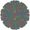
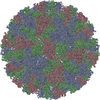
| Movie |
Movie viewer |
|---|---|
| Structure viewer | EM map: SurfViewMolmilJmol/JSmol |
| Supplemental images |

- Downloads & links
-EMDB archive
| Map data | emd_11115.map.gz | 226.7 MB | EMDB map data format | |
|---|---|---|---|---|
| Header (meta data) | emd-11115-v30.xmlemd-11115.xml | 12.6 KB 12.6 KB | Display Display | EMDB header |
| Images | 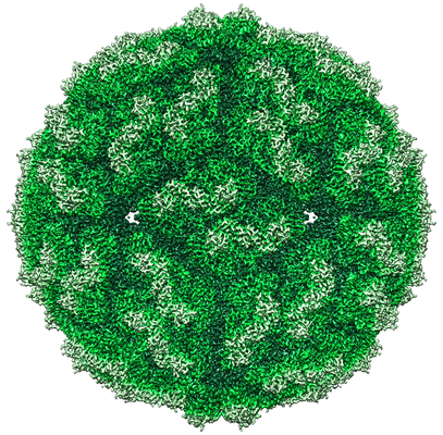 emd_11115.png emd_11115.png | 326.4 KB | ||
| Filedesc metadata | emd-11115.cif.gz | 5.7 KB | ||
| Archive directory |  http://ftp.pdbj.org/pub/emdb/structures/EMD-11115ftp://ftp.pdbj.org/pub/emdb/structures/EMD-11115 http://ftp.pdbj.org/pub/emdb/structures/EMD-11115ftp://ftp.pdbj.org/pub/emdb/structures/EMD-11115 | HTTPS FTP |
-Related structure data
| Related structure data | 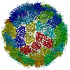 6z8dMC 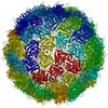 6z8eC 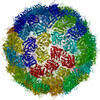 6z8fC M: atomic model generated by this map C: citing same article ( |
|---|---|
| Similar structure data |
-Links
| EMDB pages | EMDB (EBI/PDBe) / EMDataResource |
|---|---|
| Related items in Molecule of the Month |

-Map
| File | Download / File: emd_11115.map.gz / Format: CCP4 / Size: 244.1 MB / Type: IMAGE STORED AS FLOATING POINT NUMBER (4 BYTES) | ||||||||||||||||||||||||||||||||||||||||||||||||||||||||||||||||||||
|---|---|---|---|---|---|---|---|---|---|---|---|---|---|---|---|---|---|---|---|---|---|---|---|---|---|---|---|---|---|---|---|---|---|---|---|---|---|---|---|---|---|---|---|---|---|---|---|---|---|---|---|---|---|---|---|---|---|---|---|---|---|---|---|---|---|---|---|---|---|
| Annotation | HPBV CP VLP | ||||||||||||||||||||||||||||||||||||||||||||||||||||||||||||||||||||
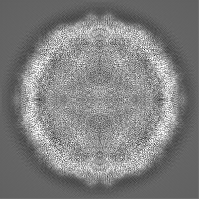
| Projections & slices | Image control
Images are generated by Spider. | ||||||||||||||||||||||||||||||||||||||||||||||||||||||||||||||||||||
| Voxel size | X=Y=Z: 1.065 Å | ||||||||||||||||||||||||||||||||||||||||||||||||||||||||||||||||||||
| Density |
| ||||||||||||||||||||||||||||||||||||||||||||||||||||||||||||||||||||
| Symmetry | Space group: 1 | ||||||||||||||||||||||||||||||||||||||||||||||||||||||||||||||||||||
| Details | EMDB XML:
CCP4 map header:
| ||||||||||||||||||||||||||||||||||||||||||||||||||||||||||||||||||||
 Z (Sec.)
 Y (Row.)
 X (Col.)


-Supplemental data
- Sample components
-Entire : Human picobirnavirus
| Entire | Name: Human picobirnavirus |
|---|---|
| Components |
|
-Supramolecule #1: Human picobirnavirus
| Supramolecule | Name: Human picobirnavirus / type: virus / ID: 1 / Parent: 0 / Macromolecule list: all / NCBI-ID: 145856 / Sci species name: Human picobirnavirus / Virus type: VIRUS-LIKE PARTICLE / Virus isolate: STRAIN / Virus enveloped: No / Virus empty: No |
|---|---|
| Host (natural) | Organism:  Homo sapiens (human) Homo sapiens (human) |
| Virus shell | Shell ID: 1 / Name: Capsid Protein / Diameter: 380.0 Å / T number (triangulation number): 1 |
-Macromolecule #1: Capsid protein precursor
| Macromolecule | Name: Capsid protein precursor / type: protein_or_peptide / ID: 1 / Number of copies: 2 / Enantiomer: LEVO |
|---|---|
| Source (natural) | Organism: Human picobirnavirus |
| Molecular weight | Theoretical: 62.084227 KDa |
| Recombinant expression | Organism:  |
| Sequence | String: MKQNDTKKTT QRRNSKKYSS KTNRGTKRAP RDQEVGTGAQ ESTRNDVAWY ARYPHILEEA TRLPFAYPIG QYYDTGYSVA SATEWSKYV DTSLTIPGVM CVNFTPTPGE SYNKNSPINI AAQNVYTYVR HMNSGHANYE QADLMMYLLA MDSLYIFHSY V RKILAISK ...String: MKQNDTKKTT QRRNSKKYSS KTNRGTKRAP RDQEVGTGAQ ESTRNDVAWY ARYPHILEEA TRLPFAYPIG QYYDTGYSVA SATEWSKYV DTSLTIPGVM CVNFTPTPGE SYNKNSPINI AAQNVYTYVR HMNSGHANYE QADLMMYLLA MDSLYIFHSY V RKILAISK LYTPVNKYFP RALLVALGVD PEDVFANQAQ WEYFVNMVAY RAGAFAAPAS MTYYERHAWM SNGLYVDQDV TR AQIYMFK PTMLWKYENL GTTGTKLVPL MMPKAGDNRK LVDFQVLFNN LVSTMLGDED FGIMSGDVFK AFGADGLVKL LAV DSTTMT LPTYDPLILA QIHSARAVGA PILETSTLTG FPGRQWQITQ NPDVNNGAII FHPSFGYDGQ DHEELSFRAM CSNM ILNLP GEAHSAEMII EATRLATMFQ VKAVPAGDTS KPVLYLPNGF GTEVVNDYTM ISVDKATPHD LTIHTFFNNI LVPNA KENY VANLELLNNI IQFDWAPQLY LTYGIAQESF GPFAQLNDWT ILTGETLARM HEVCVTSMFD VPQMGFNK UniProtKB: Capsid protein precursor |
-Experimental details
-Structure determination
| Method | cryo EM |
|---|---|
 Processing Processing | single particle reconstruction |
| Aggregation state | particle |
-Sample preparation
| Buffer | pH: 7.5 / Component:
| ||||||
|---|---|---|---|---|---|---|---|
| Vitrification | Cryogen name: ETHANE / Instrument: LEICA EM CPC |
- Electron microscopy
| Microscope | FEI TITAN KRIOS |
|---|---|
| Image recording | Film or detector model: FEI FALCON III (4k x 4k) / Detector mode: INTEGRATING / Number real images: 2360 / Average electron dose: 1.56 e/Å2 |
| Electron beam | Acceleration voltage: 300 kV / Electron source:  FIELD EMISSION GUN FIELD EMISSION GUN |
| Electron optics | C2 aperture diameter: 70.0 µm / Calibrated defocus max: 4.04 µm / Calibrated defocus min: 0.306 µm / Illumination mode: FLOOD BEAM / Imaging mode: BRIGHT FIELD / Cs: 2.7 mm / Nominal defocus max: 3.5 µm / Nominal defocus min: 0.8 µm / Nominal magnification: 75000 |
| Sample stage | Specimen holder model: FEI TITAN KRIOS AUTOGRID HOLDER / Cooling holder cryogen: NITROGEN |
| Experimental equipment |  Model: Titan Krios / Image courtesy: FEI Company |
-Image processing
| Startup model | Type of model: PDB ENTRY PDB model - PDB ID: |
|---|---|
| Final reconstruction | Applied symmetry - Point group: I (icosahedral) / Resolution.type: BY AUTHOR / Resolution: 2.63 Å / Resolution method: FSC 0.143 CUT-OFF / Software - Name: RELION (ver. 2.1) / Number images used: 102722 |
| Initial angle assignment | Type: MAXIMUM LIKELIHOOD / Software - Name: RELION (ver. 2.1) |
| Final angle assignment | Type: MAXIMUM LIKELIHOOD / Software - Name: RELION (ver. 2.1) |