 Movie
Movie Controller


+ Open data

- Basic information
| Entry | Database: EMDB / ID: EMD-10800 | ||||||||||||
|---|---|---|---|---|---|---|---|---|---|---|---|---|---|
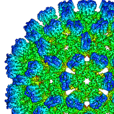
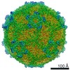
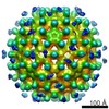
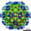
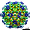
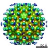
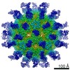
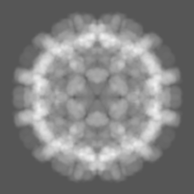
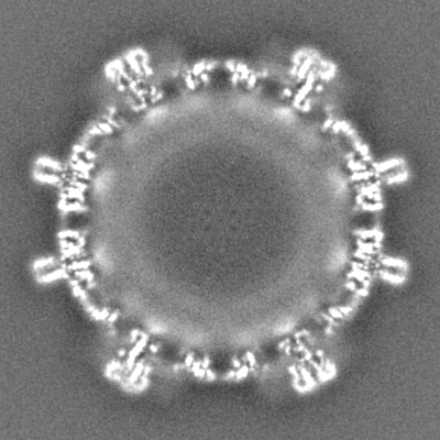
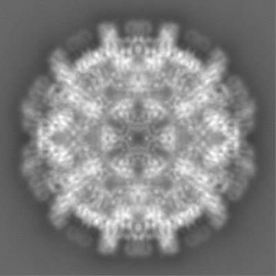
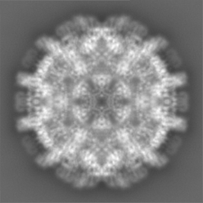
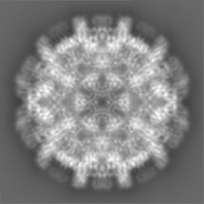
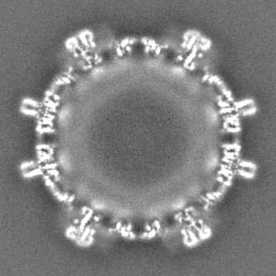
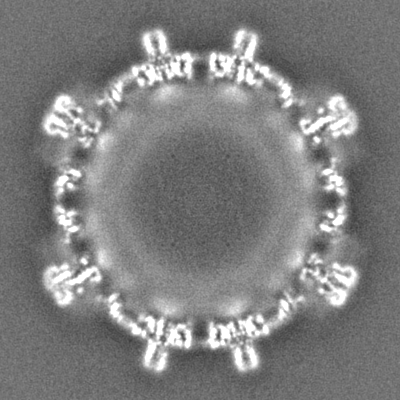
| Title | Duck hepatitis B virus capsid | ||||||||||||
 Map data Map data | |||||||||||||
 Sample Sample |
| ||||||||||||
 Keywords Keywords | duck hepatitis B core protein / extension domain / spike / slowly folding / VIRUS LIKE PARTICLE | ||||||||||||
| Function / homology |  Function and homology information Function and homology informationmicrotubule-dependent intracellular transport of viral material towards nucleus / T=4 icosahedral viral capsid / viral penetration into host nucleus / host cell / host cell cytoplasm / symbiont entry into host cell / structural molecule activity / DNA binding / RNA binding Similarity search - Function | ||||||||||||
| Biological species |  Hepatitis B virus duck/DHBV-16 Hepatitis B virus duck/DHBV-16 | ||||||||||||
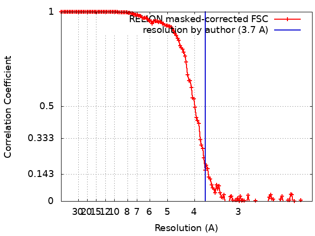
| Method | single particle reconstruction / cryo EM / Resolution: 3.7 Å | ||||||||||||
 Authors Authors | Makbul C / Bottcher B | ||||||||||||
| Funding support |  Germany, 3 items Germany, 3 items
| ||||||||||||
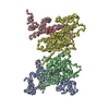
 Citation Citation | Journal: Elife / Year: 2020 Title: Slowly folding surface extension in the prototypic avian hepatitis B virus capsid governs stability. Authors: Cihan Makbul / Michael Nassal / Bettina Böttcher / Abstract: Hepatitis B virus (HBV) is an important but difficult to study human pathogen. Most basics of the hepadnaviral life-cycle were unraveled using duck HBV (DHBV) as a model although DHBV has a capsid ...Hepatitis B virus (HBV) is an important but difficult to study human pathogen. Most basics of the hepadnaviral life-cycle were unraveled using duck HBV (DHBV) as a model although DHBV has a capsid protein (CP) comprising ~260 rather than ~180 amino acids. Here we present high-resolution structures of several DHBV capsid-like particles (CLPs) determined by electron cryo-microscopy. As for HBV, DHBV CLPs consist of a dimeric α-helical frame-work with protruding spikes at the dimer interface. A fundamental new feature is a ~ 45 amino acid proline-rich extension in each monomer replacing the tip of the spikes in HBV CP. In vitro, folding of the extension takes months, implying a catalyzed process in vivo. DHBc variants lacking a folding-proficient extension produced regular CLPs in bacteria but failed to form stable nucleocapsids in hepatoma cells. We propose that the extension domain acts as a conformational switch with differential response options during viral infection. | ||||||||||||
| History |
|
- Structure visualization
| Movie |
Movie viewer |
|---|---|
| Structure viewer | EM map: SurfViewMolmilJmol/JSmol |
| Supplemental images |
- Downloads & links
-EMDB archive
| Map data | emd_10800.map.gz | 226.1 MB | EMDB map data format | |
|---|---|---|---|---|
| Header (meta data) | emd-10800-v30.xmlemd-10800.xml | 19.9 KB 19.9 KB | Display Display | EMDB header |
| FSC (resolution estimation) | emd_10800_fsc.xml | 14.2 KB | Display | FSC data file |
| Images |  emd_10800.png emd_10800.png | 364 KB | ||
| Masks | emd_10800_msk_1.map | 244.1 MB | Mask map | |
| Filedesc metadata | emd-10800.cif.gz | 6.4 KB | ||
| Others | emd_10800_half_map_1.map.gzemd_10800_half_map_2.map.gz | 222.9 MB 222.9 MB | ||
| Archive directory |  http://ftp.pdbj.org/pub/emdb/structures/EMD-10800ftp://ftp.pdbj.org/pub/emdb/structures/EMD-10800 http://ftp.pdbj.org/pub/emdb/structures/EMD-10800ftp://ftp.pdbj.org/pub/emdb/structures/EMD-10800 | HTTPS FTP |
-Related structure data
| Related structure data |  6yghMC  6ygiC M: atomic model generated by this map C: citing same article ( |
|---|---|
| Similar structure data |
-Links
| EMDB pages | EMDB (EBI/PDBe) / EMDataResource |
|---|---|
| Related items in Molecule of the Month |


-Map
| File | Download / File: emd_10800.map.gz / Format: CCP4 / Size: 244.1 MB / Type: IMAGE STORED AS FLOATING POINT NUMBER (4 BYTES) | ||||||||||||||||||||||||||||||||||||||||||||||||||||||||||||
|---|---|---|---|---|---|---|---|---|---|---|---|---|---|---|---|---|---|---|---|---|---|---|---|---|---|---|---|---|---|---|---|---|---|---|---|---|---|---|---|---|---|---|---|---|---|---|---|---|---|---|---|---|---|---|---|---|---|---|---|---|---|
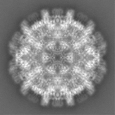
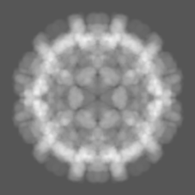
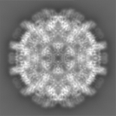
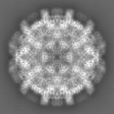
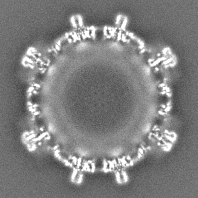
| Projections & slices | Image control
Images are generated by Spider. | ||||||||||||||||||||||||||||||||||||||||||||||||||||||||||||
| Voxel size | X=Y=Z: 1.0635 Å | ||||||||||||||||||||||||||||||||||||||||||||||||||||||||||||
| Density |
| ||||||||||||||||||||||||||||||||||||||||||||||||||||||||||||
| Symmetry | Space group: 1 | ||||||||||||||||||||||||||||||||||||||||||||||||||||||||||||
| Details | EMDB XML:
CCP4 map header:
| ||||||||||||||||||||||||||||||||||||||||||||||||||||||||||||
 Z (Sec.)
 Y (Row.)
 X (Col.)


-Supplemental data
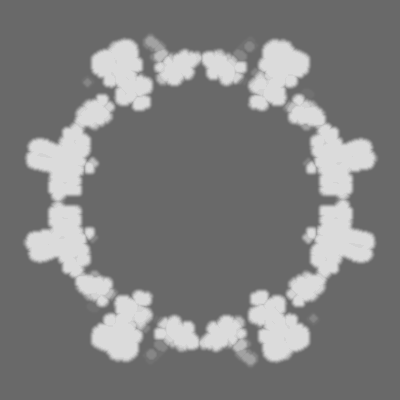
-Mask #1
| File | emd_10800_msk_1.map | ||||||||||||
|---|---|---|---|---|---|---|---|---|---|---|---|---|---|
| Projections & Slices |
| ||||||||||||
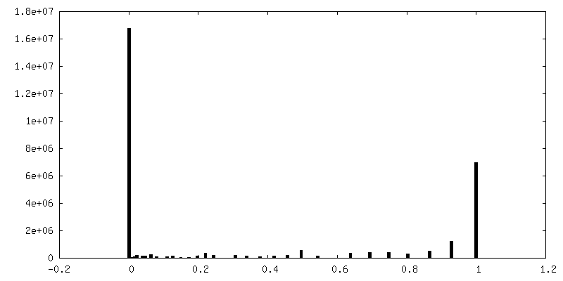
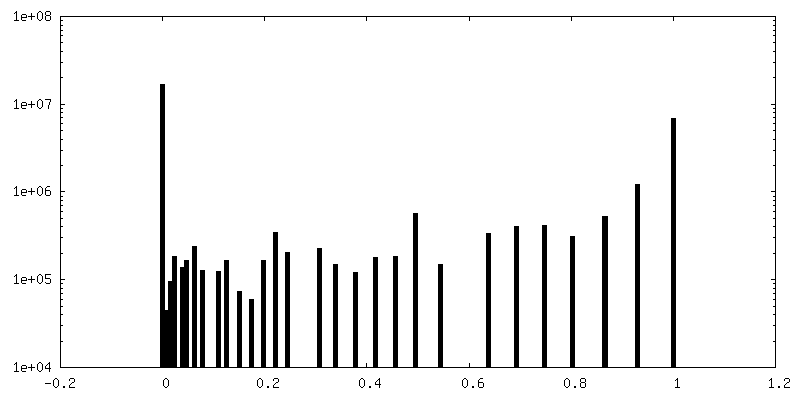
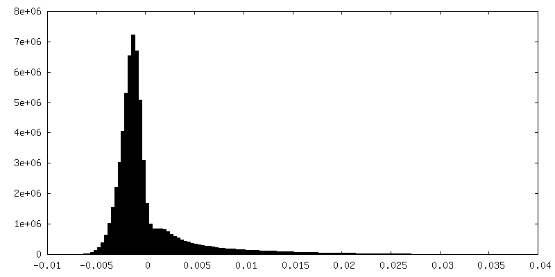
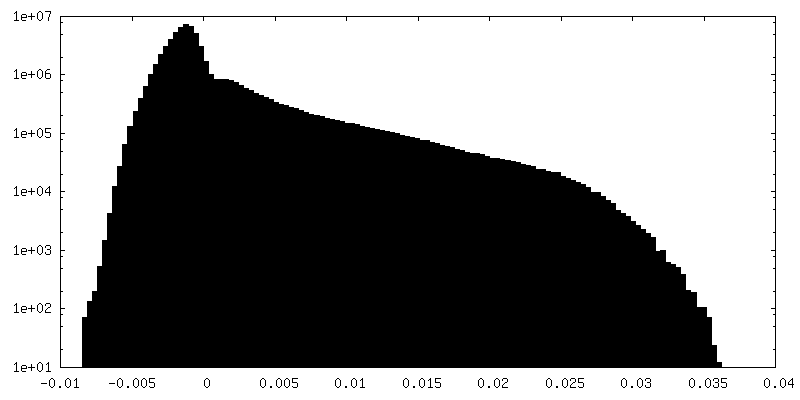
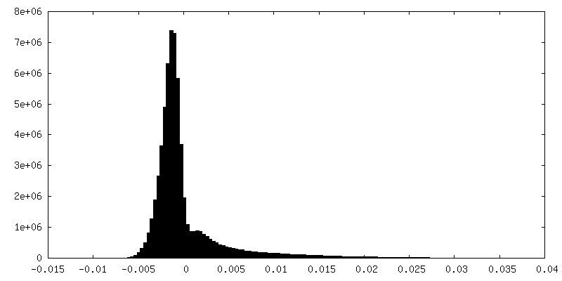
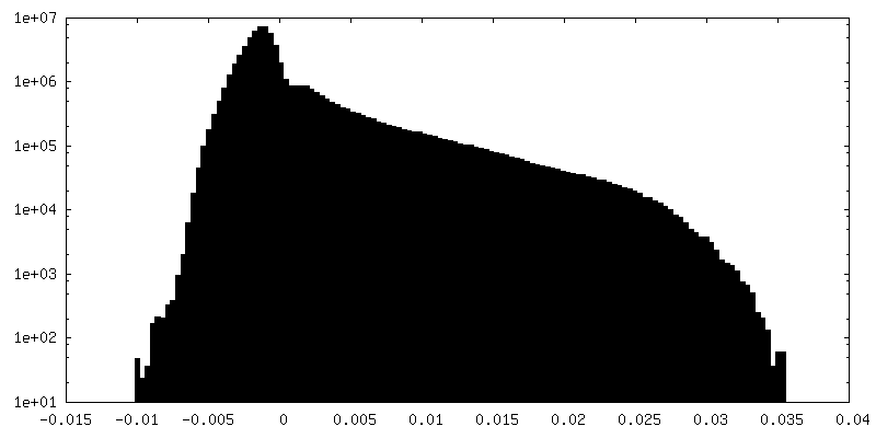
| Density Histograms |


-Half map: #1
| File | emd_10800_half_map_1.map | ||||||||||||
|---|---|---|---|---|---|---|---|---|---|---|---|---|---|
| Projections & Slices |
| ||||||||||||
| Density Histograms |


-Half map: #2
| File | emd_10800_half_map_2.map | ||||||||||||
|---|---|---|---|---|---|---|---|---|---|---|---|---|---|
| Projections & Slices |
| ||||||||||||
| Density Histograms |

- Sample components
-Entire : Hepatitis B virus duck/DHBV-16
| Entire | Name: Hepatitis B virus duck/DHBV-16 |
|---|---|
| Components |
|
-Supramolecule #1: Hepatitis B virus duck/DHBV-16
| Supramolecule | Name: Hepatitis B virus duck/DHBV-16 / type: virus / ID: 1 / Parent: 0 / Macromolecule list: all / NCBI-ID: 489543 / Sci species name: Hepatitis B virus duck/DHBV-16 / Virus type: VIRUS-LIKE PARTICLE / Virus isolate: SPECIES / Virus enveloped: No / Virus empty: No |
|---|---|
| Host (natural) | Organism:  |
| Molecular weight | Theoretical: 7.2 MDa |
| Virus shell | Shell ID: 1 / Name: duck Hepatitis B virus capsid / Diameter: 370.0 Å / T number (triangulation number): 4 |
-Macromolecule #1: Capsid protein
| Macromolecule | Name: Capsid protein / type: protein_or_peptide / ID: 1 / Number of copies: 6 / Enantiomer: LEVO |
|---|---|
| Source (natural) | Organism: Hepatitis B virus duck/DHBV-16 |
| Molecular weight | Theoretical: 30.33851 KDa |
| Recombinant expression | Organism:  |
| Sequence | String: MDINASRALA NVYDLPDDFF PKIDDLVRDA KDALEPYWKS DSIKKHVLIA THFVDLIEDF WQTTQGMHEI AESLRAVIPP TTTPVPPGY LIQHEEAEEI PLGDLFKHQE ERIVSFQPDY PITARIHAHL KAYAKINEES LDRARRLLWW HYNCLLWGEA Q VTNYISRL ...String: MDINASRALA NVYDLPDDFF PKIDDLVRDA KDALEPYWKS DSIKKHVLIA THFVDLIEDF WQTTQGMHEI AESLRAVIPP TTTPVPPGY LIQHEEAEEI PLGDLFKHQE ERIVSFQPDY PITARIHAHL KAYAKINEES LDRARRLLWW HYNCLLWGEA Q VTNYISRL RTWLSTPEKY RGRDAPTIEA ITRPIQVAQG GRKTTTGTRK PRGLEPRRRK VKTTVVYGRR RSKSRERRAP TP QRAGSPL PRSSSSHHRS PSPRK UniProtKB: Capsid protein |
-Experimental details
-Structure determination
| Method | cryo EM |
|---|---|
 Processing Processing | single particle reconstruction |
| Aggregation state | particle |
-Sample preparation
| Concentration | 3.2 mg/mL | ||||||||||||||||||
|---|---|---|---|---|---|---|---|---|---|---|---|---|---|---|---|---|---|---|---|
| Buffer | pH: 7.5 Component:
| ||||||||||||||||||
| Grid | Model: Quantifoil R1.2/1.3 / Material: COPPER / Mesh: 400 / Support film - Material: CARBON / Support film - topology: HOLEY ARRAY / Pretreatment - Type: GLOW DISCHARGE / Pretreatment - Time: 120 sec. / Pretreatment - Atmosphere: AIR / Pretreatment - Pressure: 0.029 kPa | ||||||||||||||||||
| Vitrification | Cryogen name: ETHANE / Chamber humidity: 100 % / Chamber temperature: 277 K / Instrument: FEI VITROBOT MARK IV Details: For the vitrification, grids (400 mesh copper grids (type R 1.2/1.3. Quantifoil Micro Tools, Jena/Germany) were rendered hydrophilic by glow discharging in air at a pressure of 29 Pa for 2 ...Details: For the vitrification, grids (400 mesh copper grids (type R 1.2/1.3. Quantifoil Micro Tools, Jena/Germany) were rendered hydrophilic by glow discharging in air at a pressure of 29 Pa for 2 minutes at medium power with a Plasma Cleaner (model PDC-002. Harrick Plasma Ithaca, NY/USA). Then, 3.5 ul of DHBc solution was pipetted onto the grids and they were plunge frozen in liquid ethane with a Vitrobot mark IV (FEI-Thermo Fisher Scientific). The settings for the Vitrobot were 3s blot time, 45 s wait time, blot force 0 at a temperature of 4 C and 100 % humidity. |
- Electron microscopy
| Microscope | FEI TITAN KRIOS |
|---|---|
| Image recording | Film or detector model: FEI FALCON III (4k x 4k) / Detector mode: COUNTING / Digitization - Dimensions - Width: 4096 pixel / Digitization - Dimensions - Height: 4096 pixel / Number grids imaged: 1 / Number real images: 2473 / Average exposure time: 75.0 sec. / Average electron dose: 77.0 e/Å2 Details: movie mode, 3 images per hole, 47 fractions per movie |
| Electron beam | Acceleration voltage: 300 kV / Electron source:  FIELD EMISSION GUN FIELD EMISSION GUN |
| Electron optics | C2 aperture diameter: 70.0 µm / Calibrated defocus max: 2.1 µm / Calibrated defocus min: 0.5 µm / Illumination mode: FLOOD BEAM / Imaging mode: BRIGHT FIELD / Cs: 2.7 mm / Nominal magnification: 75000 |
| Sample stage | Specimen holder model: FEI TITAN KRIOS AUTOGRID HOLDER / Cooling holder cryogen: NITROGEN |
| Experimental equipment |  Model: Titan Krios / Image courtesy: FEI Company |
+Image processing

-Atomic model buiding 1
| Refinement | Space: REAL / Protocol: AB INITIO MODEL |
|---|---|
| Output model | PDB-6ygh: |
