 ムービー
ムービー コントローラー


+ データを開く

- 基本情報
基本情報
| 登録情報 | データベース: EMDB / ID: EMD-0835 | |||||||||
|---|---|---|---|---|---|---|---|---|---|---|
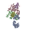
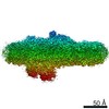
| タイトル | Structure of the PSI-FCPI supercomplex from diatom | |||||||||
 マップデータ マップデータ | ||||||||||
 試料 試料 |
| |||||||||
 キーワード キーワード | Photosystem I / ELECTRON TRANSPORT | |||||||||
| 生物種 |  Chaetoceros gracilis (珪藻) Chaetoceros gracilis (珪藻) | |||||||||
| 手法 | 単粒子再構成法 / クライオ電子顕微鏡法 / 解像度: 2.4 Å | |||||||||
 データ登録者 データ登録者 | Nagao R / Kato K | |||||||||
 引用 引用 | ジャーナル: Nat Commun / 年: 2020 タイトル: Structural basis for assembly and function of a diatom photosystem I-light-harvesting supercomplex. 著者: Ryo Nagao / Koji Kato / Kentaro Ifuku / Takehiro Suzuki / Minoru Kumazawa / Ikuo Uchiyama / Yasuhiro Kashino / Naoshi Dohmae / Seiji Akimoto / Jian-Ren Shen / Naoyuki Miyazaki / Fusamichi Akita /  要旨: Photosynthetic light-harvesting complexes (LHCs) play a pivotal role in collecting solar energy for photochemical reactions in photosynthesis. One of the major LHCs are fucoxanthin chlorophyll a/c- ...Photosynthetic light-harvesting complexes (LHCs) play a pivotal role in collecting solar energy for photochemical reactions in photosynthesis. One of the major LHCs are fucoxanthin chlorophyll a/c-binding proteins (FCPs) present in diatoms, a group of organisms having important contribution to the global carbon cycle. Here, we report a 2.40-Å resolution structure of the diatom photosystem I (PSI)-FCPI supercomplex by cryo-electron microscopy. The supercomplex is composed of 16 different FCPI subunits surrounding a monomeric PSI core. Each FCPI subunit showed different protein structures with different pigment contents and binding sites, and they form a complicated pigment-protein network together with the PSI core to harvest and transfer the light energy efficiently. In addition, two unique, previously unidentified subunits were found in the PSI core. The structure provides numerous insights into not only the light-harvesting strategy in diatom PSI-FCPI but also evolutionary dynamics of light harvesters among oxyphototrophs. | |||||||||
| 履歴 |
|
- 構造の表示
構造の表示
| ムービー |
 ムービービューア ムービービューア |
|---|---|
| 構造ビューア | EMマップ: SurfViewMolmilJmol/JSmol |
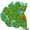
| 添付画像 |

- ダウンロードとリンク
-EMDBアーカイブ
| マップデータ | emd_0835.map.gz | 33.4 MB | EMDBマップデータ形式 | |
|---|---|---|---|---|
| ヘッダ (付随情報) | emd-0835-v30.xmlemd-0835.xml | 52.4 KB 52.4 KB | 表示 表示 | EMDBヘッダ |
| FSC (解像度算出) | emd_0835_fsc.xml | 17.7 KB | 表示 | FSCデータファイル |
| 画像 | 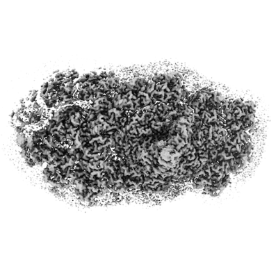 emd_0835.png emd_0835.png | 65.2 KB | ||
| Filedesc metadata | emd-0835.cif.gz | 11.8 KB | ||
| アーカイブディレクトリ |  http://ftp.pdbj.org/pub/emdb/structures/EMD-0835ftp://ftp.pdbj.org/pub/emdb/structures/EMD-0835 http://ftp.pdbj.org/pub/emdb/structures/EMD-0835ftp://ftp.pdbj.org/pub/emdb/structures/EMD-0835 | HTTPS FTP |
-関連構造データ


-リンク
| EMDBのページ | EMDB (EBI/PDBe) / EMDataResource |
|---|
-マップ
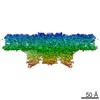
| ファイル | ダウンロード / ファイル: emd_0835.map.gz / 形式: CCP4 / 大きさ: 488.4 MB / タイプ: IMAGE STORED AS FLOATING POINT NUMBER (4 BYTES) | ||||||||||||||||||||||||||||||||||||||||||||||||||||||||||||
|---|---|---|---|---|---|---|---|---|---|---|---|---|---|---|---|---|---|---|---|---|---|---|---|---|---|---|---|---|---|---|---|---|---|---|---|---|---|---|---|---|---|---|---|---|---|---|---|---|---|---|---|---|---|---|---|---|---|---|---|---|---|
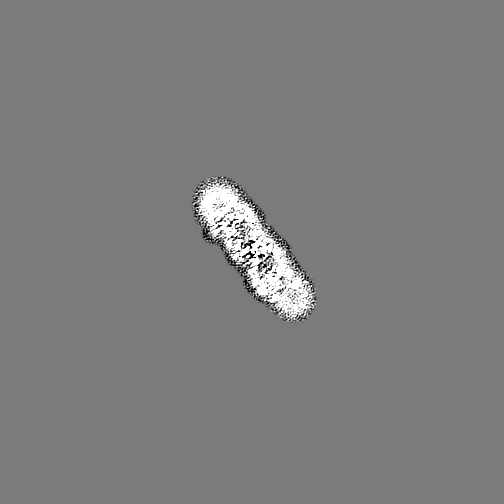
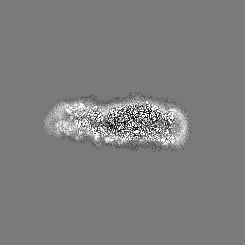
| 投影像・断面図 | 画像のコントロール
画像は Spider により作成 | ||||||||||||||||||||||||||||||||||||||||||||||||||||||||||||
| ボクセルのサイズ | X=Y=Z: 1.113 Å | ||||||||||||||||||||||||||||||||||||||||||||||||||||||||||||
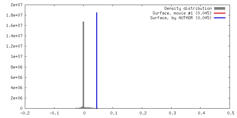
| 密度 |
| ||||||||||||||||||||||||||||||||||||||||||||||||||||||||||||
| 対称性 | 空間群: 1 | ||||||||||||||||||||||||||||||||||||||||||||||||||||||||||||
| 詳細 | EMDB XML:
CCP4マップ ヘッダ情報:
| ||||||||||||||||||||||||||||||||||||||||||||||||||||||||||||
 Z (Sec.)
 Y (Row.)
 X (Col.)


-添付データ
- 試料の構成要素
+全体 : PSI-FCPI
+超分子 #1: PSI-FCPI
+分子 #1: Photosystem I P700 chlorophyll a apoprotein A1
+分子 #2: Photosystem I P700 chlorophyll a apoprotein A2
+分子 #3: Photosystem I iron-sulfur center
+分子 #4: Photosystem I reaction center subunit II
+分子 #5: Photosystem I reaction center subunit IV
+分子 #6: Photosystem I reaction center subunit III
+分子 #7: Photosystem I reaction center subunit VIII
+分子 #8: Photosystem I reaction center subunit IX
+分子 #9: Photosystem I reaction center subunit XI
+分子 #10: Photosystem I reaction center subunit XII
+分子 #11: Unknown protein 1
+分子 #12: Photosystem I reaction center subunit Psa28
+分子 #13: Fucoxanthin chlorophyll a/c-binding protein Lhcr15
+分子 #14: Fucoxanthin chlorophyll a/c-binding protein Lhcr8
+分子 #15: Fucoxanthin chlorophyll a/c-binding protein Lhcr2
+分子 #16: Fucoxanthin chlorophyll a/c-binding protein Lhcr9
+分子 #17: Fucoxanthin chlorophyll a/c-binding protein Lhcr11
+分子 #18: Fucoxanthin chlorophyll a/c-binding protein Lhcr12
+分子 #19: Fucoxanthin chlorophyll a/c-binding protein Lhcr10
+分子 #20: Fucoxanthin chlorophyll a/c-binding protein Lhcr4
+分子 #21: Fucoxanthin chlorophyll a/c-binding protein Lhcf6
+分子 #22: Fucoxanthin chlorophyll a/c-binding protein Lhcr3
+分子 #23: Fucoxanthin chlorophyll a/c-binding protein Lhcq13
+分子 #24: Fucoxanthin chlorophyll a/c-binding protein Lhcq3
+分子 #25: Fucoxanthin chlorophyll a/c-binding protein Lhcq11
+分子 #26: Fucoxanthin chlorophyll a/c-binding protein Lhcq10
+分子 #27: Fucoxanthin chlorophyll a/c-binding protein Lhcq8
+分子 #28: Fucoxanthin chlorophyll a/c-binding protein Lhcq5
+分子 #29: CHLOROPHYLL A ISOMER

+分子 #30: CHLOROPHYLL A

+分子 #31: PHYLLOQUINONE

+分子 #32: IRON/SULFUR CLUSTER

+分子 #33: BETA-CAROTENE

+分子 #34: 1,2-DIPALMITOYL-PHOSPHATIDYL-GLYCEROLE

+分子 #35: DODECYL-BETA-D-MALTOSIDE

+分子 #36: 1,2-DISTEAROYL-MONOGALACTOSYL-DIGLYCERIDE

+分子 #37: (3S,3'S,5R,5'R,6S,6'R,8'R)-3,5'-dihydroxy-8-oxo-6',7'-didehydro-5...
+分子 #38: Chlorophyll c1
+分子 #39: (3S,3'R,5R,6S,7cis)-7',8'-didehydro-5,6-dihydro-5,6-epoxy-beta,be...

+分子 #40: water

-実験情報
-構造解析
| 手法 | クライオ電子顕微鏡法 |
|---|---|
 解析 解析 | 単粒子再構成法 |
| 試料の集合状態 | particle |
-試料調製
| 濃度 | 2 mg/mL | |||||||||
|---|---|---|---|---|---|---|---|---|---|---|
| 緩衝液 | pH: 6.5 構成要素:
| |||||||||
| グリッド | モデル: Quantifoil R1.2/1.3 / 材質: MOLYBDENUM / メッシュ: 300 / 支持フィルム - 材質: CARBON / 支持フィルム - トポロジー: HOLEY ARRAY / 前処理 - タイプ: PLASMA CLEANING / 前処理 - 時間: 30 sec. | |||||||||
| 凍結 | 凍結剤: ETHANE / チャンバー内湿度: 100 % / チャンバー内温度: 277 K / 装置: FEI VITROBOT MARK IV |
- 電子顕微鏡法
| 顕微鏡 | FEI TITAN KRIOS |
|---|---|
| 撮影 | フィルム・検出器のモデル: FEI FALCON III (4k x 4k) 検出モード: COUNTING / 平均電子線量: 50.0 e/Å2 |
| 電子線 | 加速電圧: 300 kV / 電子線源:  FIELD EMISSION GUN FIELD EMISSION GUN |
| 電子光学系 | 照射モード: FLOOD BEAM / 撮影モード: BRIGHT FIELD |
| 実験機器 |  モデル: Titan Krios / 画像提供: FEI Company |
+画像解析

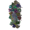
-原子モデル構築 1
| 精密化 | 空間: REAL / プロトコル: FLEXIBLE FIT / 当てはまり具合の基準: Correlation coefficient |
|---|---|
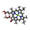
| 得られたモデル |  PDB-6l4u: |
