 ムービー
ムービー コントローラー


+ データを開く

- 基本情報
基本情報
| 登録情報 | データベース: EMDB / ID: EMD-6846 | ||||||||||||||||||
|---|---|---|---|---|---|---|---|---|---|---|---|---|---|---|---|---|---|---|---|
| タイトル | Volta phase contrast cryotomogram of a cryosectioned prometaphase S. pombe cell, strain nda3-KM311 | ||||||||||||||||||
 マップデータ マップデータ | Volta phase contrast cryotomogram of a cryosectioned prometaphase S. pombe cell, strain nda3-KM311 | ||||||||||||||||||
 試料 試料 |
| ||||||||||||||||||
| 生物種 |  | ||||||||||||||||||
| 手法 | 電子線トモグラフィー法 / クライオ電子顕微鏡法 | ||||||||||||||||||
 データ登録者 データ登録者 | Cai S / Chen C / Tan Z / Huang Y / Shi J / Gan L | ||||||||||||||||||
| 資金援助 |  シンガポール, 5件 シンガポール, 5件
| ||||||||||||||||||
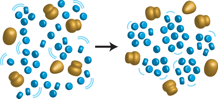
 引用 引用 | ジャーナル: Proc Natl Acad Sci U S A / 年: 2018 タイトル: Cryo-ET reveals the macromolecular reorganization of mitotic chromosomes in vivo. 著者: Shujun Cai / Chen Chen / Zhi Yang Tan / Yinyi Huang / Jian Shi / Lu Gan / 要旨: Chromosomes condense during mitosis in most eukaryotes. This transformation involves rearrangements at the nucleosome level and has consequences for transcription. Here, we use cryo-electron ...Chromosomes condense during mitosis in most eukaryotes. This transformation involves rearrangements at the nucleosome level and has consequences for transcription. Here, we use cryo-electron tomography (cryo-ET) to determine the 3D arrangement of nuclear macromolecular complexes, including nucleosomes, in frozen-hydrated cells. Using 3D classification analysis, we did not find evidence that nucleosomes resembling the crystal structure are abundant. This observation and those from other groups support the notion that a subset of fission yeast nucleosomes may be partially unwrapped in vivo. In both interphase and mitotic cells, there is also no evidence of monolithic structures the size of Hi-C domains. The chromatin is mingled with two features: pockets, which are positions free of macromolecular complexes; and "megacomplexes," which are multimegadalton globular complexes like preribosomes. Mitotic chromatin is more crowded than interphase chromatin in subtle ways. Nearest-neighbor distance analyses show that mitotic chromatin is more compacted at the oligonucleosome than the dinucleosome level. Like interphase, mitotic chromosomes contain megacomplexes and pockets. This uneven chromosome condensation helps explain a longstanding enigma of mitosis: a subset of genes is up-regulated. | ||||||||||||||||||
| 履歴 |
|
- 構造の表示
構造の表示
| ムービー |
 ムービービューア ムービービューア |
|---|---|
| 添付画像 |
- ダウンロードとリンク
-EMDBアーカイブ
| マップデータ | emd_6846.map.gz | 3.4 GB | EMDBマップデータ形式 | |
|---|---|---|---|---|
| ヘッダ (付随情報) | emd-6846-v30.xmlemd-6846.xml | 11.2 KB 11.2 KB | 表示 表示 | EMDBヘッダ |
| 画像 |  emd_6846.png emd_6846.png | 212.6 KB | ||
| アーカイブディレクトリ |  http://ftp.pdbj.org/pub/emdb/structures/EMD-6846ftp://ftp.pdbj.org/pub/emdb/structures/EMD-6846 http://ftp.pdbj.org/pub/emdb/structures/EMD-6846ftp://ftp.pdbj.org/pub/emdb/structures/EMD-6846 | HTTPS FTP |
-検証レポート
| 文書・要旨 | emd_6846_validation.pdf.gz | 78.4 KB | 表示 | EMDB検証レポート |
|---|---|---|---|---|
| 文書・詳細版 | emd_6846_full_validation.pdf.gz | 77.6 KB | 表示 | |
| XML形式データ | emd_6846_validation.xml.gz | 499 B | 表示 | |
| アーカイブディレクトリ | https://ftp.pdbj.org/pub/emdb/validation_reports/EMD-6846ftp://ftp.pdbj.org/pub/emdb/validation_reports/EMD-6846 | HTTPS FTP |
-関連構造データ
| 電子顕微鏡画像生データ | EMPIAR-10125 (タイトル: Cryo-ET reveals the macromolecular reorganization of S. pombe mitotic chromosomes in vivo Data size: 35.1 Data #1: Tilt series of fission yeast cells and chromatin lysates in various cell cycle states [tilt series]) |
|---|
-リンク
| EMDBのページ | EMDB (EBI/PDBe) / EMDataResource |
|---|
-マップ
| ファイル | ダウンロード / ファイル: emd_6846.map.gz / 形式: CCP4 / 大きさ: 4.5 GB / タイプ: IMAGE STORED AS SIGNED INTEGER (2 BYTES) | ||||||||||||||||||||||||||||||||||||||||||||||||||||||||||||||||||||
|---|---|---|---|---|---|---|---|---|---|---|---|---|---|---|---|---|---|---|---|---|---|---|---|---|---|---|---|---|---|---|---|---|---|---|---|---|---|---|---|---|---|---|---|---|---|---|---|---|---|---|---|---|---|---|---|---|---|---|---|---|---|---|---|---|---|---|---|---|---|
| 注釈 | Volta phase contrast cryotomogram of a cryosectioned prometaphase S. pombe cell, strain nda3-KM311 | ||||||||||||||||||||||||||||||||||||||||||||||||||||||||||||||||||||
| 投影像・断面図 | 画像のコントロール
画像は Spider により作成 これらの図は立方格子座標系で作成されたものです | ||||||||||||||||||||||||||||||||||||||||||||||||||||||||||||||||||||
| ボクセルのサイズ | X=Y=Z: 4.6 Å | ||||||||||||||||||||||||||||||||||||||||||||||||||||||||||||||||||||
| 密度 |
| ||||||||||||||||||||||||||||||||||||||||||||||||||||||||||||||||||||
| 対称性 | 空間群: 1 | ||||||||||||||||||||||||||||||||||||||||||||||||||||||||||||||||||||
| 詳細 | EMDB XML:
CCP4マップ ヘッダ情報:
| ||||||||||||||||||||||||||||||||||||||||||||||||||||||||||||||||||||
 Z (Sec.)
 Y (Row.)
 X (Col.)


-添付データ
- 試料の構成要素
-全体 : S. pombe cell, arrested in prometaphase
| 全体 | 名称: S. pombe cell, arrested in prometaphase |
|---|---|
| 要素 |
|
-超分子 #1: S. pombe cell, arrested in prometaphase
| 超分子 | 名称: S. pombe cell, arrested in prometaphase / タイプ: cell / ID: 1 / 親要素: 0 |
|---|---|
| 由来(天然) | 生物種: |
-実験情報
-構造解析
| 手法 | クライオ電子顕微鏡法 |
|---|---|
 解析 解析 | 電子線トモグラフィー法 |
| 試料の集合状態 | cell |
-試料調製
| 緩衝液 | pH: 7 / 詳細: yeast-extract supplemented |
|---|---|
| グリッド | モデル: C-flat-1/1 / 材質: COPPER / メッシュ: 200 / 支持フィルム - 材質: CARBON / 支持フィルム - トポロジー: HOLEY |
| 凍結 | 凍結剤: ETHANE |
| 加圧凍結法 | 装置: OTHER 詳細: Self-pressurized freezing. The value given for _emd_high_pressure_freezing.instrument is Vitrobot cup. This is not in a list of allowed values set(['LEICA EM PACT2', 'LEICA EM PACT', 'EMS-002 ...詳細: Self-pressurized freezing. The value given for _emd_high_pressure_freezing.instrument is Vitrobot cup. This is not in a list of allowed values set(['LEICA EM PACT2', 'LEICA EM PACT', 'EMS-002 RAPID IMMERSION FREEZER', 'OTHER', 'LEICA EM HPM100', 'BAL-TEC HPM 010']) so OTHER is written into the XML file. |
| Cryo protectant | 30% dextran |
| 切片作成 | ウルトラミクロトーム - 装置: Leica UC7/FC7 / ウルトラミクロトーム - 温度: 123 K / ウルトラミクロトーム - 最終 厚さ: 100 nm |
| 位置合わせマーカー | Manufacturer: BBI / 直径: 10 nm |
- 電子顕微鏡法
| 顕微鏡 | FEI TITAN KRIOS |
|---|---|
| 特殊光学系 | 位相板: VOLTA PHASE PLATE |
| 撮影 | フィルム・検出器のモデル: FEI FALCON II (4k x 4k) 検出モード: INTEGRATING / デジタル化 - サイズ - 横: 4096 pixel / デジタル化 - サイズ - 縦: 4096 pixel / デジタル化 - サンプリング間隔: 14.0 µm / 撮影したグリッド数: 1 / 実像数: 61 / 平均電子線量: 1.6 e/Å2 |
| 電子線 | 加速電圧: 300 kV / 電子線源:  FIELD EMISSION GUN FIELD EMISSION GUN |
| 電子光学系 | 倍率(補正後): 30400 / 照射モード: FLOOD BEAM / 撮影モード: BRIGHT FIELD / 最小 デフォーカス(公称値): 0.5 µm / 倍率(公称値): 18000 |
| 試料ステージ | 試料ホルダーモデル: FEI TITAN KRIOS AUTOGRID HOLDER ホルダー冷却材: NITROGEN |
| 実験機器 |  モデル: Titan Krios / 画像提供: FEI Company |
-画像解析
| 最終 再構成 | アルゴリズム: BACK PROJECTION / ソフトウェア - 名称: IMOD (ver. 4.9) / 使用した粒子像数: 61 |
|---|
