Japan Agency for Medical Research and Development (AMED)
JP21gm161003
Japan
Japan Science and Technology
JPMJFR214K
Japan
Japan Science and Technology
JPMJMS2024
Japan
Citation
Journal: iScience / Year: 2025 Title: Cryo-ET of actin cytoskeleton and membrane structure in lamellipodia formation using optogenetics. Authors: Hironori Inaba / Tsuyoshi Imasaki / Kazuhiro Aoyama / Shogo Yoshihara / Hiroko Takazaki / Takayuki Kato / Hidemasa Goto / Kaoru Mitsuoka / Ryo Nitta / Takao Nakata / Abstract: Lamellipodia are sheet-like protrusions essential for cell migration and endocytosis, but their ultrastructural dynamics remain poorly understood because conventional electron microscopy lacks ...Lamellipodia are sheet-like protrusions essential for cell migration and endocytosis, but their ultrastructural dynamics remain poorly understood because conventional electron microscopy lacks temporal resolution. Here, we combined optogenetics with cryo-electron tomography (cryo-ET) to visualize the actin cytoskeleton and membrane structures during lamellipodia formation with temporal precision. Using photoactivatable-Rac1 (PA-Rac1) in COS-7 cells, we induced lamellipodia formation with a 2-min blue light irradiation, rapidly vitrified samples, and analyzed their ultrastructure with cryo-ET. We obtained 16 tomograms of lamellipodia at different degrees of extension from three cells. These revealed small protrusions with unbundled actin filaments, "mini filopodia" composed of short, bundled actin filaments at the leading edge, and actin bundles running nearly parallel to the leading edge within inner regions of lamellipodia, suggesting dynamic reorganizations of the actin cytoskeleton. This approach provides a powerful framework for elucidating the ultrastructural dynamics of cellular processes with precise temporal control.
In the structure databanks used in Yorodumi, some data are registered as the other names, "COVID-19 virus" and "2019-nCoV". Here are the details of the virus and the list of structure data.
Jan 31, 2019. EMDB accession codes are about to change! (news from PDBe EMDB page)
EMDB accession codes are about to change! (news from PDBe EMDB page)
The allocation of 4 digits for EMDB accession codes will soon come to an end. Whilst these codes will remain in use, new EMDB accession codes will include an additional digit and will expand incrementally as the available range of codes is exhausted. The current 4-digit format prefixed with “EMD-” (i.e. EMD-XXXX) will advance to a 5-digit format (i.e. EMD-XXXXX), and so on. It is currently estimated that the 4-digit codes will be depleted around Spring 2019, at which point the 5-digit format will come into force.
The EM Navigator/Yorodumi systems omit the EMD- prefix.
Related info.:Q: What is EMD? / ID/Accession-code notation in Yorodumi/EM Navigator
Yorodumi is a browser for structure data from EMDB, PDB, SASBDB, etc.
This page is also the successor to EM Navigator detail page, and also detail information page/front-end page for Omokage search.
The word "yorodu" (or yorozu) is an old Japanese word meaning "ten thousand". "mi" (miru) is to see.
Related info.:EMDB / PDB / SASBDB / Comparison of 3 databanks / Yorodumi Search / Aug 31, 2016. New EM Navigator & Yorodumi / Yorodumi Papers / Jmol/JSmol / Function and homology information / Changes in new EM Navigator and Yorodumi
 Movie
Movie Controller

 Yorodumi
 Open data
 Basic information
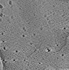
 Map data
 Sample
 Keywords
 Chlorocebus aethiops (grivet monkey)
 Authors
 Japan, 13 items
  Citation
 Structure visualization
 Downloads & links
 EMDB map data format
 emd_61093.png
 http://ftp.pdbj.org/pub/emdb/structures/EMD-61093
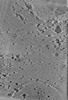

 Z (Sec.)
 Y (Row.)
 X (Col.)


 Sample components
 Processing
 Electron microscopy
 FIELD EMISSION GUN
