 Movie
Movie Controller

[English] 日本語
 Yorodumi
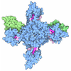
- EMDB-54978: Cryo-EM structure of H. neapolitanus CsoSCA C283A/C284A inactive ... -
+ Open data

- Basic information
| Entry |  | ||||||||||||
|---|---|---|---|---|---|---|---|---|---|---|---|---|---|
| Title | Cryo-EM structure of H. neapolitanus CsoSCA C283A/C284A inactive mutant, hexamer | ||||||||||||
 Map data Map data | Sharpened map from final non-uniform refinement in CryoSPARC. Sharpening B-factor: -72.7 A^2. | ||||||||||||
 Sample Sample |
| ||||||||||||
 Keywords Keywords | Carbonic anhydrase / carboxysome / CO2 concentration mechanism / LYASE | ||||||||||||
| Function / homology |  Function and homology information Function and homology informationcarboxysome / carbon fixation / detection of maltose stimulus / carbohydrate transport / carbohydrate transmembrane transporter activity / maltose binding / maltose transport / maltodextrin transmembrane transport / ATP-binding cassette (ABC) transporter complex, substrate-binding subunit-containing / ATP-binding cassette (ABC) transporter complex ...carboxysome / carbon fixation / detection of maltose stimulus / carbohydrate transport / carbohydrate transmembrane transporter activity / maltose binding / maltose transport / maltodextrin transmembrane transport / ATP-binding cassette (ABC) transporter complex, substrate-binding subunit-containing / ATP-binding cassette (ABC) transporter complex / carbonic anhydrase / carbonate dehydratase activity / cell chemotaxis / outer membrane-bounded periplasmic space / periplasmic space / DNA damage response / metal ion binding / membrane Similarity search - Function | ||||||||||||
| Biological species |  Halothiobacillus neapolitanus c2 (bacteria) Halothiobacillus neapolitanus c2 (bacteria) | ||||||||||||
| Method | single particle reconstruction / cryo EM / Resolution: 2.08 Å | ||||||||||||
 Authors Authors | Gaullier G / Vogiatzi N / Blikstad C | ||||||||||||
| Funding support |  Sweden, 3 items Sweden, 3 items
| ||||||||||||
 Citation Citation | Journal: Biorxiv / Year: 2026 Title: Molecular mechanism of redox regulation of the alpha-carboxysomal carbonic anhydrase CsoSCA Authors: Vogiatzi N / Gaullier G / Leufstadius J / Andersson T / Scherbauer T / Blikstad C | ||||||||||||
| History |
|
- Structure visualization
| Supplemental images |
|---|

- Downloads & links
-EMDB archive
| Map data | emd_54978.map.gz | 398 MB | EMDB map data format | |
|---|---|---|---|---|
| Header (meta data) | emd-54978-v30.xmlemd-54978.xml | 24.1 KB 24.1 KB | Display Display | EMDB header |
| FSC (resolution estimation) | emd_54978_fsc.xml | 15.8 KB | Display | FSC data file |
| Images |  emd_54978.png emd_54978.png | 63.5 KB | ||
| Masks | emd_54978_msk_1.mapemd_54978_msk_2.mapemd_54978_msk_3.map | 421.9 MB 421.9 MB 421.9 MB | Mask map | |
| Filedesc metadata | emd-54978.cif.gz | 7.8 KB | ||
| Others | emd_54978_half_map_1.map.gzemd_54978_half_map_2.map.gz | 391.5 MB 391.5 MB | ||
| Archive directory |  https://data.pdbj.org/pub/emdb/structures/EMD-54978ftp://data.pdbj.org/pub/emdb/structures/EMD-54978 https://data.pdbj.org/pub/emdb/structures/EMD-54978ftp://data.pdbj.org/pub/emdb/structures/EMD-54978 | HTTPS FTP |
-Related structure data
| Related structure data |  9skxMC  9skrC  9sksC  9sktC  9skuC  9skvC  9skwC  9skyC  9skzC M: atomic model generated by this map C: citing same article ( |
|---|---|
| Similar structure data |


-Links
| EMDB pages | EMDB (EBI/PDBe) / EMDataResource |
|---|---|
| Related items in Molecule of the Month |


-Map
| File | Download / File: emd_54978.map.gz / Format: CCP4 / Size: 421.9 MB / Type: IMAGE STORED AS FLOATING POINT NUMBER (4 BYTES) | ||||||||||||||||||||||||||||||||||||
|---|---|---|---|---|---|---|---|---|---|---|---|---|---|---|---|---|---|---|---|---|---|---|---|---|---|---|---|---|---|---|---|---|---|---|---|---|---|
| Annotation | Sharpened map from final non-uniform refinement in CryoSPARC. Sharpening B-factor: -72.7 A^2. | ||||||||||||||||||||||||||||||||||||
| Projections & slices | Image control
Images are generated by Spider. | ||||||||||||||||||||||||||||||||||||
| Voxel size | X=Y=Z: 0.65 Å | ||||||||||||||||||||||||||||||||||||
| Density |
| ||||||||||||||||||||||||||||||||||||
| Symmetry | Space group: 1 | ||||||||||||||||||||||||||||||||||||
| Details | EMDB XML:
|
 Z (Sec.)
 Y (Row.)
 X (Col.)


-Supplemental data
-Mask #1
| File | emd_54978_msk_1.map | ||||||||||||
|---|---|---|---|---|---|---|---|---|---|---|---|---|---|
| Projections & Slices |
| ||||||||||||
| Density Histograms |


-Mask #2
| File | emd_54978_msk_2.map | ||||||||||||
|---|---|---|---|---|---|---|---|---|---|---|---|---|---|
| Projections & Slices |
| ||||||||||||
| Density Histograms |


-Mask #3
| File | emd_54978_msk_3.map | ||||||||||||
|---|---|---|---|---|---|---|---|---|---|---|---|---|---|
| Projections & Slices |
| ||||||||||||
| Density Histograms |


-Half map: Half-map A from final non-uniform refinement in CryoSPARC.
| File | emd_54978_half_map_1.map | ||||||||||||
|---|---|---|---|---|---|---|---|---|---|---|---|---|---|
| Annotation | Half-map A from final non-uniform refinement in CryoSPARC. | ||||||||||||
| Projections & Slices |
| ||||||||||||
| Density Histograms |


-Half map: Half-map B from final non-uniform refinement in CryoSPARC.
| File | emd_54978_half_map_2.map | ||||||||||||
|---|---|---|---|---|---|---|---|---|---|---|---|---|---|
| Annotation | Half-map B from final non-uniform refinement in CryoSPARC. | ||||||||||||
| Projections & Slices |
| ||||||||||||
| Density Histograms |


- Sample components
-Entire : HnCsoSCA_C283A-C284A
| Entire | Name: HnCsoSCA_C283A-C284A |
|---|---|
| Components |
|
-Supramolecule #1: HnCsoSCA_C283A-C284A
| Supramolecule | Name: HnCsoSCA_C283A-C284A / type: complex / ID: 1 / Parent: 0 / Macromolecule list: #1 |
|---|---|
| Source (natural) | Organism: Halothiobacillus neapolitanus c2 (bacteria) |
| Molecular weight | Theoretical: 611.1651 KDa |
-Macromolecule #1: Maltose/maltodextrin-binding periplasmic protein,Carboxysome shel...
| Macromolecule | Name: Maltose/maltodextrin-binding periplasmic protein,Carboxysome shell carbonic anhydrase type: protein_or_peptide / ID: 1 Details: Double mutation C283A, C284A, engineered to test redox dependence. This is a fusion protein with an N-terminal Strep-MBP tag and a TEV protease site between the MBP tag and CsoSCA. The MBP ...Details: Double mutation C283A, C284A, engineered to test redox dependence. This is a fusion protein with an N-terminal Strep-MBP tag and a TEV protease site between the MBP tag and CsoSCA. The MBP tag and the disordered N-terminus of CsoSCA are not visible in the map.,Double mutation C283A, C284A, engineered to test redox dependence. This is a fusion protein with an N-terminal Strep-MBP tag and a TEV protease site between the MBP tag and CsoSCA. The MBP tag and the disordered N-terminus of CsoSCA are not visible in the map. Number of copies: 2 / Enantiomer: LEVO / EC number: carbonic anhydrase |
|---|---|
| Source (natural) | Organism: Halothiobacillus neapolitanus c2 (bacteria) |
| Molecular weight | Theoretical: 101.980648 KDa |
| Recombinant expression | Organism:  Escherichia coli BL21 (bacteria) Escherichia coli BL21 (bacteria) |
| Sequence | String: MWSHPQFEKG SSMKIEEGKL VIWINGDKGY NGLAEVGKKF EKDTGIKVTV EHPDKLEEKF PQVAATGDGP DIIFWAHDRF GGYAQSGLL AEITPDKAFQ DKLYPFTWDA VRYNGKLIAY PIAVEALSLI YNKDLLPNPP KTWEEIPALD KELKAKGKSA L MFNLQEPY ...String: MWSHPQFEKG SSMKIEEGKL VIWINGDKGY NGLAEVGKKF EKDTGIKVTV EHPDKLEEKF PQVAATGDGP DIIFWAHDRF GGYAQSGLL AEITPDKAFQ DKLYPFTWDA VRYNGKLIAY PIAVEALSLI YNKDLLPNPP KTWEEIPALD KELKAKGKSA L MFNLQEPY FTWPLIAADG GYAFKYENGK YDIKDVGVDN AGAKAGLTFL VDLIKNKHMN ADTDYSIAEA AFNKGETAMT IN GPWAWSN IDTSKVNYGV TVLPTFKGQP SKPFVGVLSA GINAASPNKE LAKEFLENYL LTDEGLEAVN KDKPLGAVAL KSY EEELAK DPRIAATMEN AQKGEIMPNI PQMSAFWYAV RTAVINAASG RQTVDEALKD AQTNSSSNNN NNNNNNNLGI EENL YFQSN ANTRNTRSKQ RAPFGVSSSV KPRLDLIEQA PNPAYDRHPA CITLPERTCR HPLTDLEANE QLGRCEDSVK NRFDR VIPF LQVVAGIPLG LDYVTRVQEL AQSSLGHTLP EELLKDNWIS GHNLKGIFGY ATAKALTAAT EQFSRKIMSE KDDSAS AIG FFLDCGFHAV DISPCADGRL KGLLPYILRL PLTAFTYRKA YAGSMFDIED DLAQWEKNEL RRYREGVPNT ADQPTRY LK IAVYHFSTSD PTHSGCAAHG SNDRAALEAA LTQLMKFREA VENAHAAGAS IDILLIGVDT DTDAIRVHIP DSKGFLNP Y RYVDNTVTYA QTLHLAPDEA RVIIHEAILN ANRSDGWAKG NGVASEGMRR FIGQLLINNL SQIDYVVNRH GGRYPPNDI GHAERYISVG DGFDEVQIRN LAYYAHLDTV EENAIDVDVG IKIFTKLNLS RGLPIPIAIH YRYDPNVPGS RERTVVKARR IYNAIKERF SSLDEQNLLQ FRLSVQAQDI GSPIEEVASA UniProtKB: Maltose/maltodextrin-binding periplasmic protein, Carboxysome shell carbonic anhydrase |
-Macromolecule #2: ZINC ION
| Macromolecule | Name: ZINC ION / type: ligand / ID: 2 / Number of copies: 2 / Formula: ZN |
|---|---|
| Molecular weight | Theoretical: 65.409 Da |
-Macromolecule #3: water
| Macromolecule | Name: water / type: ligand / ID: 3 / Number of copies: 253 / Formula: HOH |
|---|---|
| Molecular weight | Theoretical: 18.015 Da |
| Chemical component information |  ChemComp-HOH: |
-Experimental details
-Structure determination
| Method | cryo EM |
|---|---|
 Processing Processing | single particle reconstruction |
| Aggregation state | particle |
-Sample preparation
| Concentration | 0.428 mg/mL | |||||||||
|---|---|---|---|---|---|---|---|---|---|---|
| Buffer | pH: 7.5 Component:
| |||||||||
| Grid | Model: Quantifoil R1.2/1.3 / Material: COPPER / Mesh: 300 / Support film - Material: CARBON / Support film - topology: HOLEY ARRAY / Pretreatment - Type: GLOW DISCHARGE / Pretreatment - Time: 60 sec. / Pretreatment - Atmosphere: OTHER / Pretreatment - Pressure: 0.04 kPa Details: Performed with a Pelco easiGlow with a current of 20 mA. | |||||||||
| Vitrification | Cryogen name: ETHANE / Chamber humidity: 100 % / Chamber temperature: 277.15 K / Instrument: FEI VITROBOT MARK IV Details: 4C, 100% relative humidity, delay time 0s, blot time 3s, blot force 0. |
- Electron microscopy
| Microscope | TFS KRIOS |
|---|---|
| Specialist optics | Energy filter - Name: GIF Bioquantum / Energy filter - Slit width: 20 eV |
| Image recording | Film or detector model: GATAN K3 BIOQUANTUM (6k x 4k) / Number grids imaged: 1 / Number real images: 10000 / Average exposure time: 1.6 sec. / Average electron dose: 68.973 e/Å2 |
| Electron beam | Acceleration voltage: 300 kV / Electron source:  FIELD EMISSION GUN FIELD EMISSION GUN |
| Electron optics | C2 aperture diameter: 50.0 µm / Illumination mode: FLOOD BEAM / Imaging mode: BRIGHT FIELD / Cs: 2.7 mm / Nominal defocus max: 1.8 µm / Nominal defocus min: 0.6 µm / Nominal magnification: 130000 |
| Sample stage | Specimen holder model: FEI TITAN KRIOS AUTOGRID HOLDER / Cooling holder cryogen: NITROGEN |
| Experimental equipment |  Model: Titan Krios / Image courtesy: FEI Company |


