Movie
 Controller


[English] 日本語
 Yorodumi
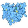
- EMDB-52843: Cryo-EM structure of the kinetoplastid trans-spliceosome activate... -
+ Open data

- Basic information
| Entry |  | |||||||||
|---|---|---|---|---|---|---|---|---|---|---|
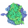
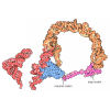
| Title | Cryo-EM structure of the kinetoplastid trans-spliceosome activated for catalytic step II (C* complex) | |||||||||
 Map data | Composite map | |||||||||
 Sample |
| |||||||||
 Keywords | Trans-spliceosome / Spliceosome / SPLICING | |||||||||
| Function / homology |  Function and homology information U2-type post-spliceosomal complex / spliceosomal complex disassembly / exon-exon junction complex / pre-mRNA 3'-splice site binding / Lsm1-7-Pat1 complex / U6 snRNP / generation of catalytic spliceosome for first transesterification step / U12-type spliceosomal complex / pICln-Sm protein complex / snRNP binding ...U2-type post-spliceosomal complex / spliceosomal complex disassembly / exon-exon junction complex / pre-mRNA 3'-splice site binding / Lsm1-7-Pat1 complex / U6 snRNP / generation of catalytic spliceosome for first transesterification step / U12-type spliceosomal complex / pICln-Sm protein complex / snRNP binding / small nuclear ribonucleoprotein complex / SMN-Sm protein complex / spliceosomal tri-snRNP complex / P granule / post-mRNA release spliceosomal complex / U2-type catalytic step 2 spliceosome / U1 snRNP / U2 snRNP / U4 snRNP / U2-type prespliceosome / U2-type catalytic step 1 spliceosome / generation of catalytic spliceosome for second transesterification step / spliceosomal complex assembly / pre-mRNA intronic binding / precatalytic spliceosome / Prp19 complex / U2 snRNA binding / U5 snRNA binding / spliceosomal tri-snRNP complex assembly / U5 snRNP / U1 snRNA binding / RNA splicing / U6 snRNA binding / mRNA cis splicing, via spliceosome / nuclear-transcribed mRNA catabolic process / U4/U6 x U5 tri-snRNP complex / RNA processing / spliceosomal snRNP assembly / catalytic step 2 spliceosome / protein K63-linked ubiquitination / mRNA processing / spliceosomal complex / peptidylprolyl isomerase / peptidyl-prolyl cis-trans isomerase activity / mRNA splicing, via spliceosome / RING-type E3 ubiquitin transferase / regulation of gene expression / metallopeptidase activity / ubiquitin protein ligase activity / nucleic acid binding / RNA helicase activity / RNA helicase / mRNA binding / chromatin binding / GTPase activity / GTP binding / RNA binding / ATP binding / nucleus / cytosol / cytoplasm Similarity search - Function | |||||||||
| Biological species |  Leishmania tarentolae (eukaryote) | |||||||||
| Method | single particle reconstruction / cryo EM / Resolution: 2.7 Å | |||||||||
 Authors | Nadenoen T / Vanden Broeck A | |||||||||
| Funding support |  Belgium, European Union, 2 items
| |||||||||
 Citation | Journal: Nat Commun / Year: 2026 Title: Structural basis of step II spliced leader RNA trans-splicing in trypanosomatid parasites. Authors: Théo Nadenoen / Franco Agustín Biglione / Marylène Vandevenne / Arnaud Vanden Broeck /  Abstract: Messenger RNA (mRNA) synthesis is fundamental to eukaryotic gene expression. In kinetoplastid parasites, including the human pathogens Trypanosoma and Leishmania, mRNAs are produced through a ...Messenger RNA (mRNA) synthesis is fundamental to eukaryotic gene expression. In kinetoplastid parasites, including the human pathogens Trypanosoma and Leishmania, mRNAs are produced through a mechanism called Spliced Leader (SL) RNA trans-splicing. In this process, a short SL exon from a small noncoding RNA is joined to the 5' end of every mRNA, ensuring transcript stability and translation. Despite decades of study, the structural and mechanistic basis of SL trans-splicing remains elusive. Here, we report cryogenic electron microscopy structures of step II SL trans-splicing machineries from Leishmania tarentolae. These structures reveal the molecular mechanism of SL exon ligation and uncover lineage-specific adaptations that remodel the trypanosomatid trans-spliceosome for SL trans-splicing while preserving fundamental spliceosomal chemistry. Altogether, our results establish a mechanistic framework for SL trans-splicing and illuminate the evolutionary diversification of RNA processing in deeply diverged eukaryotes. | |||||||||
| History |
|
- Structure visualization
| Supplemental images |
|---|

- Downloads & links
-EMDB archive
| Map data | emd_52843.map.gz | 119.5 MB | EMDB map data format | |
|---|---|---|---|---|
| Header (meta data) | emd-52843-v30.xmlemd-52843.xml | 88 KB 88 KB | Display Display | EMDB header |
| Images |  emd_52843.png | 247.1 KB | ||
| Filedesc metadata | emd-52843.cif.gz | 23.6 KB | ||
| Archive directory |  https://data.pdbj.org/pub/emdb/structures/EMD-52843ftp://data.pdbj.org/pub/emdb/structures/EMD-52843 | HTTPS FTP |
-Related structure data
| Related structure data |  9if7MC  9ibcC  9ibdC  9if8C C: citing same article ( M: atomic model generated by this map |
|---|---|
| Similar structure data |


-Links
| EMDB pages | EMDB (EBI/PDBe) / EMDataResource |
|---|---|
| Related items in Molecule of the Month |
-Map
| File | Download / File: emd_52843.map.gz / Format: CCP4 / Size: 244.1 MB / Type: IMAGE STORED AS FLOATING POINT NUMBER (4 BYTES) | ||||||||||||||||||||||||||||||||||||
|---|---|---|---|---|---|---|---|---|---|---|---|---|---|---|---|---|---|---|---|---|---|---|---|---|---|---|---|---|---|---|---|---|---|---|---|---|---|
| Annotation | Composite map | ||||||||||||||||||||||||||||||||||||
| Projections & slices | Image control
Images are generated by Spider. | ||||||||||||||||||||||||||||||||||||
| Voxel size | X=Y=Z: 1.065 Å | ||||||||||||||||||||||||||||||||||||
| Density |
| ||||||||||||||||||||||||||||||||||||
| Symmetry | Space group: 1 | ||||||||||||||||||||||||||||||||||||
| Details | EMDB XML:
|
 Z (Sec.)
 Y (Row.)
 X (Col.)


-Supplemental data
- Sample components
+Entire : Leishmania tarentolae trans-spliceosome activated for catalytic s...
+Supramolecule #1: Leishmania tarentolae trans-spliceosome activated for catalytic s...
+Macromolecule #1: Probable eukaryotic initiation factor 4A
+Macromolecule #2: RRM domain-containing protein
+Macromolecule #3: Mago nashi-like protein,putative
+Macromolecule #4: Prp8 protein homologue, putative
+Macromolecule #5: ATP-dependent RNA helicase, putative
+Macromolecule #6: Small nuclear ribonucleoprotein component-like protein
+Macromolecule #7: PRKR-interacting protein 1
+Macromolecule #8: Splicing factor Cactin C-terminal domain-containing protein
+Macromolecule #9: RNA-binding motif protein
+Macromolecule #10: Splicing regulator SDE2-like protein
+Macromolecule #11: Guanine nucleotide-binding protein subunit beta-like protein
+Macromolecule #12: G10 protein
+Macromolecule #13: Guanine nucleotide-binding protein subunit beta-like protein
+Macromolecule #14: Spliceosome-associated CWC15-like protein
+Macromolecule #15: Cell division control protein
+Macromolecule #16: CWF21 domain-containing protein
+Macromolecule #17: Crooked neck-like protein 1
+Macromolecule #18: Pre-mRNA-splicing factor SYF1
+Macromolecule #19: RNA helicase
+Macromolecule #20: U2 small nuclear ribonucleoprotein 40K, putative
+Macromolecule #21: RRM domain-containing protein
+Macromolecule #22: NF-kappa-B-activating protein C-terminal domain-containing protein
+Macromolecule #23: MI domain-containing protein
+Macromolecule #24: Pre-mRNA-splicing factor SLU7
+Macromolecule #25: Pre-mRNA-splicing factor 18
+Macromolecule #26: Uncharacterized protein
+Macromolecule #27: PRP19-related complex protein 3
+Macromolecule #28: Unspecified product
+Macromolecule #29: SNW domain-containing protein 1
+Macromolecule #30: peptidylprolyl isomerase
+Macromolecule #31: Guanine nucleotide-binding protein subunit beta-like protein
+Macromolecule #32: Pre-mRNA-splicing factor ISY1 homolog
+Macromolecule #33: Zinc knuckle
+Macromolecule #34: Pre-mRNA-splicing factor SPF27
+Macromolecule #35: Pre-mRNA-processing factor 19
+Macromolecule #36: Probable RNA helicase
+Macromolecule #37: Unidentified protein
+Macromolecule #38: LSM domain-containing protein
+Macromolecule #39: Sm domain-containing protein
+Macromolecule #40: U6 snRNA-associated Sm-like protein LSm4
+Macromolecule #41: LSM domain-containing protein
+Macromolecule #42: Sm domain-containing protein
+Macromolecule #43: Lsm7p protein, putative
+Macromolecule #44: Sm domain-containing protein
+Macromolecule #46: Small nuclear ribonucleoprotein, putative
+Macromolecule #47: Small nuclear ribonucleoprotein Sm D2
+Macromolecule #48: Lsm5p, putative
+Macromolecule #49: Small nuclear ribonucleaoprotein, putative
+Macromolecule #50: Small nuclear ribonucleoprotein E
+Macromolecule #51: Sm protein F
+Macromolecule #52: Sm protein G
+Macromolecule #53: Small nuclear ribonucleoprotein Sm D3
+Macromolecule #54: Sm protein B
+Macromolecule #45: Y-branched pre-mRNA
+Macromolecule #55: SL RNA exon
+Macromolecule #56: SL RNA outron
+Macromolecule #57: U2 snRNA
+Macromolecule #58: U5 snRNA
+Macromolecule #59: U6 snRNA
+Macromolecule #60: MAGNESIUM ION
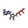
+Macromolecule #61: ADENOSINE-5'-DIPHOSPHATE
+Macromolecule #62: POTASSIUM ION
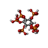
+Macromolecule #63: INOSITOL HEXAKISPHOSPHATE
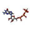
+Macromolecule #64: GUANOSINE-5'-TRIPHOSPHATE
+Macromolecule #65: ZINC ION
+Macromolecule #66: 7-METHYL-GUANOSINE-5'-TRIPHOSPHATE-5'-GUANOSINE

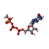
+Macromolecule #67: 5'-O-[(S)-hydroxy{[(R)-hydroxy{[(S)-hydroxy(methoxy)phosphoryl]ox...
-Experimental details
-Structure determination
| Method | cryo EM |
|---|---|
 Processing | single particle reconstruction |
| Aggregation state | particle |
-Sample preparation
| Buffer | pH: 7.8 |
|---|---|
| Grid | Model: Quantifoil R3.5/1 / Material: GOLD / Support film - Material: CARBON / Support film - Film thickness: 2 / Pretreatment - Type: GLOW DISCHARGE / Pretreatment - Time: 30 sec. |
| Vitrification | Cryogen name: ETHANE / Chamber humidity: 100 % / Chamber temperature: 283 K / Instrument: FEI VITROBOT MARK IV |
- Electron microscopy
| Microscope | TFS KRIOS |
|---|---|
| Specialist optics | Energy filter - Slit width: 20 eV |
| Image recording | Film or detector model: GATAN K3 BIOQUANTUM (6k x 4k) / Number grids imaged: 2 / Number real images: 31410 / Average exposure time: 2.0 sec. / Average electron dose: 50.0 e/Å2 |
| Electron beam | Acceleration voltage: 300 kV / Electron source:  FIELD EMISSION GUN |
| Electron optics | Illumination mode: FLOOD BEAM / Imaging mode: BRIGHT FIELD / Nominal defocus max: 2.5 µm / Nominal defocus min: 0.5 µm / Nominal magnification: 64000 |
| Experimental equipment | 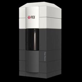 Model: Titan Krios / Image courtesy: FEI Company |