 Movie
Movie Controller

[English] 日本語
 Yorodumi
- EMDB-52548: Cryo-electron tomogram #01 of thylakoids in spinach chloroplast. ... -
+ Open data

- Basic information
| Entry |  | |||||||||
|---|---|---|---|---|---|---|---|---|---|---|
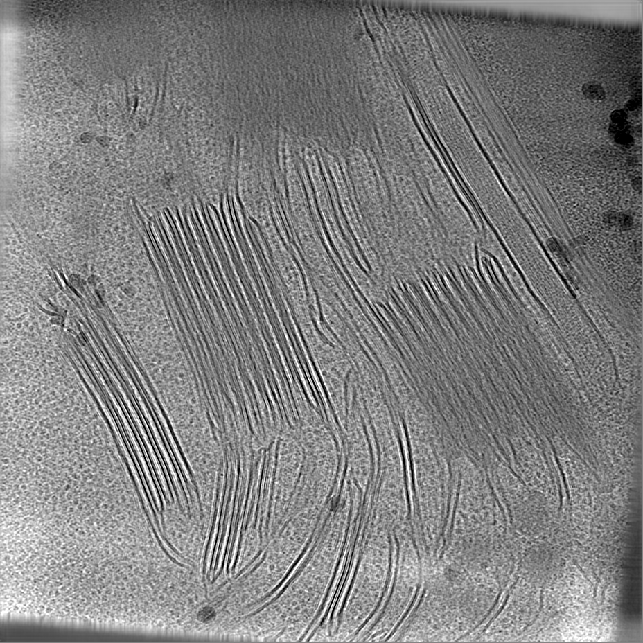
| Title | Cryo-electron tomogram #01 of thylakoids in spinach chloroplast. Denoised with cryoCARE | |||||||||
 Map data Map data | Cryo-electron tomogram #01 of thylakoids in spinach chloroplast. Denoised with cryoCARE. | |||||||||
 Sample Sample |
| |||||||||
 Keywords Keywords | chloroplast / spinach / thylakoids / plastoglobules / PHOTOSYNTHESIS | |||||||||
| Biological species |  Spinacia oleracea (spinach) Spinacia oleracea (spinach) | |||||||||
| Method | electron tomography / cryo EM | |||||||||
 Authors Authors | Wietrzynski W / Lamm L / Engel BD | |||||||||
| Funding support |  France, 1 items France, 1 items
| |||||||||
 Citation Citation | Journal: Elife / Year: 2025 Title: Molecular architecture of thylakoid membranes within intact spinach chloroplasts. Authors: Wojciech Wietrzynski / Lorenz Lamm / William H J Wood / Matina-Jasemi Loukeri / Lorna Malone / Tingying Peng / Matthew P Johnson / Benjamin D Engel /    Abstract: Thylakoid membranes coordinate the light reactions of photosynthesis across multiple scales, coupling the architecture of an elaborate membrane network to the spatial organization of individual ...Thylakoid membranes coordinate the light reactions of photosynthesis across multiple scales, coupling the architecture of an elaborate membrane network to the spatial organization of individual protein complexes embedded within this network. Previously, we used in situ cryo-electron tomography (cryo-ET) to reveal the native thylakoid architecture of the green alga (Engel et al., 2015) and then map the molecular organization of these thylakoids with single-molecule precision (Wietrzynski et al., 2020). However, it remains to be shown how generalizable this green algal blueprint is to the thylakoids of vascular plants, which possess distinct membrane architecture subdivided into grana stacks interconnected by non-stacked stromal lamellae. Here, we continue our cryo-ET investigation to reveal the molecular architecture of thylakoids within intact chloroplasts isolated from spinach (). We visualize the fine ultrastructural details of grana membranes, as well as interactions between thylakoids and plastoglobules. We apply AI-based computational approaches (Lamm et al., 2024) to quantify the organization of photosynthetic complexes within the plane of the thylakoid membrane and across adjacent stacked membranes. Our analysis reveals that the molecular organization of thylakoid membranes in vascular plants and green algae is strikingly similar. We find that PSII organization is non-crystalline and has uniform concentration both within the membrane plane and across stacked grana membranes. Similar to , we observe strict lateral heterogeneity of PSII and PSI at the boundary between appressed and non-appressed thylakoid domains, with no evidence for a distinct grana margin region where these complexes have been proposed to intermix. Based on these measurements, we support a simple two-domain model for the molecular organization of thylakoid membranes in both green algae and plants. #1: Journal: Elife / Year: 2025Title: Molecular architecture of thylakoid membranes within intact spinach chloroplasts Authors: Wietrzynski W / Lamm L / Wood WH / Loukeri MJ / Malone L / Peng T / Johnson MP / Engel BD | |||||||||
| History |
|
- Structure visualization
| Supplemental images |
|---|

- Downloads & links
-EMDB archive
| Map data | emd_52548.map.gz | 1.4 GB |  EMDB map data format EMDB map data format | |
|---|---|---|---|---|
| Header (meta data) | emd-52548-v30.xmlemd-52548.xml | 14 KB 14 KB | Display Display | EMDB header |
| Images |  emd_52548.png emd_52548.png | 188.9 KB | ||
| Filedesc metadata | emd-52548.cif.gz | 4.3 KB | ||
| Others | emd_52548_additional_1.map.gz | 4.2 MB | ||
| Archive directory |  http://ftp.pdbj.org/pub/emdb/structures/EMD-52548ftp://ftp.pdbj.org/pub/emdb/structures/EMD-52548 http://ftp.pdbj.org/pub/emdb/structures/EMD-52548ftp://ftp.pdbj.org/pub/emdb/structures/EMD-52548 | HTTPS FTP |
-Related structure data


-Links
| EMDB pages | EMDB (EBI/PDBe) / EMDataResource |
|---|
-Map
| File | Download / File: emd_52548.map.gz / Format: CCP4 / Size: 1.5 GB / Type: IMAGE STORED AS FLOATING POINT NUMBER (4 BYTES) | ||||||||||||||||||||||||||||||||
|---|---|---|---|---|---|---|---|---|---|---|---|---|---|---|---|---|---|---|---|---|---|---|---|---|---|---|---|---|---|---|---|---|---|
| Annotation | Cryo-electron tomogram #01 of thylakoids in spinach chloroplast. Denoised with cryoCARE. | ||||||||||||||||||||||||||||||||
| Projections & slices | Image control
Images are generated by Spider. generated in cubic-lattice coordinate | ||||||||||||||||||||||||||||||||
| Voxel size | X=Y=Z: 14.08 Å | ||||||||||||||||||||||||||||||||
| Density |
| ||||||||||||||||||||||||||||||||
| Symmetry | Space group: 1 | ||||||||||||||||||||||||||||||||
| Details | EMDB XML:
|
 Z (Sec.)
 Y (Row.)
 X (Col.)


-Supplemental data
-Additional map: Segmentation of membranes from the tomogram #01 containing...
| File | emd_52548_additional_1.map | ||||||||||||
|---|---|---|---|---|---|---|---|---|---|---|---|---|---|
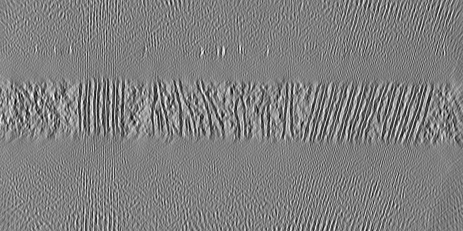
| Annotation | Segmentation of membranes from the tomogram #01 containing thylakoids in spinach chloroplast | ||||||||||||
| Projections & Slices |
| ||||||||||||
| Density Histograms |


- Sample components
-Entire : Chloroplast
| Entire | Name: Chloroplast |
|---|---|
| Components |
|
-Supramolecule #1: Chloroplast
| Supramolecule | Name: Chloroplast / type: organelle_or_cellular_component / ID: 1 / Parent: 0 Details: Isolated chloroplast of Spinach (Spinacia oleracea) visualised by cryo-ET |
|---|---|
| Source (natural) | Organism: Spinacia oleracea (spinach) / Organ: leaf / Organelle: chloroplast |
-Experimental details
-Structure determination
| Method | cryo EM |
|---|---|
 Processing Processing | electron tomography |
| Aggregation state | cell |
-Sample preparation
| Buffer | pH: 7.2 Details: 0.45 M Sorbitol, 20 mM Tricine-KOH, 10 mM EDTA, 10 mM NaHCO3, 5 mM MgCl2, 0.1% BSA, 0.2% D-Ascorbate |
|---|---|
| Grid | Model: Quantifoil R2/1 / Material: COPPER / Mesh: 200 / Support film - Material: CARBON / Support film - topology: HOLEY |
| Vitrification | Cryogen name: ETHANE-PROPANE / Chamber humidity: 75 % / Chamber temperature: 295 K / Instrument: FEI VITROBOT MARK IV |
| Cryo protectant | no cryoprotectant |
| Sectioning | Focused ion beam - Instrument: OTHER / Focused ion beam - Ion: OTHER / Focused ion beam - Voltage: 30 / Focused ion beam - Current: 0.1 / Focused ion beam - Duration: 1800 / Focused ion beam - Temperature: 77 K / Focused ion beam - Initial thickness: 1000 / Focused ion beam - Final thickness: 120 Focused ion beam - Details: Clumps of chloroplast were milled in a step-wise manner according to the routine protocols.. The value given for _em_focused_ion_beam.instrument is TFS Aquilos. This is ...Focused ion beam - Details: Clumps of chloroplast were milled in a step-wise manner according to the routine protocols.. The value given for _em_focused_ion_beam.instrument is TFS Aquilos. This is not in a list of allowed values {'OTHER', 'DB235'} so OTHER is written into the XML file. |
- Electron microscopy
| Microscope | TFS KRIOS |
|---|---|
| Image recording | Film or detector model: GATAN K2 SUMMIT (4k x 4k) / Detector mode: COUNTING / Number real images: 60 / Average electron dose: 120.0 e/Å2 |
| Electron beam | Acceleration voltage: 300 kV / Electron source:  FIELD EMISSION GUN FIELD EMISSION GUN |
| Electron optics | Illumination mode: FLOOD BEAM / Imaging mode: BRIGHT FIELD / Nominal defocus max: 5.0 µm / Nominal defocus min: 2.5 µm |
| Experimental equipment |  Model: Titan Krios / Image courtesy: FEI Company |
-Image processing
| Final reconstruction | Software - Name: IMOD / Number images used: 60 |
|---|---|
| CTF correction | Type: NONE |
