 Movie
Movie Controller

[English] 日本語
 Yorodumi
- EMDB-50359: Rhodobacter microvirus Ebor attached to B10 host cell reconstruct... -
+ Open data

- Basic information
| Entry |  | ||||||||||||
|---|---|---|---|---|---|---|---|---|---|---|---|---|---|
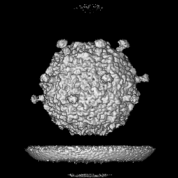
| Title | Rhodobacter microvirus Ebor attached to B10 host cell reconstructed by single particle analysis with applied C5 symmetry | ||||||||||||
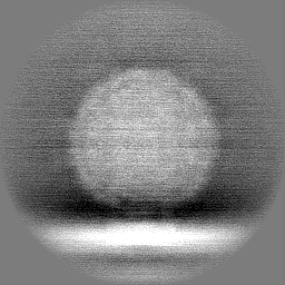
 Map data Map data | Ebor attached to B10 host cell, class 1 | ||||||||||||
 Sample Sample |
| ||||||||||||
 Keywords Keywords | ssDNA virus / Microviridae / Tainavirinae / Alphaproteobacteria / Rhodobacter capsulatus / aquatic virus / jelly-roll fold / VIRUS | ||||||||||||
| Biological species |  Rhodobacter capsulatus SB 1003 (bacteria) / Rhodobacter capsulatus SB 1003 (bacteria) /  Rhodobacter phage Ebor (virus) Rhodobacter phage Ebor (virus) | ||||||||||||
| Method | single particle reconstruction / cryo EM / Resolution: 7.84 Å | ||||||||||||
 Authors Authors | Bardy P / MacDonald CIW / Jenkins HT / Chechik M / Hart SJ / Turkenburg JP / Blaza JN / Fogg PCM / Antson AA | ||||||||||||
| Funding support |  United Kingdom, 3 items United Kingdom, 3 items
| ||||||||||||
 Citation Citation | Journal: bioRxiv / Year: 2024 Title: A stargate mechanism of genome delivery unveiled by cryogenic electron tomography. Authors: Pavol Bardy / Conor I W MacDonald / Paul C Kirchberger / Huw T Jenkins / Tibor Botka / Lewis Byrom / Nawshin T B Alim / Daouda A K Traore / Hannah C König / Tristan R Nicholas / Maria ...Authors: Pavol Bardy / Conor I W MacDonald / Paul C Kirchberger / Huw T Jenkins / Tibor Botka / Lewis Byrom / Nawshin T B Alim / Daouda A K Traore / Hannah C König / Tristan R Nicholas / Maria Chechik / Samuel J Hart / Johan P Turkenburg / James N Blaza / J Thomas Beatty / Paul C M Fogg / Alfred A Antson /    Abstract: Single-stranded DNA bacteriophages of the family are major components of the global virosphere. Microviruses are highly abundant in aquatic ecosystems and are prominent members of the mammalian gut ...Single-stranded DNA bacteriophages of the family are major components of the global virosphere. Microviruses are highly abundant in aquatic ecosystems and are prominent members of the mammalian gut microbiome, where their diversity has been linked to various chronic health disorders. Despite the clear importance of microviruses, little is known about the molecular mechanism of host infection. Here, we have characterized an exceptionally large microvirus, Ebor, and provide crucial insights into long-standing mechanistic questions. Cryogenic electron microscopy of Ebor revealed a capsid with trimeric protrusions that recognise lipopolysaccharides on the host surface. Cryogenic electron tomography of the host cell colonized with virus particles demonstrated that the virus initially attaches to the cell via five such protrusions, located at the corners of a single pentamer. This interaction triggers a stargate mechanism of capsid opening along the 5-fold symmetry axis, enabling delivery of the virus genome. Despite variations in specific virus-host interactions among different family viruses, structural data indicate that the stargate mechanism of infection is universally employed by all members of the family. Startlingly, our data reveal a mechanistic link for the opening of relatively small capsids made out of a single jelly-roll fold with the structurally unrelated giant viruses. | ||||||||||||
| History |
|
- Structure visualization
| Supplemental images |
|---|

- Downloads & links
-EMDB archive
| Map data | emd_50359.map.gz | 58 MB |  EMDB map data format EMDB map data format | |
|---|---|---|---|---|
| Header (meta data) | emd-50359-v30.xmlemd-50359.xml | 35.7 KB 35.7 KB | Display Display | EMDB header |
| Images |  emd_50359.png emd_50359.png | 83.1 KB | ||
| Filedesc metadata | emd-50359.cif.gz | 6.5 KB | ||
| Others | emd_50359_additional_1.map.gzemd_50359_additional_2.map.gzemd_50359_additional_3.map.gzemd_50359_additional_4.map.gzemd_50359_additional_5.map.gzemd_50359_additional_6.map.gzemd_50359_additional_7.map.gzemd_50359_half_map_1.map.gzemd_50359_half_map_2.map.gz | 58 MB 58 MB 116.8 KB 49.7 MB 49.5 MB 49.5 MB 49.6 MB 49.5 MB 49.5 MB | ||
| Archive directory |  http://ftp.pdbj.org/pub/emdb/structures/EMD-50359ftp://ftp.pdbj.org/pub/emdb/structures/EMD-50359 http://ftp.pdbj.org/pub/emdb/structures/EMD-50359ftp://ftp.pdbj.org/pub/emdb/structures/EMD-50359 | HTTPS FTP |
-Related structure data


-Links
| EMDB pages | EMDB (EBI/PDBe) / EMDataResource |
|---|
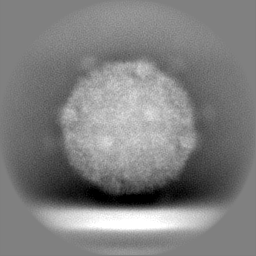
-Map
| File | Download / File: emd_50359.map.gz / Format: CCP4 / Size: 64 MB / Type: IMAGE STORED AS FLOATING POINT NUMBER (4 BYTES) | ||||||||||||||||||||||||||||||||||||
|---|---|---|---|---|---|---|---|---|---|---|---|---|---|---|---|---|---|---|---|---|---|---|---|---|---|---|---|---|---|---|---|---|---|---|---|---|---|
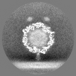
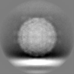
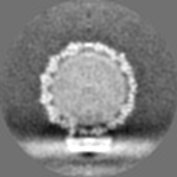
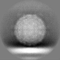
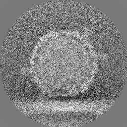
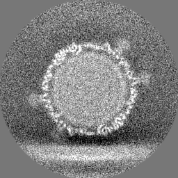
| Annotation | Ebor attached to B10 host cell, class 1 | ||||||||||||||||||||||||||||||||||||
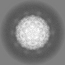
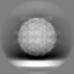
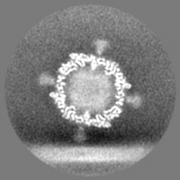
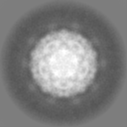
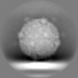
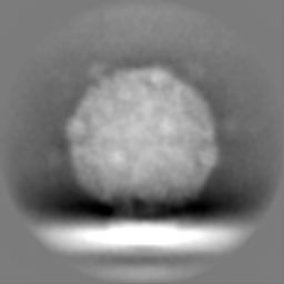
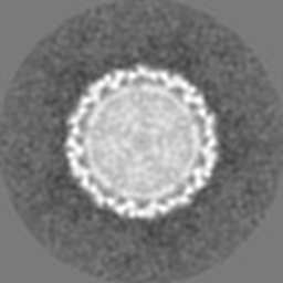
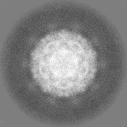
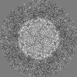
| Projections & slices | Image control
Images are generated by Spider. | ||||||||||||||||||||||||||||||||||||
| Voxel size | X=Y=Z: 2.143 Å | ||||||||||||||||||||||||||||||||||||
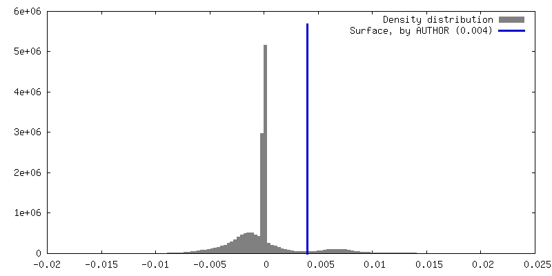
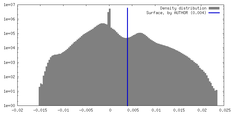
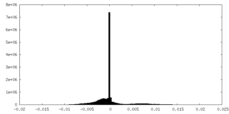
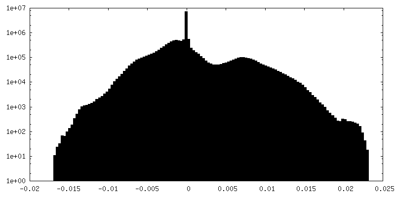
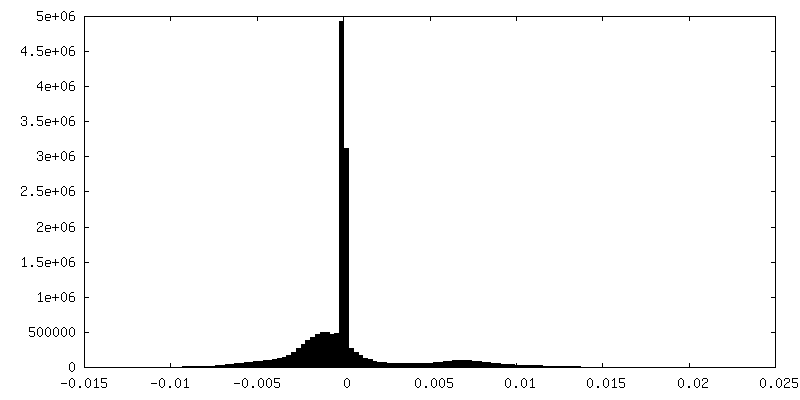
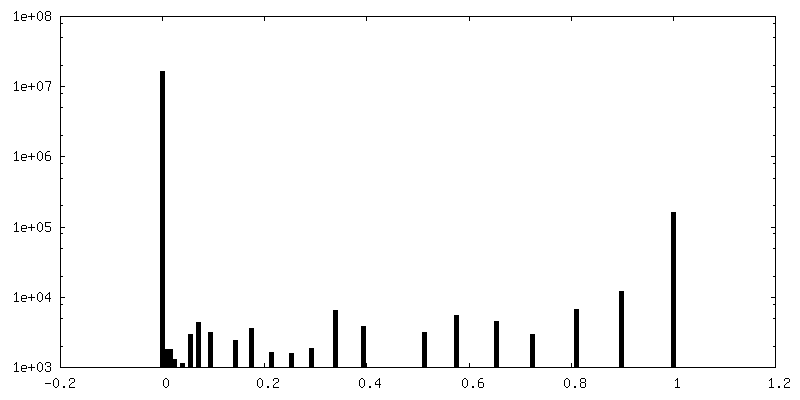
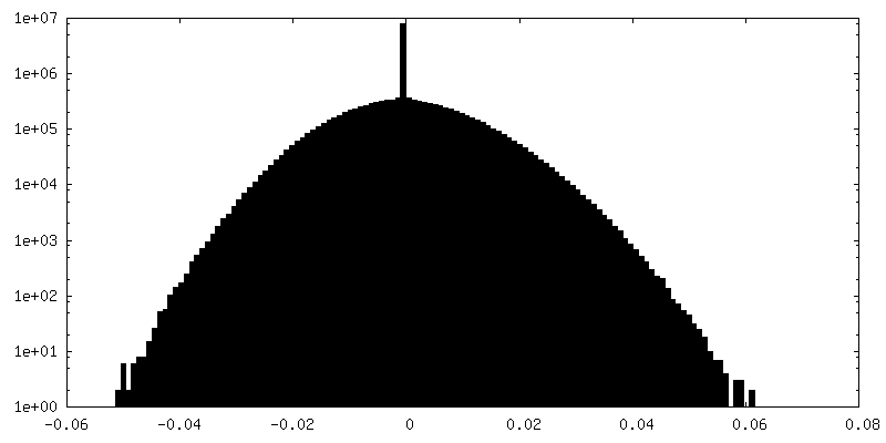
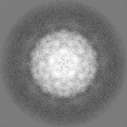
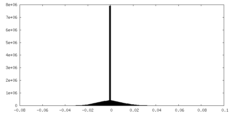
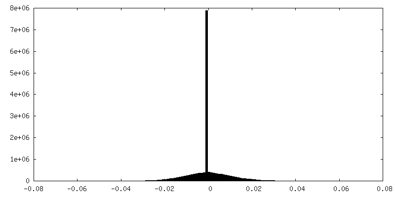
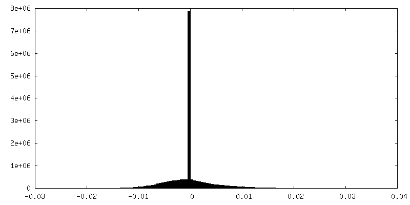
| Density |
| ||||||||||||||||||||||||||||||||||||
| Symmetry | Space group: 1 | ||||||||||||||||||||||||||||||||||||
| Details | EMDB XML:
|
 Z (Sec.)
 Y (Row.)
 X (Col.)


-Supplemental data
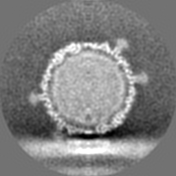
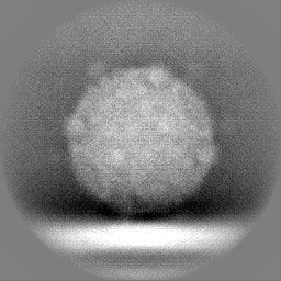
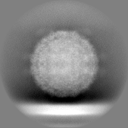
-Additional map: Ebor attached to B10 host cell, class 2
| File | emd_50359_additional_1.map | ||||||||||||
|---|---|---|---|---|---|---|---|---|---|---|---|---|---|
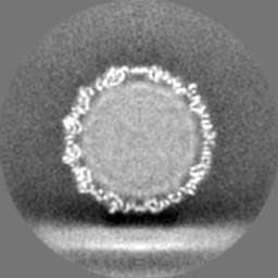
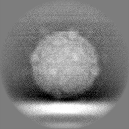
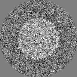
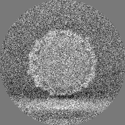
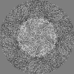
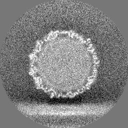
| Annotation | Ebor attached to B10 host cell, class 2 | ||||||||||||
| Projections & Slices |
| ||||||||||||
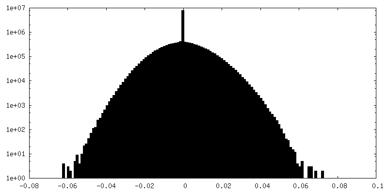
| Density Histograms |

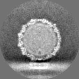
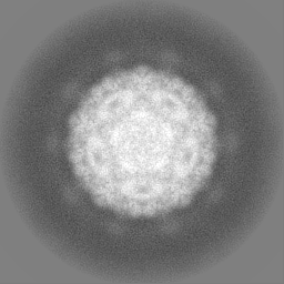
-Additional map: Ebor attached to B10 host cell, class 3
| File | emd_50359_additional_2.map | ||||||||||||
|---|---|---|---|---|---|---|---|---|---|---|---|---|---|
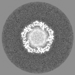
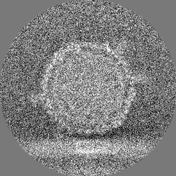
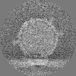
| Annotation | Ebor attached to B10 host cell, class 3 | ||||||||||||
| Projections & Slices |
| ||||||||||||
| Density Histograms |


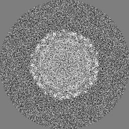
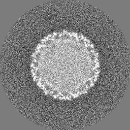
-Additional map: mask used for 3D classification
| File | emd_50359_additional_3.map | ||||||||||||
|---|---|---|---|---|---|---|---|---|---|---|---|---|---|
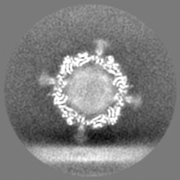
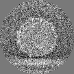
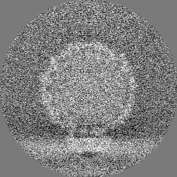
| Annotation | mask used for 3D classification | ||||||||||||
| Projections & Slices |
| ||||||||||||
| Density Histograms |


-Additional map: half map 1 for class 2
| File | emd_50359_additional_4.map | ||||||||||||
|---|---|---|---|---|---|---|---|---|---|---|---|---|---|
| Annotation | half map 1 for class 2 | ||||||||||||
| Projections & Slices |
| ||||||||||||
| Density Histograms |


-Additional map: half map 2 for class 2
| File | emd_50359_additional_5.map | ||||||||||||
|---|---|---|---|---|---|---|---|---|---|---|---|---|---|
| Annotation | half map 2 for class 2 | ||||||||||||
| Projections & Slices |
| ||||||||||||
| Density Histograms |


-Additional map: half map 1 for class 3
| File | emd_50359_additional_6.map | ||||||||||||
|---|---|---|---|---|---|---|---|---|---|---|---|---|---|
| Annotation | half map 1 for class 3 | ||||||||||||
| Projections & Slices |
| ||||||||||||
| Density Histograms |

-Additional map: half map 2 for class 3
| File | emd_50359_additional_7.map | ||||||||||||
|---|---|---|---|---|---|---|---|---|---|---|---|---|---|
| Annotation | half map 2 for class 3 | ||||||||||||
| Projections & Slices |
| ||||||||||||
| Density Histograms |


-Half map: half map 2 for class 1
| File | emd_50359_half_map_1.map | ||||||||||||
|---|---|---|---|---|---|---|---|---|---|---|---|---|---|
| Annotation | half map 2 for class 1 | ||||||||||||
| Projections & Slices |
| ||||||||||||
| Density Histograms |


-Half map: half map 1 for class 1
| File | emd_50359_half_map_2.map | ||||||||||||
|---|---|---|---|---|---|---|---|---|---|---|---|---|---|
| Annotation | half map 1 for class 1 | ||||||||||||
| Projections & Slices |
| ||||||||||||
| Density Histograms |


- Sample components
-Entire : Rhodobacter phage Ebor
| Entire | Name: Rhodobacter phage Ebor (virus) |
|---|---|
| Components |
|
-Supramolecule #1: Rhodobacter phage Ebor
| Supramolecule | Name: Rhodobacter phage Ebor / type: virus / ID: 1 / Parent: 0 / Macromolecule list: all Details: Phage, purified from Rhodobacter capsulatus strain SB1003 by ion-exchange chromatography, attached to host cell (B10 strain) NCBI-ID: 3144506 / Sci species name: Rhodobacter phage Ebor / Sci species strain: variant R120 / Virus type: VIRION / Virus isolate: OTHER / Virus enveloped: No / Virus empty: No |
|---|---|
| Host (natural) | Organism: Rhodobacter capsulatus (bacteria) / Strain: B10 |
| Molecular weight | Theoretical: 5.7 MDa |
| Virus shell | Shell ID: 1 / Name: Genome-releasing capsid / Diameter: 460.0 Å / T number (triangulation number): 1 |
-Macromolecule #1: Major capsid protein
| Macromolecule | Name: Major capsid protein / type: protein_or_peptide / ID: 1 / Enantiomer: LEVO |
|---|---|
| Source (natural) | Organism: Rhodobacter capsulatus SB 1003 (bacteria) / Strain: SB1003 |
| Sequence | String: MSYQTKRNVR REARTLMGRF KAGKLAPVMA VPVKGSEGGM LSQSVSFELD PIAGRMATPI TAEMCAVFVP VQACDALKNP EADYAGMTEI VREKLLSGNP LFVLEPETDV SKRCGVNPRR NNGLMRVNEI VRLAHNCAVN FLRRRRYVDA VQLTAANHST TPAILSQTVL ...String: MSYQTKRNVR REARTLMGRF KAGKLAPVMA VPVKGSEGGM LSQSVSFELD PIAGRMATPI TAEMCAVFVP VQACDALKNP EADYAGMTEI VREKLLSGNP LFVLEPETDV SKRCGVNPRR NNGLMRVNEI VRLAHNCAVN FLRRRRYVDA VQLTAANHST TPAILSQTVL DRFNGALDPD PNVNGAVQLS MPDMRLPVAS DAEKAVTAPL TVKRGNENRR LDAGISRLAA AEVAAATDAA LYAVFGGREA GNVSLTDFYN AQKMDELTRV MRKICDDNPE YGEEMVLRWA HGLSVDPGRV PFLLAEKSVV LGRQIIGATD TAGVEDGVKR SDMAAQLSFT VPIPTTELGG IIVTFACIKP DETLSSQPHP ILADHWRLDN FVADELALDP QPVMARELDY KVAQANETTV VFYTGLNELK KTYVSYGLCR ALDPNTVESK NAVWQLEVPL SVTPETVLYP ADLPQYPFAD QQAEVCTYVV QSTAVMPTPM IFGPSPVEQL AVIETEDLFE D |
-Experimental details
-Structure determination
| Method | cryo EM |
|---|---|
 Processing Processing | single particle reconstruction |
| Aggregation state | particle |
-Sample preparation
| Buffer | pH: 7.75 Component:
| ||||||||||||
|---|---|---|---|---|---|---|---|---|---|---|---|---|---|
| Grid | Model: Quantifoil R2/1 / Material: COPPER / Mesh: 200 / Support film - Material: CARBON / Support film - topology: HOLEY / Pretreatment - Type: GLOW DISCHARGE / Pretreatment - Time: 30 sec. / Pretreatment - Atmosphere: AIR | ||||||||||||
| Vitrification | Cryogen name: ETHANE / Chamber humidity: 100 % / Chamber temperature: 295.15 K / Instrument: FEI VITROBOT MARK IV / Details: blot time 2 sec, blot force 0, wait time 8 sec. | ||||||||||||
| Details | concentration of 7x10^10 PFU/ml |
- Electron microscopy
| Microscope | TFS KRIOS |
|---|---|
| Specialist optics | Energy filter - Name: GIF Bioquantum |
| Image recording | Film or detector model: GATAN K3 BIOQUANTUM (6k x 4k) / Number grids imaged: 1 / Number real images: 2750 / Average electron dose: 27.9 e/Å2 / Details: acquired using lacey carbon setup in EPU |
| Electron beam | Acceleration voltage: 300 kV / Electron source:  FIELD EMISSION GUN FIELD EMISSION GUN |
| Electron optics | Illumination mode: FLOOD BEAM / Imaging mode: BRIGHT FIELD / Nominal defocus max: 3.0 µm / Nominal defocus min: 0.75 µm |
| Sample stage | Specimen holder model: FEI TITAN KRIOS AUTOGRID HOLDER / Cooling holder cryogen: NITROGEN |
| Experimental equipment |  Model: Titan Krios / Image courtesy: FEI Company |
