 Movie
Movie Controller

[English] 日本語
 Yorodumi
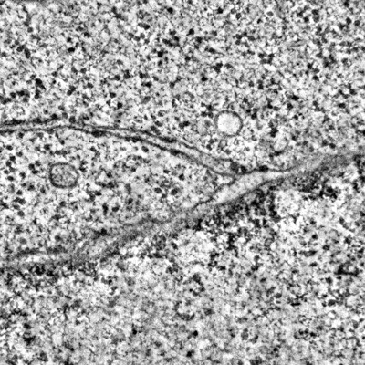
- EMDB-50068: Electron tomogram of ER-nuclear envelope junction of HeLa cell in... -
+ Open data

- Basic information
| Entry |  | |||||||||
|---|---|---|---|---|---|---|---|---|---|---|
| Title | Electron tomogram of ER-nuclear envelope junction of HeLa cell in interphase | |||||||||
 Map data Map data | ||||||||||
 Sample Sample |
| |||||||||
 Keywords Keywords | Endoplasmic reticulum / Nuclear envelope / Organelle contact site / Membrane junction / CELL CYCLE | |||||||||
| Biological species |  Homo sapiens (human) Homo sapiens (human) | |||||||||
| Method | electron tomography / cryo EM / negative staining | |||||||||
 Authors Authors | Bragulat-Teixidor H / Otsuka S | |||||||||
| Funding support |  Austria, 2 items Austria, 2 items
| |||||||||
 Citation Citation | Journal: EMBO Rep / Year: 2024 Title: The endoplasmic reticulum connects to the nucleus by constricted junctions that mature after mitosis. Authors: Helena Bragulat-Teixidor / Keisuke Ishihara / Gréta Martina Szücs / Shotaro Otsuka /  Abstract: Junctions between the endoplasmic reticulum (ER) and the outer membrane of the nuclear envelope (NE) physically connect both organelles. These ER-NE junctions are essential for supplying the NE with ...Junctions between the endoplasmic reticulum (ER) and the outer membrane of the nuclear envelope (NE) physically connect both organelles. These ER-NE junctions are essential for supplying the NE with lipids and proteins synthesized in the ER. However, little is known about the structure of these ER-NE junctions. Here, we systematically study the ultrastructure of ER-NE junctions in cryo-fixed mammalian cells staged in anaphase, telophase, and interphase by correlating live cell imaging with three-dimensional electron microscopy. Our results show that ER-NE junctions in interphase cells have a pronounced hourglass shape with a constricted neck of 7-20 nm width. This morphology is significantly distinct from that of junctions within the ER network, and their morphology emerges as early as telophase. The highly constricted ER-NE junctions are seen in several mammalian cell types, but not in budding yeast. We speculate that the unique and highly constricted ER-NE junctions are regulated via novel mechanisms that contribute to ER-to-NE lipid and protein traffic in higher eukaryotes. | |||||||||
| History |
|
- Structure visualization
| Supplemental images |
|---|
- Downloads & links
-EMDB archive
| Map data | emd_50068.map.gz | 12.8 GB |  EMDB map data format EMDB map data format | |
|---|---|---|---|---|
| Header (meta data) | emd-50068-v30.xmlemd-50068.xml | 9.5 KB 9.5 KB | Display Display | EMDB header |
| Images |  emd_50068.png emd_50068.png | 231.4 KB | ||
| Filedesc metadata | emd-50068.cif.gz | 4.1 KB | ||
| Archive directory |  http://ftp.pdbj.org/pub/emdb/structures/EMD-50068ftp://ftp.pdbj.org/pub/emdb/structures/EMD-50068 http://ftp.pdbj.org/pub/emdb/structures/EMD-50068ftp://ftp.pdbj.org/pub/emdb/structures/EMD-50068 | HTTPS FTP |
-Validation report
| Summary document | emd_50068_validation.pdf.gz | 508.2 KB | Display | EMDB validaton report |
|---|---|---|---|---|
| Full document | emd_50068_full_validation.pdf.gz | 507.7 KB | Display | |
| Data in XML | emd_50068_validation.xml.gz | 4 KB | Display | |
| Data in CIF | emd_50068_validation.cif.gz | 4.5 KB | Display | |
| Arichive directory | https://ftp.pdbj.org/pub/emdb/validation_reports/EMD-50068ftp://ftp.pdbj.org/pub/emdb/validation_reports/EMD-50068 | HTTPS FTP |
-Related structure data


-Links
| EMDB pages | EMDB (EBI/PDBe) / EMDataResource |
|---|
-Map
| File | Download / File: emd_50068.map.gz / Format: CCP4 / Size: 17.8 GB / Type: IMAGE STORED AS SIGNED INTEGER (2 BYTES) | ||||||||||||||||||||
|---|---|---|---|---|---|---|---|---|---|---|---|---|---|---|---|---|---|---|---|---|---|
| Voxel size | X=Y=Z: 4.51 Å | ||||||||||||||||||||
| Density |
| ||||||||||||||||||||
| Symmetry | Space group: 1 | ||||||||||||||||||||
| Details | EMDB XML:
|
-Supplemental data
- Sample components
-Entire : HeLa Kyoto cell
| Entire | Name: HeLa Kyoto cell |
|---|---|
| Components |
|
-Supramolecule #1: HeLa Kyoto cell
| Supramolecule | Name: HeLa Kyoto cell / type: cell / ID: 1 / Parent: 0 |
|---|---|
| Source (natural) | Organism: Homo sapiens (human) / Strain: HeLa |
-Experimental details
-Structure determination
| Method | negative staining, cryo EM |
|---|---|
 Processing Processing | electron tomography |
| Aggregation state | cell |
-Sample preparation
| Buffer | pH: 7.4 Details: DMEM without Riboflavin and Phenol Red, containing 10% FBS, 1% Pen/Strep, and 50 nM SiR-DNA |
|---|---|
| Staining | Type: NEGATIVE / Material: Uranyl acetate and lead citrate |
| Sugar embedding | Material: Agar 100 Epoxy resin Details: Frozen cells were substituted in 0.1% uranyl acetate (UA), 2% Osmium tetroxide and 5% H2O in acetone following this temperature ramp: -90 C to -80 C for 10 hours, -80 C to -30 C for 10 ...Details: Frozen cells were substituted in 0.1% uranyl acetate (UA), 2% Osmium tetroxide and 5% H2O in acetone following this temperature ramp: -90 C to -80 C for 10 hours, -80 C to -30 C for 10 hours, -30 C for 4 hours, -30 C to 0 C for 6 hours, 0 C to 20 C for 4 hours, 20 C for 5-6 hours. Afterwards, samples were washed three times in pure acetone for at least 10 minutes each, and subsequently infiltrated with Agar 100 Epoxy resin. The resin infiltration was done progressively at room temperature with increasing concentrations of resin in acetone (3:1 for 2-3 hours, 1:1 for 2-3 hours, and 1:3 overnight). Infiltration with pure resin was done at room temperature for at least 5 hours. Resin was polymerized at 60 C for 72 hours. |
| Vitrification | Cryogen name: NITROGEN |
| High pressure freezing | Instrument: OTHER Details: High pressure freezing chamber was 1 mm thick, 6.0 mm diameter, with central cavities 5.0 mm x 5.0 mm x 25 um deep. The chamber had been in contact with 1-hexadecene.. The value given for _ ...Details: High pressure freezing chamber was 1 mm thick, 6.0 mm diameter, with central cavities 5.0 mm x 5.0 mm x 25 um deep. The chamber had been in contact with 1-hexadecene.. The value given for _em_high_pressure_freezing.instrument is Leica EM ICE. This is not in a list of allowed values {'OTHER', 'LEICA EM PACT', 'LEICA EM HPM100', 'EMS-002 RAPID IMMERSION FREEZER', 'BAL-TEC HPM 010', 'LEICA EM PACT2'} so OTHER is written into the XML file. |
| Cryo protectant | 20% Ficoll-PM400 |
| Sectioning | Ultramicrotomy - Instrument: Leica Ultracut UCT / Ultramicrotomy - Temperature: 298 K / Ultramicrotomy - Final thickness: 250 |
| Fiducial marker | Manufacturer: Cytodiagnostics / Diameter: 15 nm |
- Electron microscopy
| Microscope | FEI TECNAI F20 |
|---|---|
| Image recording | Film or detector model: FEI EAGLE (4k x 4k) / Average electron dose: 40.0 e/Å2 |
| Electron beam | Acceleration voltage: 200 kV / Electron source:  FIELD EMISSION GUN FIELD EMISSION GUN |
| Electron optics | Illumination mode: FLOOD BEAM / Imaging mode: BRIGHT FIELD / Nominal defocus max: 0.5 µm / Nominal defocus min: 0.2 µm |
| Experimental equipment |  Model: Tecnai F20 / Image courtesy: FEI Company |
-Image processing
| Final reconstruction | Algorithm: BACK PROJECTION / Number images used: 110 |
|---|
