 Movie
Movie Controller

[English] 日本語
 Yorodumi
- EMDB-44745: 2.65 Angstrom Apoferritin single particle cryo-EM reconstruction ... -
+ Open data

- Basic information
| Entry |  | |||||||||
|---|---|---|---|---|---|---|---|---|---|---|
| Title | 2.65 Angstrom Apoferritin single particle cryo-EM reconstruction from a standard 120-keV LaB6 electron microscope (Tecnai G2 spirit) fitted with Gatan Alpine camera (a 100keV optimised direct electron detector). | |||||||||
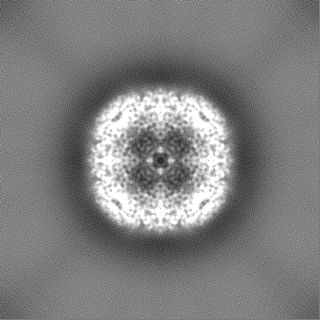
 Map data Map data | B-Factor Sharpened Map | |||||||||
 Sample Sample |
| |||||||||
 Keywords Keywords | Apoferritin / High resolution / LaB6 / Tecnai. / METAL TRANSPORT | |||||||||
| Biological species |  | |||||||||
| Method | single particle reconstruction / cryo EM / Resolution: 2.65 Å | |||||||||
 Authors Authors | Venugopal H / Ramm G | |||||||||
| Funding support |  Australia, Australia,  United States, 2 items United States, 2 items
| |||||||||
 Citation Citation | Journal: Sci Adv / Year: 2025 Title: High-resolution cryo-EM using a common LaB 120-keV electron microscope equipped with a sub-200-keV direct electron detector. Authors: Hariprasad Venugopal / Jesse Mobbs / Cyntia Taveneau / Daniel R Fox / Ziva Vuckovic / Sahil Gulati / Gavin Knott / Rhys Grinter / David Thal / Stephen Mick / Cory Czarnik / Georg Ramm /  Abstract: High-resolution cryo-electron microscopy (cryo-EM) requires costly 200- to 300-keV cryo-transmission electron microscopes (cryo-TEMs) with field emission gun (FEG) sources, stable columns, constant- ...High-resolution cryo-electron microscopy (cryo-EM) requires costly 200- to 300-keV cryo-transmission electron microscopes (cryo-TEMs) with field emission gun (FEG) sources, stable columns, constant-powered lenses, autoloader, and direct electron detectors (DED). Recent advances in 100-keV imaging with the emergence of sub-200-keV optimized DED technology promises the development of more affordable cryo-TEMs. So far, 100-keV imaging has required microscopes with FEG sources. We here explored whether a standard 120-keV TEMs with thermionic lanthanum hexaboride (LaB) source can be upgraded with a sub-200-keV DED for high-resolution cryo-EM. Using this imaging configuration, we successfully obtained a 2.65 Å reconstruction for apoferritin, 4.33 Å for 64-kDa hemoglobin, and 4.4 Å for an asymmetric 153kDa membrane protein GPCR. All results were achieved using standard automated data collection with SerialEM, demonstrating the feasibility to collect large cryo-EM datasets with a side-entry cryo-holder. These results showcase a widely accessible solution to obtaining interpretable cryo-EM structures at low cost and contribute to the "democratization" of cryo-EM. | |||||||||
| History |
|
- Structure visualization
| Supplemental images |
|---|

- Downloads & links
-EMDB archive
| Map data | emd_44745.map.gz | 117.8 MB |  EMDB map data format EMDB map data format | |
|---|---|---|---|---|
| Header (meta data) | emd-44745-v30.xmlemd-44745.xml | 17 KB 17 KB | Display Display | EMDB header |
| FSC (resolution estimation) | emd_44745_fsc.xml | 11.2 KB | Display | FSC data file |
| Images |  emd_44745.png emd_44745.png | 49.6 KB | ||
| Masks | emd_44745_msk_1.map | 125 MB | Mask map | |
| Filedesc metadata | emd-44745.cif.gz | 4.5 KB | ||
| Others | emd_44745_additional_1.map.gzemd_44745_half_map_1.map.gzemd_44745_half_map_2.map.gz | 62.2 MB 115.7 MB 115.7 MB | ||
| Archive directory |  http://ftp.pdbj.org/pub/emdb/structures/EMD-44745ftp://ftp.pdbj.org/pub/emdb/structures/EMD-44745 http://ftp.pdbj.org/pub/emdb/structures/EMD-44745ftp://ftp.pdbj.org/pub/emdb/structures/EMD-44745 | HTTPS FTP |
-Links
| EMDB pages | EMDB (EBI/PDBe) / EMDataResource |
|---|
-Map
| File | Download / File: emd_44745.map.gz / Format: CCP4 / Size: 125 MB / Type: IMAGE STORED AS FLOATING POINT NUMBER (4 BYTES) | ||||||||||||||||||||||||||||||||||||
|---|---|---|---|---|---|---|---|---|---|---|---|---|---|---|---|---|---|---|---|---|---|---|---|---|---|---|---|---|---|---|---|---|---|---|---|---|---|
| Annotation | B-Factor Sharpened Map | ||||||||||||||||||||||||||||||||||||
| Projections & slices | Image control
Images are generated by Spider. | ||||||||||||||||||||||||||||||||||||
| Voxel size | X=Y=Z: 0.811 Å | ||||||||||||||||||||||||||||||||||||
| Density |
| ||||||||||||||||||||||||||||||||||||
| Symmetry | Space group: 1 | ||||||||||||||||||||||||||||||||||||
| Details | EMDB XML:
|
 Z (Sec.)
 Y (Row.)
 X (Col.)


-Supplemental data
-Mask #1
| File | emd_44745_msk_1.map | ||||||||||||
|---|---|---|---|---|---|---|---|---|---|---|---|---|---|
| Projections & Slices |
| ||||||||||||
| Density Histograms |


-Additional map: Raw Map
| File | emd_44745_additional_1.map | ||||||||||||
|---|---|---|---|---|---|---|---|---|---|---|---|---|---|
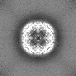
| Annotation | Raw Map | ||||||||||||
| Projections & Slices |
| ||||||||||||
| Density Histograms |


-Half map: Half2
| File | emd_44745_half_map_1.map | ||||||||||||
|---|---|---|---|---|---|---|---|---|---|---|---|---|---|
| Annotation | Half2 | ||||||||||||
| Projections & Slices |
| ||||||||||||
| Density Histograms |


-Half map: Half1
| File | emd_44745_half_map_2.map | ||||||||||||
|---|---|---|---|---|---|---|---|---|---|---|---|---|---|
| Annotation | Half1 | ||||||||||||
| Projections & Slices |
| ||||||||||||
| Density Histograms |


- Sample components
-Entire : Apoferritin
| Entire | Name: Apoferritin |
|---|---|
| Components |
|
-Supramolecule #1: Apoferritin
| Supramolecule | Name: Apoferritin / type: complex / ID: 1 / Parent: 0 |
|---|---|
| Source (natural) | Organism: |
| Molecular weight | Theoretical: 480 kDa/nm |
-Experimental details
-Structure determination
| Method | cryo EM |
|---|---|
 Processing Processing | single particle reconstruction |
| Aggregation state | particle |
-Sample preparation
| Buffer | pH: 8 / Details: 50 mM Tris-HCl (pH 8.0), 150 mM NaCl |
|---|---|
| Vitrification | Cryogen name: ETHANE / Chamber humidity: 100 % / Chamber temperature: 277.15 K / Instrument: FEI VITROBOT MARK IV |
- Electron microscopy
| Microscope | FEI TECNAI SPIRIT |
|---|---|
| Image recording | Film or detector model: GATAN ALPINE (2.3k x 3.2k) / Digitization - Dimensions - Width: 2304 pixel / Digitization - Dimensions - Height: 3240 pixel / Number grids imaged: 1 / Number real images: 923 / Average exposure time: 6.55 sec. / Average electron dose: 50.3 e/Å2 |
| Electron beam | Acceleration voltage: 120 kV / Electron source: LAB6 |
| Electron optics | C2 aperture diameter: 50.0 µm / Illumination mode: FLOOD BEAM / Imaging mode: BRIGHT FIELD / Cs: 2.2 mm / Nominal defocus max: 0.9 µm / Nominal defocus min: 0.3 µm / Nominal magnification: 42000 |
| Sample stage | Specimen holder model: GATAN 626 SINGLE TILT LIQUID NITROGEN CRYO TRANSFER HOLDER Cooling holder cryogen: NITROGEN |
| Experimental equipment |  Model: Tecnai Spirit / Image courtesy: FEI Company |

