 Movie
Movie Controller

[English] 日本語
 Yorodumi
- EMDB-43812: Cryo-EM Structure of the Glycosyltransferase ArnC from Salmonella... -
+ Open data

- Basic information
| Entry |  | |||||||||
|---|---|---|---|---|---|---|---|---|---|---|
| Title | Cryo-EM Structure of the Glycosyltransferase ArnC from Salmonella enterica in the UDP-bound State Determined on Talos Arctica microscope | |||||||||
 Map data Map data | ArnC/UDP sharpened map | |||||||||
 Sample Sample |
| |||||||||
 Keywords Keywords | Glycosyltransferase / undecaprenyl phosphate / aminoarabinose / polymyxin resistance / GT-A / TRANSFERASE | |||||||||
| Function / homology |  Function and homology information Function and homology informationundecaprenyl-phosphate 4-deoxy-4-formamido-L-arabinose transferase / undecaprenyl-phosphate 4-deoxy-4-formamido-L-arabinose transferase activity / 4-amino-4-deoxy-alpha-L-arabinopyranosyl undecaprenyl phosphate biosynthetic process / phosphotransferase activity, for other substituted phosphate groups / lipopolysaccharide biosynthetic process / lipid A biosynthetic process / response to antibiotic / plasma membrane Similarity search - Function | |||||||||
| Biological species |  Salmonella enterica subsp. enterica serovar Typhimurium str. LT2 (bacteria) Salmonella enterica subsp. enterica serovar Typhimurium str. LT2 (bacteria) | |||||||||
| Method | single particle reconstruction / cryo EM / Resolution: 2.96 Å | |||||||||
 Authors Authors | Ashraf KU / Punetha A / Petrou VI | |||||||||
| Funding support |  United States, 2 items United States, 2 items
| |||||||||
 Citation Citation | Journal: bioRxiv / Year: 2025 Title: Structural basis of undecaprenyl phosphate glycosylation leading to polymyxin resistance in Gram-negative bacteria. Authors: Khuram U Ashraf / Mariana Bunoro-Batista / T Bertie Ansell / Ankita Punetha / Stephannie Rosario-Garrido / Emre Firlar / Jason T Kaelber / Phillip J Stansfeld / Vasileios I Petrou /  Abstract: In Gram-negative bacteria, the enzymatic modification of Lipid A with aminoarabinose (L-Ara4N) leads to resistance against polymyxin antibiotics and cationic antimicrobial peptides. ArnC, an integral ...In Gram-negative bacteria, the enzymatic modification of Lipid A with aminoarabinose (L-Ara4N) leads to resistance against polymyxin antibiotics and cationic antimicrobial peptides. ArnC, an integral membrane glycosyltransferase, attaches a formylated form of aminoarabinose to the lipid undecaprenyl phosphate, enabling its association with the bacterial inner membrane. Here, we present cryo-electron microscopy structures of ArnC from in and nucleotide-bound conformations. These structures reveal a conformational transition that takes place upon binding of the partial donor substrate. Using coarse-grained and atomistic simulations, we provide insights into substrate coordination before and during catalysis, and we propose a catalytic mechanism that may operate on all similar metal-dependent polyprenyl phosphate glycosyltransferases. The reported structures provide a new target for drug design aiming to combat polymyxin resistance. | |||||||||
| History |
|
- Structure visualization
| Supplemental images |
|---|

- Downloads & links
-EMDB archive
| Map data | emd_43812.map.gz | 46.9 MB | EMDB map data format | |
|---|---|---|---|---|
| Header (meta data) | emd-43812-v30.xmlemd-43812.xml | 22.7 KB 22.7 KB | Display Display | EMDB header |
| FSC (resolution estimation) | emd_43812_fsc.xml | 10.8 KB | Display | FSC data file |
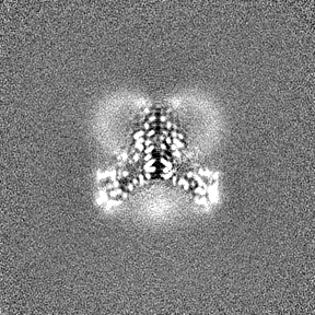
| Images |  emd_43812.png emd_43812.png | 213.5 KB | ||
| Filedesc metadata | emd-43812.cif.gz | 7.4 KB | ||
| Others | emd_43812_half_map_1.map.gzemd_43812_half_map_2.map.gz | 84.3 MB 84.3 MB | ||
| Archive directory |  http://ftp.pdbj.org/pub/emdb/structures/EMD-43812ftp://ftp.pdbj.org/pub/emdb/structures/EMD-43812 http://ftp.pdbj.org/pub/emdb/structures/EMD-43812ftp://ftp.pdbj.org/pub/emdb/structures/EMD-43812 | HTTPS FTP |
-Related structure data
| Related structure data |  9ascMC  8vxhC 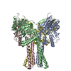 9b77C M: atomic model generated by this map C: citing same article ( |
|---|---|
| Similar structure data |


-Links
| EMDB pages | EMDB (EBI/PDBe) / EMDataResource |
|---|---|
| Related items in Molecule of the Month |


-Map
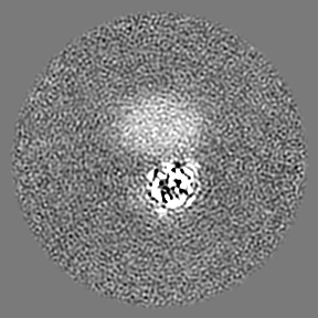
| File | Download / File: emd_43812.map.gz / Format: CCP4 / Size: 91.1 MB / Type: IMAGE STORED AS FLOATING POINT NUMBER (4 BYTES) | ||||||||||||||||||||||||||||||||||||
|---|---|---|---|---|---|---|---|---|---|---|---|---|---|---|---|---|---|---|---|---|---|---|---|---|---|---|---|---|---|---|---|---|---|---|---|---|---|
| Annotation | ArnC/UDP sharpened map | ||||||||||||||||||||||||||||||||||||
| Projections & slices | Image control
Images are generated by Spider. | ||||||||||||||||||||||||||||||||||||
| Voxel size | X=Y=Z: 0.82 Å | ||||||||||||||||||||||||||||||||||||
| Density |
| ||||||||||||||||||||||||||||||||||||
| Symmetry | Space group: 1 | ||||||||||||||||||||||||||||||||||||
| Details | EMDB XML:
|
 Z (Sec.)
 Y (Row.)
 X (Col.)


-Supplemental data
-Half map: ArnC/UDP half-map A
| File | emd_43812_half_map_1.map | ||||||||||||
|---|---|---|---|---|---|---|---|---|---|---|---|---|---|
| Annotation | ArnC/UDP half-map A | ||||||||||||
| Projections & Slices |
| ||||||||||||
| Density Histograms |


-Half map: ArnC/UDP half-map B
| File | emd_43812_half_map_2.map | ||||||||||||
|---|---|---|---|---|---|---|---|---|---|---|---|---|---|
| Annotation | ArnC/UDP half-map B | ||||||||||||
| Projections & Slices |
| ||||||||||||
| Density Histograms |


- Sample components
-Entire : Salmonella enterica ArnC in MSP1E3D1 nanodisc bound to UDP and Mn2+
| Entire | Name: Salmonella enterica ArnC in MSP1E3D1 nanodisc bound to UDP and Mn2+ |
|---|---|
| Components |
|
-Supramolecule #1: Salmonella enterica ArnC in MSP1E3D1 nanodisc bound to UDP and Mn2+
| Supramolecule | Name: Salmonella enterica ArnC in MSP1E3D1 nanodisc bound to UDP and Mn2+ type: complex / ID: 1 / Parent: 0 / Macromolecule list: #1 |
|---|---|
| Source (natural) | Organism: Salmonella enterica subsp. enterica serovar Typhimurium str. LT2 (bacteria) |
| Molecular weight | Theoretical: 162.7 KDa |
-Macromolecule #1: Undecaprenyl-phosphate 4-deoxy-4-formamido-L-arabinose transferase
| Macromolecule | Name: Undecaprenyl-phosphate 4-deoxy-4-formamido-L-arabinose transferase type: protein_or_peptide / ID: 1 / Number of copies: 4 / Enantiomer: LEVO EC number: undecaprenyl-phosphate 4-deoxy-4-formamido-L-arabinose transferase |
|---|---|
| Source (natural) | Organism: Salmonella enterica subsp. enterica serovar Typhimurium str. LT2 (bacteria) |
| Molecular weight | Theoretical: 40.725539 KDa |
| Recombinant expression | Organism: |
| Sequence | String: MDYKDDDDKH HHHHHHHHHE NLYFQSYVGG GSGGGSMFDA APIKKVSVVI PVYNEQESLP ELIRRTTTAC ESLGKAWEIL LIDDGSSDS SAELMVKASQ EADSHIISIL LNRNYGQHAA IMAGFSHVSG DLIITLDADL QNPPEEIPRL VAKADEGFDV V GTVRQNRQ ...String: MDYKDDDDKH HHHHHHHHHE NLYFQSYVGG GSGGGSMFDA APIKKVSVVI PVYNEQESLP ELIRRTTTAC ESLGKAWEIL LIDDGSSDS SAELMVKASQ EADSHIISIL LNRNYGQHAA IMAGFSHVSG DLIITLDADL QNPPEEIPRL VAKADEGFDV V GTVRQNRQ DSLFRKSASK IINLLIQRTT GKAMGDYGCM LRAYRRPIID TMLRCHERST FIPILANIFA RRATEIPVHH AE REFGDSK YSFMRLINLM YDLVTCLTTT PLRLLSLLGS VIAIGGFSLS VLLIVLRLAL GPQWAAEGVF MLFAVLFTFI GAQ FIGMGL LGEYIGRIYN DVRARPRYFV QQVIYPESTP FTEESHQ UniProtKB: Undecaprenyl-phosphate 4-deoxy-4-formamido-L-arabinose transferase |
-Macromolecule #2: URIDINE-5'-DIPHOSPHATE
| Macromolecule | Name: URIDINE-5'-DIPHOSPHATE / type: ligand / ID: 2 / Number of copies: 4 / Formula: UDP |
|---|---|
| Molecular weight | Theoretical: 404.161 Da |
| Chemical component information |  ChemComp-UDP: |
-Macromolecule #3: MANGANESE (II) ION
| Macromolecule | Name: MANGANESE (II) ION / type: ligand / ID: 3 / Number of copies: 4 / Formula: MN |
|---|---|
| Molecular weight | Theoretical: 54.938 Da |
-Experimental details
-Structure determination
| Method | cryo EM |
|---|---|
 Processing Processing | single particle reconstruction |
| Aggregation state | particle |
-Sample preparation
| Concentration | 1.5 mg/mL | |||||||||||||||
|---|---|---|---|---|---|---|---|---|---|---|---|---|---|---|---|---|
| Buffer | pH: 7.5 Component:
| |||||||||||||||
| Grid | Model: UltrAuFoil R1.2/1.3 / Material: GOLD / Mesh: 300 / Support film - Material: GOLD / Support film - topology: HOLEY / Pretreatment - Type: GLOW DISCHARGE / Pretreatment - Time: 30 sec. / Pretreatment - Atmosphere: AIR | |||||||||||||||
| Vitrification | Cryogen name: ETHANE / Chamber humidity: 100 % / Chamber temperature: 277.15 K / Instrument: FEI VITROBOT MARK IV Details: 3 microliters of ArnC incorporated into nanodiscs was applied to a glow-discharged UltraAuFoil (1.2/1.3) 300 mesh grids (Quantifoil), blotted with filter paper for 3.5 s, and flash-frozen by ...Details: 3 microliters of ArnC incorporated into nanodiscs was applied to a glow-discharged UltraAuFoil (1.2/1.3) 300 mesh grids (Quantifoil), blotted with filter paper for 3.5 s, and flash-frozen by plunging in liquid ethane cooled with liquid nitrogen. Grids were stored in liquid nitrogen.. |
- Electron microscopy
| Microscope | FEI TALOS ARCTICA |
|---|---|
| Specialist optics | Energy filter - Name: GIF Bioquantum / Energy filter - Slit width: 20 eV |
| Image recording | Film or detector model: GATAN K2 SUMMIT (4k x 4k) / Detector mode: COUNTING / Digitization - Dimensions - Width: 3838 pixel / Digitization - Dimensions - Height: 3710 pixel / Digitization - Frames/image: 1-53 / Number grids imaged: 1 / Number real images: 7925 / Average exposure time: 5.3 sec. / Average electron dose: 39.88 e/Å2 |
| Electron beam | Acceleration voltage: 200 kV / Electron source:  FIELD EMISSION GUN FIELD EMISSION GUN |
| Electron optics | C2 aperture diameter: 50.0 µm / Illumination mode: FLOOD BEAM / Imaging mode: BRIGHT FIELD / Cs: 2.7 mm / Nominal defocus max: 2.5 µm / Nominal defocus min: 0.5 µm / Nominal magnification: 165000 |
| Sample stage | Specimen holder model: FEI TITAN KRIOS AUTOGRID HOLDER / Cooling holder cryogen: NITROGEN |
| Experimental equipment |  Model: Talos Arctica / Image courtesy: FEI Company |
+Image processing

-Atomic model buiding 1
| Initial model | Chain - Source name: AlphaFold / Chain - Initial model type: in silico model |
|---|---|
| Refinement | Space: REAL / Protocol: AB INITIO MODEL |
| Output model | PDB-9asc: |
