 Movie
Movie Controller

[English] 日本語
 Yorodumi
- EMDB-4305: Correlative cryo-FM and Cryo-ET of vitreous sections of yeast cells -
+ Open data

- Basic information
| Entry | Database: EMDB / ID: EMD-4305 | |||||||||
|---|---|---|---|---|---|---|---|---|---|---|
| Title | Correlative cryo-FM and Cryo-ET of vitreous sections of yeast cells | |||||||||
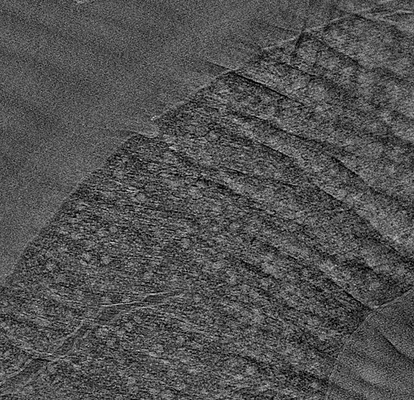
 Map data Map data | This is a tomogram of a vitreous section of yeast cells expressing Pil1-GFP. There are two eisosomal sites seen. | |||||||||
 Sample Sample |
| |||||||||
| Biological species |  | |||||||||
| Method | electron tomography | |||||||||
 Authors Authors | Bharat TAM / Hoffmann PC / Kukulski W | |||||||||
 Citation Citation | Journal: Structure / Year: 2018 Title: Correlative Microscopy of Vitreous Sections Provides Insights into BAR-Domain Organization In Situ. Authors: Tanmay A M Bharat / Patrick C Hoffmann / Wanda Kukulski /  Abstract: Electron microscopy imaging of macromolecular complexes in their native cellular context is limited by the inherent difficulty to acquire high-resolution tomographic data from thick cells and to ...Electron microscopy imaging of macromolecular complexes in their native cellular context is limited by the inherent difficulty to acquire high-resolution tomographic data from thick cells and to specifically identify elusive structures within crowded cellular environments. Here, we combined cryo-fluorescence microscopy with electron cryo-tomography of vitreous sections into a coherent correlative microscopy workflow, ideal for detection and structural analysis of elusive protein assemblies in situ. We used this workflow to address an open question on BAR-domain coating of yeast plasma membrane compartments known as eisosomes. BAR domains can sense or induce membrane curvature, and form scaffold-like membrane coats in vitro. Our results demonstrate that in cells, the BAR protein Pil1 localizes to eisosomes of varying membrane curvature. Sub-tomogram analysis revealed a dense protein coat on curved eisosomes, which was not present on shallow eisosomes, indicating that while BAR domains can assemble at shallow membranes in vivo, scaffold formation is tightly coupled to curvature generation. | |||||||||
| History |
|
- Structure visualization
| Movie |
 Movie viewer Movie viewer |
|---|---|
| Supplemental images |
- Downloads & links
-EMDB archive
| Map data | emd_4305.map.gz | 155.2 MB | EMDB map data format | |
|---|---|---|---|---|
| Header (meta data) | emd-4305-v30.xmlemd-4305.xml | 8.6 KB 8.6 KB | Display Display | EMDB header |
| Images |  emd_4305.png emd_4305.png | 140.3 KB | ||
| Archive directory |  http://ftp.pdbj.org/pub/emdb/structures/EMD-4305ftp://ftp.pdbj.org/pub/emdb/structures/EMD-4305 http://ftp.pdbj.org/pub/emdb/structures/EMD-4305ftp://ftp.pdbj.org/pub/emdb/structures/EMD-4305 | HTTPS FTP |
-Related structure data
| EM raw data | EMPIAR-10161 (Title: Correlative microscopy of vitreous sections provides insights into BAR-domain organisation in situ Data size: 27.9 Data #1: 9 folders each containing raw tilt series of vitreous sections of yeast cells and the corresponding correlative microscopy data [tilt series]) |
|---|
-Links
| EMDB pages | EMDB (EBI/PDBe) / EMDataResource |
|---|
-Map
| File | Download / File: emd_4305.map.gz / Format: CCP4 / Size: 221.7 MB / Type: IMAGE STORED AS SIGNED BYTE | ||||||||||||||||||||||||||||||||||||||||||||||||||||||||||||
|---|---|---|---|---|---|---|---|---|---|---|---|---|---|---|---|---|---|---|---|---|---|---|---|---|---|---|---|---|---|---|---|---|---|---|---|---|---|---|---|---|---|---|---|---|---|---|---|---|---|---|---|---|---|---|---|---|---|---|---|---|---|
| Annotation | This is a tomogram of a vitreous section of yeast cells expressing Pil1-GFP. There are two eisosomal sites seen. | ||||||||||||||||||||||||||||||||||||||||||||||||||||||||||||
| Voxel size | X=Y=Z: 14.34 Å | ||||||||||||||||||||||||||||||||||||||||||||||||||||||||||||
| Density |
| ||||||||||||||||||||||||||||||||||||||||||||||||||||||||||||
| Symmetry | Space group: 1 | ||||||||||||||||||||||||||||||||||||||||||||||||||||||||||||
| Details | EMDB XML:
CCP4 map header:
| ||||||||||||||||||||||||||||||||||||||||||||||||||||||||||||
-Supplemental data
- Sample components
-Entire : Saccharomyces cerevisiae
| Entire | Name: |
|---|---|
| Components |
|
-Supramolecule #1: Saccharomyces cerevisiae
| Supramolecule | Name: Saccharomyces cerevisiae / type: cell / ID: 1 / Parent: 0 Details: genotype: MATalpha, his3delta200, leu2-3,112, ura3-52, lys2-801, PIL1-EGFP::HIS3MX6 |
|---|---|
| Source (natural) | Organism: |
-Experimental details
-Structure determination
 Processing Processing | electron tomography |
|---|---|
| Aggregation state | cell |
-Sample preparation
| Buffer | pH: 7.4 |
|---|---|
| High pressure freezing | Instrument: OTHER Details: The value given for _emd_high_pressure_freezing.instrument is Leica HPM100. This is not in a list of allowed values set(['LEICA EM PACT2', 'LEICA EM PACT', 'EMS-002 RAPID IMMERSION FREEZER', ...Details: The value given for _emd_high_pressure_freezing.instrument is Leica HPM100. This is not in a list of allowed values set(['LEICA EM PACT2', 'LEICA EM PACT', 'EMS-002 RAPID IMMERSION FREEZER', 'OTHER', 'LEICA EM HPM100', 'BAL-TEC HPM 010']) so OTHER is written into the XML file. |
| Sectioning | Ultramicrotomy - Instrument: Leica / Ultramicrotomy - Temperature: 100 K / Ultramicrotomy - Final thickness: 150 |
| Fiducial marker | Manufacturer: CMC Utrecht / Diameter: 10 nm |
- Electron microscopy
| Microscope | FEI TITAN KRIOS |
|---|---|
| Specialist optics | Energy filter - Name: GIF Quantum LS / Energy filter - Slit width: 20 eV / Energy filter - Lower energy threshold: 0 eV / Energy filter - Upper energy threshold: 20 eV |
| Image recording | Film or detector model: GATAN K2 SUMMIT (4k x 4k) / Detector mode: COUNTING / Average electron dose: 0.9 e/Å2 |
| Electron beam | Acceleration voltage: 300 kV / Electron source:  FIELD EMISSION GUN FIELD EMISSION GUN |
| Electron optics | Illumination mode: FLOOD BEAM / Imaging mode: BRIGHT FIELD |
| Experimental equipment |  Model: Titan Krios / Image courtesy: FEI Company |
-Image processing
| Final reconstruction | Algorithm: SIMULTANEOUS ITERATIVE (SIRT) / Software - Name: IMOD / Number images used: 121 |
|---|---|
| CTF correction | Software - Name: IMOD |
