+ データを開く
- 基本情報
基本情報
| 登録情報 |  | |||||||||
|---|---|---|---|---|---|---|---|---|---|---|
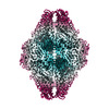
| タイトル | Structure of E.coli beta-Galactosidase from a multi-species dataset | |||||||||
 マップデータ マップデータ | Final map sharp | |||||||||
 試料 試料 |
| |||||||||
 キーワード キーワード | hydrolase / D2 / beta-gal | |||||||||
| 生物種 |  | |||||||||
| 手法 | 単粒子再構成法 / クライオ電子顕微鏡法 / 解像度: 2.73 Å | |||||||||
 データ登録者 データ登録者 | Kopylov M / Bobe D / Eng ET | |||||||||
| 資金援助 |  米国, 1件 米国, 1件
| |||||||||
 引用 引用 |  ジャーナル: Acta Crystallogr F Struct Biol Commun / 年: 2024 ジャーナル: Acta Crystallogr F Struct Biol Commun / 年: 2024タイトル: Multi-species cryoEM calibration and workflow verification standard. 著者: Daija Bobe / Mykhailo Kopylov / Jessalyn Miller / Aaron P Owji / Edward T Eng /  要旨: Cryogenic electron microscopy (cryoEM) is a rapidly growing structural biology modality that has been successful in revealing molecular details of biological systems. However, unlike established ...Cryogenic electron microscopy (cryoEM) is a rapidly growing structural biology modality that has been successful in revealing molecular details of biological systems. However, unlike established biophysical and analytical techniques with calibration standards, cryoEM has lacked comprehensive biological test samples. Here, a cryoEM calibration sample consisting of a mixture of compatible macromolecules is introduced that can not only be used for resolution optimization, but also provides multiple reference points for evaluating instrument performance, data quality and image-processing workflows in a single experiment. This combined test specimen provides researchers with a reference point for validating their cryoEM pipeline, benchmarking their methodologies and testing new algorithms. | |||||||||
| 履歴 |
|
- 構造の表示
構造の表示
| 添付画像 |
|---|
- ダウンロードとリンク
-EMDBアーカイブ
| マップデータ |  emd_41919.map.gz emd_41919.map.gz | 167.9 MB |  EMDBマップデータ形式 EMDBマップデータ形式 | |
|---|---|---|---|---|
| ヘッダ (付随情報) |  emd-41919-v30.xml emd-41919-v30.xml emd-41919.xml emd-41919.xml | 15.7 KB 15.7 KB | 表示 表示 |  EMDBヘッダ EMDBヘッダ |
| FSC (解像度算出) |  emd_41919_fsc.xml emd_41919_fsc.xml | 11.8 KB | 表示 |  FSCデータファイル FSCデータファイル |
| 画像 |  emd_41919.png emd_41919.png | 143.4 KB | ||
| マスクデータ |  emd_41919_msk_1.map emd_41919_msk_1.map | 178 MB |  マスクマップ マスクマップ | |
| Filedesc metadata |  emd-41919.cif.gz emd-41919.cif.gz | 4.3 KB | ||
| その他 |  emd_41919_half_map_1.map.gz emd_41919_half_map_1.map.gz emd_41919_half_map_2.map.gz emd_41919_half_map_2.map.gz | 165 MB 165 MB | ||
| アーカイブディレクトリ |  http://ftp.pdbj.org/pub/emdb/structures/EMD-41919 http://ftp.pdbj.org/pub/emdb/structures/EMD-41919 ftp://ftp.pdbj.org/pub/emdb/structures/EMD-41919 ftp://ftp.pdbj.org/pub/emdb/structures/EMD-41919 | HTTPS FTP |
-検証レポート
| 文書・要旨 |  emd_41919_validation.pdf.gz emd_41919_validation.pdf.gz | 1.1 MB | 表示 |  EMDB検証レポート EMDB検証レポート |
|---|---|---|---|---|
| 文書・詳細版 |  emd_41919_full_validation.pdf.gz emd_41919_full_validation.pdf.gz | 1.1 MB | 表示 | |
| XML形式データ |  emd_41919_validation.xml.gz emd_41919_validation.xml.gz | 20.9 KB | 表示 | |
| CIF形式データ |  emd_41919_validation.cif.gz emd_41919_validation.cif.gz | 27 KB | 表示 | |
| アーカイブディレクトリ |  https://ftp.pdbj.org/pub/emdb/validation_reports/EMD-41919 https://ftp.pdbj.org/pub/emdb/validation_reports/EMD-41919 ftp://ftp.pdbj.org/pub/emdb/validation_reports/EMD-41919 ftp://ftp.pdbj.org/pub/emdb/validation_reports/EMD-41919 | HTTPS FTP |
-関連構造データ
- リンク
リンク
| EMDBのページ |  EMDB (EBI/PDBe) / EMDB (EBI/PDBe) /  EMDataResource EMDataResource |
|---|
- マップ
マップ
| ファイル |  ダウンロード / ファイル: emd_41919.map.gz / 形式: CCP4 / 大きさ: 178 MB / タイプ: IMAGE STORED AS FLOATING POINT NUMBER (4 BYTES) ダウンロード / ファイル: emd_41919.map.gz / 形式: CCP4 / 大きさ: 178 MB / タイプ: IMAGE STORED AS FLOATING POINT NUMBER (4 BYTES) | ||||||||||||||||||||||||||||||||||||
|---|---|---|---|---|---|---|---|---|---|---|---|---|---|---|---|---|---|---|---|---|---|---|---|---|---|---|---|---|---|---|---|---|---|---|---|---|---|
| 注釈 | Final map sharp | ||||||||||||||||||||||||||||||||||||
| 投影像・断面図 | 画像のコントロール
画像は Spider により作成 | ||||||||||||||||||||||||||||||||||||
| ボクセルのサイズ | X=Y=Z: 1.096 Å | ||||||||||||||||||||||||||||||||||||
| 密度 |
| ||||||||||||||||||||||||||||||||||||
| 対称性 | 空間群: 1 | ||||||||||||||||||||||||||||||||||||
| 詳細 | EMDB XML:
|
-添付データ
-マスク #1
| ファイル |  emd_41919_msk_1.map emd_41919_msk_1.map | ||||||||||||
|---|---|---|---|---|---|---|---|---|---|---|---|---|---|
| 投影像・断面図 |
| ||||||||||||
| 密度ヒストグラム |
-ハーフマップ: Halfmap A
| ファイル | emd_41919_half_map_1.map | ||||||||||||
|---|---|---|---|---|---|---|---|---|---|---|---|---|---|
| 注釈 | Halfmap A | ||||||||||||
| 投影像・断面図 |
| ||||||||||||
| 密度ヒストグラム |
-ハーフマップ: Halfmap B
| ファイル | emd_41919_half_map_2.map | ||||||||||||
|---|---|---|---|---|---|---|---|---|---|---|---|---|---|
| 注釈 | Halfmap B | ||||||||||||
| 投影像・断面図 |
| ||||||||||||
| 密度ヒストグラム |
- 試料の構成要素
-全体 : beta-Galactosidase from Escherichia coli in complex with 2-phenyl...
| 全体 | 名称: beta-Galactosidase from Escherichia coli in complex with 2-phenylethyl beta-D-thiogalactoside |
|---|---|
| 要素 |
|
-超分子 #1: beta-Galactosidase from Escherichia coli in complex with 2-phenyl...
| 超分子 | 名称: beta-Galactosidase from Escherichia coli in complex with 2-phenylethyl beta-D-thiogalactoside タイプ: complex / ID: 1 / 親要素: 0 |
|---|---|
| 由来(天然) | 生物種:  |
| 分子量 | 理論値: 2.5 MDa |
-実験情報
-構造解析
| 手法 | クライオ電子顕微鏡法 |
|---|---|
 解析 解析 | 単粒子再構成法 |
| 試料の集合状態 | particle |
- 試料調製
試料調製
| 濃度 | 1 mg/mL |
|---|---|
| 緩衝液 | pH: 7.5 |
| 凍結 | 凍結剤: ETHANE |
- 電子顕微鏡法
| 顕微鏡 | TFS KRIOS |
|---|---|
| 撮影 | フィルム・検出器のモデル: GATAN K2 SUMMIT (4k x 4k) 平均電子線量: 70.0 e/Å2 |
| 電子線 | 加速電圧: 300 kV / 電子線源:  FIELD EMISSION GUN FIELD EMISSION GUN |
| 電子光学系 | 照射モード: FLOOD BEAM / 撮影モード: BRIGHT FIELD / 最大 デフォーカス(公称値): 2.5 µm / 最小 デフォーカス(公称値): 1.5 µm |
| 実験機器 |  モデル: Titan Krios / 画像提供: FEI Company |
 ムービー
ムービー コントローラー


 Z (Sec.)
 Y (Row.)
 X (Col.)


