 Movie
Movie Controller

[English] 日本語
 Yorodumi
- EMDB-39478: Structure of Aquifex aeolicus Lumazine Synthase by Cryo-Electron ... -
+ Open data

- Basic information
| Entry |  | |||||||||
|---|---|---|---|---|---|---|---|---|---|---|
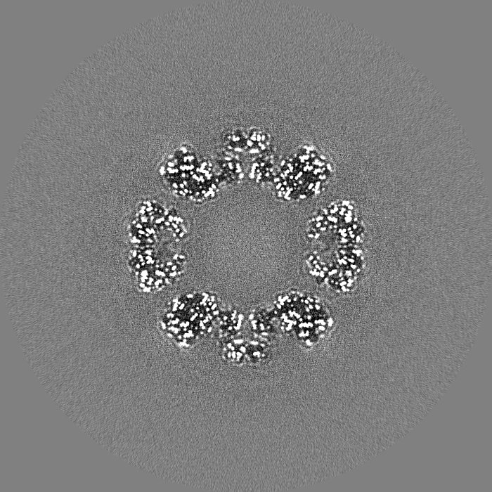
| Title | Structure of Aquifex aeolicus Lumazine Synthase by Cryo-Electron Microscopy to 1.42 Angstrom Resolution | |||||||||
 Map data Map data | Post processed map | |||||||||
 Sample Sample |
| |||||||||
 Keywords Keywords | Enzyme involved in riboflavin biosynthesis / BIOSYNTHETIC PROTEIN | |||||||||
| Function / homology |  Function and homology information Function and homology information6,7-dimethyl-8-ribityllumazine synthase / 6,7-dimethyl-8-ribityllumazine synthase activity / riboflavin synthase complex / riboflavin biosynthetic process / cytosol / cytoplasm Similarity search - Function | |||||||||
| Biological species |   Aquifex aeolicus (bacteria) Aquifex aeolicus (bacteria) | |||||||||
| Method | single particle reconstruction / cryo EM / Resolution: 1.42 Å | |||||||||
 Authors Authors | Savva CG / Sobhy M / De Biasio A / Hamdan SM | |||||||||
| Funding support | 1 items
| |||||||||
 Citation Citation | Journal: IUCrJ / Year: 2024 Title: Structure of Aquifex aeolicus lumazine synthase by cryo-electron microscopy to 1.42 Å resolution. Authors: Christos G Savva / Mohamed A Sobhy / Alfredo De Biasio / Samir M Hamdan /  Abstract: Single-particle cryo-electron microscopy (cryo-EM) has become an essential structural determination technique with recent hardware developments making it possible to reach atomic resolution, at which ...Single-particle cryo-electron microscopy (cryo-EM) has become an essential structural determination technique with recent hardware developments making it possible to reach atomic resolution, at which individual atoms, including hydrogen atoms, can be resolved. In this study, we used the enzyme involved in the penultimate step of riboflavin biosynthesis as a test specimen to benchmark a recently installed microscope and determine if other protein complexes could reach a resolution of 1.5 Å or better, which so far has only been achieved for the iron carrier ferritin. Using state-of-the-art microscope and detector hardware as well as the latest software techniques to overcome microscope and sample limitations, a 1.42 Å map of Aquifex aeolicus lumazine synthase (AaLS) was obtained from a 48 h microscope session. In addition to water molecules and ligands involved in the function of AaLS, we can observe positive density for ∼50% of the hydrogen atoms. A small improvement in the resolution was achieved by Ewald sphere correction which was expected to limit the resolution to ∼1.5 Å for a molecule of this diameter. Our study confirms that other protein complexes can be solved to near-atomic resolution. Future improvements in specimen preparation and protein complex stabilization may allow more flexible macromolecules to reach this level of resolution and should become a priority of study in the field. | |||||||||
| History |
|
- Structure visualization
| Supplemental images |
|---|

- Downloads & links
-EMDB archive
| Map data | emd_39478.map.gz | 165.5 MB | EMDB map data format | |
|---|---|---|---|---|
| Header (meta data) | emd-39478-v30.xmlemd-39478.xml | 16 KB 16 KB | Display Display | EMDB header |
| FSC (resolution estimation) | emd_39478_fsc.xml | 24.6 KB | Display | FSC data file |
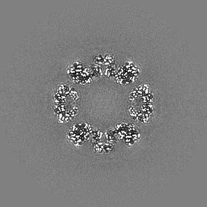
| Images |  emd_39478.png emd_39478.png | 229.5 KB | ||
| Masks | emd_39478_msk_1.map | 1.3 GB | Mask map | |
| Filedesc metadata | emd-39478.cif.gz | 5.3 KB | ||
| Others | emd_39478_half_map_1.map.gzemd_39478_half_map_2.map.gz | 1 GB 1 GB | ||
| Archive directory |  http://ftp.pdbj.org/pub/emdb/structures/EMD-39478ftp://ftp.pdbj.org/pub/emdb/structures/EMD-39478 http://ftp.pdbj.org/pub/emdb/structures/EMD-39478ftp://ftp.pdbj.org/pub/emdb/structures/EMD-39478 | HTTPS FTP |
-Related structure data
| Related structure data |  8yt4MC M: atomic model generated by this map C: citing same article ( |
|---|---|
| Similar structure data |
-Links
| EMDB pages | EMDB (EBI/PDBe) / EMDataResource |
|---|
-Map
| File | Download / File: emd_39478.map.gz / Format: CCP4 / Size: 1.3 GB / Type: IMAGE STORED AS FLOATING POINT NUMBER (4 BYTES) | ||||||||||||||||||||||||||||||||||||
|---|---|---|---|---|---|---|---|---|---|---|---|---|---|---|---|---|---|---|---|---|---|---|---|---|---|---|---|---|---|---|---|---|---|---|---|---|---|
| Annotation | Post processed map | ||||||||||||||||||||||||||||||||||||
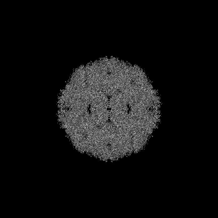
| Projections & slices | Image control
Images are generated by Spider. | ||||||||||||||||||||||||||||||||||||
| Voxel size | X=Y=Z: 0.4553 Å | ||||||||||||||||||||||||||||||||||||
| Density |
| ||||||||||||||||||||||||||||||||||||
| Symmetry | Space group: 1 | ||||||||||||||||||||||||||||||||||||
| Details | EMDB XML:
|
 Z (Sec.)
 Y (Row.)
 X (Col.)


-Supplemental data
-Mask #1
| File | emd_39478_msk_1.map | ||||||||||||
|---|---|---|---|---|---|---|---|---|---|---|---|---|---|
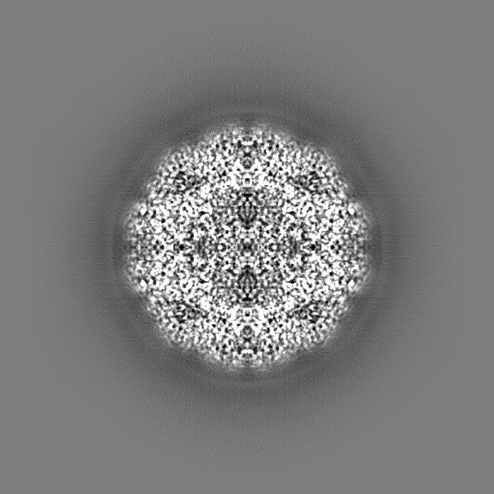
| Projections & Slices |
| ||||||||||||
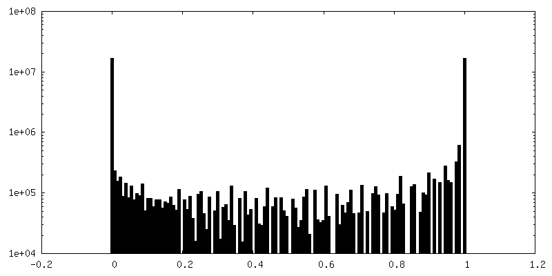
| Density Histograms |


-Half map: Half Map 2
| File | emd_39478_half_map_1.map | ||||||||||||
|---|---|---|---|---|---|---|---|---|---|---|---|---|---|
| Annotation | Half Map 2 | ||||||||||||
| Projections & Slices |
| ||||||||||||
| Density Histograms |


-Half map: Half Map 1
| File | emd_39478_half_map_2.map | ||||||||||||
|---|---|---|---|---|---|---|---|---|---|---|---|---|---|
| Annotation | Half Map 1 | ||||||||||||
| Projections & Slices |
| ||||||||||||
| Density Histograms |


- Sample components
-Entire : Aquifex aeolicus Lumazine Synthase
| Entire | Name: Aquifex aeolicus Lumazine Synthase |
|---|---|
| Components |
|
-Supramolecule #1: Aquifex aeolicus Lumazine Synthase
| Supramolecule | Name: Aquifex aeolicus Lumazine Synthase / type: complex / ID: 1 / Parent: 0 / Macromolecule list: all |
|---|---|
| Source (natural) | Organism: Aquifex aeolicus (bacteria) |
| Molecular weight | Theoretical: 1.065 MDa |
-Macromolecule #1: Lumazine synthase
| Macromolecule | Name: Lumazine synthase / type: protein_or_peptide / ID: 1 / Enantiomer: LEVO |
|---|---|
| Source (natural) | Organism: Aquifex aeolicus (bacteria) |
| Recombinant expression | Organism: |
| Sequence | String: MQIYEGKLTA EGLRFGIVAS RFNHALVDRL VEGAIDCIVR HGGREEDITL VRVPGSWEIP VAAGELARK EDIDAVIAIG VLIRGATPHF DYIASEVSKG LANLSLELRK PITFGVITAD T LEQAIERA GTKHGNKGWE AALSAIEMAN LFKSLRWSHP QFEK UniProtKB: 6,7-dimethyl-8-ribityllumazine synthase |
-Experimental details
-Structure determination
| Method | cryo EM |
|---|---|
 Processing Processing | single particle reconstruction |
| Aggregation state | particle |
-Sample preparation
| Concentration | 2.75 mg/mL |
|---|---|
| Buffer | pH: 7.5 / Details: 20mM Tris pH 7.5, 150mM NaCl and 1mM DTT |
| Grid | Model: UltrAuFoil R1.2/1.3 / Material: GOLD / Mesh: 300 / Support film - Material: GOLD / Support film - topology: HOLEY ARRAY / Pretreatment - Type: GLOW DISCHARGE / Pretreatment - Time: 30 sec. / Pretreatment - Atmosphere: AIR / Details: 30 mA |
| Vitrification | Cryogen name: ETHANE / Chamber humidity: 100 % / Chamber temperature: 277 K / Instrument: FEI VITROBOT MARK IV / Details: Blot time: 3 sec Wait time: 0 sec. |
| Details | In buffer 20mM Tris pH 7.5, 150mM NaCl and 1mM DTT |
- Electron microscopy
| Microscope | FEI TITAN KRIOS |
|---|---|
| Specialist optics | Energy filter - Name: TFS Selectris X / Energy filter - Slit width: 10 eV |
| Image recording | Film or detector model: FEI FALCON IV (4k x 4k) / Number grids imaged: 1 / Number real images: 12657 / Average exposure time: 3.0 sec. / Average electron dose: 46.0 e/Å2 |
| Electron beam | Acceleration voltage: 300 kV / Electron source:  FIELD EMISSION GUN FIELD EMISSION GUN |
| Electron optics | C2 aperture diameter: 50.0 µm / Illumination mode: FLOOD BEAM / Imaging mode: BRIGHT FIELD / Cs: 2.7 mm / Nominal defocus max: 1.2 µm / Nominal defocus min: 0.4 µm / Nominal magnification: 165000 |
| Sample stage | Specimen holder model: FEI TITAN KRIOS AUTOGRID HOLDER / Cooling holder cryogen: NITROGEN |
| Experimental equipment |  Model: Titan Krios / Image courtesy: FEI Company |
+Image processing

-Atomic model buiding 1
| Refinement | Space: RECIPROCAL / Protocol: FLEXIBLE FIT |
|---|---|
| Output model | PDB-8yt4: |
