+ データを開く
- 基本情報
基本情報
| 登録情報 | データベース: EMDB / ID: EMD-3820 | |||||||||
|---|---|---|---|---|---|---|---|---|---|---|
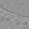
| タイトル | Electron tomographic slices of the nuclear envelope of HeLa cell in interphase | |||||||||
 マップデータ マップデータ | None | |||||||||
 試料 試料 |
| |||||||||
| 生物種 |  Homo sapiens (ヒト) Homo sapiens (ヒト) | |||||||||
| 手法 | 電子線トモグラフィー法 / クライオ電子顕微鏡法 / ネガティブ染色法 | |||||||||
 データ登録者 データ登録者 | Otsuka S / Ellenberg J | |||||||||
 引用 引用 |  ジャーナル: Nat Struct Mol Biol / 年: 2018 ジャーナル: Nat Struct Mol Biol / 年: 2018タイトル: Postmitotic nuclear pore assembly proceeds by radial dilation of small membrane openings. 著者: Shotaro Otsuka / Anna M Steyer / Martin Schorb / Jean-Karim Hériché / M Julius Hossain / Suruchi Sethi / Moritz Kueblbeck / Yannick Schwab / Martin Beck / Jan Ellenberg /  要旨: The nuclear envelope has to be reformed after mitosis to create viable daughter cells with closed nuclei. How membrane sealing of DNA and assembly of nuclear pore complexes (NPCs) are achieved and ...The nuclear envelope has to be reformed after mitosis to create viable daughter cells with closed nuclei. How membrane sealing of DNA and assembly of nuclear pore complexes (NPCs) are achieved and coordinated is poorly understood. Here, we reconstructed nuclear membrane topology and the structures of assembling NPCs in a correlative 3D EM time course of dividing human cells. Our quantitative ultrastructural analysis shows that nuclear membranes form from highly fenestrated ER sheets whose holes progressively shrink. NPC precursors are found in small membrane holes and dilate radially during assembly of the inner ring complex, forming thousands of transport channels within minutes. This mechanism is fundamentally different from that of interphase NPC assembly and explains how mitotic cells can rapidly establish a closed nuclear compartment while making it transport competent. | |||||||||
| 履歴 |
|
- 構造の表示
構造の表示
| ムービー |
 ムービービューア ムービービューア |
|---|---|
| 構造ビューア | EMマップ:  SurfView SurfView Molmil Molmil Jmol/JSmol Jmol/JSmol |
| 添付画像 |
- ダウンロードとリンク
-EMDBアーカイブ
| マップデータ |  emd_3820.map.gz emd_3820.map.gz | 7.5 GB |  EMDBマップデータ形式 EMDBマップデータ形式 | |
|---|---|---|---|---|
| ヘッダ (付随情報) |  emd-3820-v30.xml emd-3820-v30.xml emd-3820.xml emd-3820.xml | 10.7 KB 10.7 KB | 表示 表示 |  EMDBヘッダ EMDBヘッダ |
| 画像 |  emd_3820.png emd_3820.png | 139.3 KB | ||
| アーカイブディレクトリ |  http://ftp.pdbj.org/pub/emdb/structures/EMD-3820 http://ftp.pdbj.org/pub/emdb/structures/EMD-3820 ftp://ftp.pdbj.org/pub/emdb/structures/EMD-3820 ftp://ftp.pdbj.org/pub/emdb/structures/EMD-3820 | HTTPS FTP |
-検証レポート
| 文書・要旨 |  emd_3820_validation.pdf.gz emd_3820_validation.pdf.gz | 274.7 KB | 表示 |  EMDB検証レポート EMDB検証レポート |
|---|---|---|---|---|
| 文書・詳細版 |  emd_3820_full_validation.pdf.gz emd_3820_full_validation.pdf.gz | 274.2 KB | 表示 | |
| XML形式データ |  emd_3820_validation.xml.gz emd_3820_validation.xml.gz | 3.5 KB | 表示 | |
| アーカイブディレクトリ |  https://ftp.pdbj.org/pub/emdb/validation_reports/EMD-3820 https://ftp.pdbj.org/pub/emdb/validation_reports/EMD-3820 ftp://ftp.pdbj.org/pub/emdb/validation_reports/EMD-3820 ftp://ftp.pdbj.org/pub/emdb/validation_reports/EMD-3820 | HTTPS FTP |
-関連構造データ
| 電子顕微鏡画像生データ |  EMPIAR-10116 (タイトル: Raw 2d tomographic tilt series of a dividing cell EMPIAR-10116 (タイトル: Raw 2d tomographic tilt series of a dividing cellData size: 245.4 Data #1: Raw 2D tilt series for all the electron tomograms [tilt series]) |
|---|
- リンク
リンク
| EMDBのページ |  EMDB (EBI/PDBe) / EMDB (EBI/PDBe) /  EMDataResource EMDataResource |
|---|
- マップ
マップ
| ファイル |  ダウンロード / ファイル: emd_3820.map.gz / 形式: CCP4 / 大きさ: 1.8 GB / タイプ: IMAGE STORED AS SIGNED INTEGER (2 BYTES) ダウンロード / ファイル: emd_3820.map.gz / 形式: CCP4 / 大きさ: 1.8 GB / タイプ: IMAGE STORED AS SIGNED INTEGER (2 BYTES) | ||||||||||||||||||||||||||||||||||||||||||||||||||||||||||||
|---|---|---|---|---|---|---|---|---|---|---|---|---|---|---|---|---|---|---|---|---|---|---|---|---|---|---|---|---|---|---|---|---|---|---|---|---|---|---|---|---|---|---|---|---|---|---|---|---|---|---|---|---|---|---|---|---|---|---|---|---|---|
| 注釈 | None | ||||||||||||||||||||||||||||||||||||||||||||||||||||||||||||
| 投影像・断面図 | 画像のコントロール
画像は Spider により作成 これらの図は立方格子座標系で作成されたものです | ||||||||||||||||||||||||||||||||||||||||||||||||||||||||||||
| ボクセルのサイズ | X=Y=Z: 7.5 Å | ||||||||||||||||||||||||||||||||||||||||||||||||||||||||||||
| 密度 |
| ||||||||||||||||||||||||||||||||||||||||||||||||||||||||||||
| 対称性 | 空間群: 1 | ||||||||||||||||||||||||||||||||||||||||||||||||||||||||||||
| 詳細 | EMDB XML:
CCP4マップ ヘッダ情報:
| ||||||||||||||||||||||||||||||||||||||||||||||||||||||||||||
-添付データ
- 試料の構成要素
-全体 : HeLa cell
| 全体 | 名称: HeLa cell |
|---|---|
| 要素 |
|
-超分子 #1: HeLa cell
| 超分子 | 名称: HeLa cell / タイプ: cell / ID: 1 / 親要素: 0 詳細: Cells were high-pressure frozen and freeze-substituted into Lowicryl resin. |
|---|---|
| 由来(天然) | 生物種:  Homo sapiens (ヒト) / 株: HeLa Homo sapiens (ヒト) / 株: HeLa |
-実験情報
-構造解析
| 手法 | ネガティブ染色法, クライオ電子顕微鏡法 |
|---|---|
 解析 解析 | 電子線トモグラフィー法 |
| 試料の集合状態 | cell |
- 試料調製
試料調製
| 緩衝液 | pH: 7.4 詳細: CO2-independent medium without phenol red (Invitrogen), containing 20% FCS, 20% Ficoll PM400 2 mM l-glutamine, and 100 ug/ml penicillin and streptomycin |
|---|---|
| 染色 | タイプ: NEGATIVE / 材質: uranyl acetate and lead citrate |
| 糖包埋 | 材質: Lowicryl resin 詳細: Frozen cells were incubated with 0.1% uranyl acetate in acetone at -90C for 20-24 hr and, after infiltration into Lowicryl resin and UV-polymerization, samples were further polymerized by sunlight for 3-4 days. |
| グリッド | モデル: Grid / 支持フィルム - 材質: FORMVAR / 支持フィルム - トポロジー: CONTINUOUS |
| 凍結 | 凍結剤: NITROGEN |
| 加圧凍結法 | 装置: OTHER 詳細: High pressure freezing chamber was 1.0 mm thick in total, 3.0 mm diameter, with central cavities 50 um deep.. The value given for _emd_high_pressure_freezing.instrument is HPM 010. This is ...詳細: High pressure freezing chamber was 1.0 mm thick in total, 3.0 mm diameter, with central cavities 50 um deep.. The value given for _emd_high_pressure_freezing.instrument is HPM 010. This is not in a list of allowed values set(['LEICA EM PACT2', 'LEICA EM PACT', 'EMS-002 RAPID IMMERSION FREEZER', 'OTHER', 'LEICA EM HPM100', 'BAL-TEC HPM 010']) so OTHER is written into the XML file. |
| Cryo protectant | 20% FBS and Ficoll PM400 |
| 切片作成 | ウルトラミクロトーム - 装置: Leica Ultracut UCT / ウルトラミクロトーム - 温度: 25 K / ウルトラミクロトーム - 最終 厚さ: 300 |
| 位置合わせマーカー | Manufacturer: CMC university Medical Center Utrecht / 直径: 15 nm |
- 電子顕微鏡法
| 顕微鏡 | FEI TECNAI F30 |
|---|---|
| 撮影 | フィルム・検出器のモデル: FEI EAGLE (4k x 4k) / デジタル化 - サイズ - 横: 4096 pixel / デジタル化 - サイズ - 縦: 4096 pixel / 実像数: 121 / 平均電子線量: 200.0 e/Å2 |
| 電子線 | 加速電圧: 300 kV / 電子線源:  FIELD EMISSION GUN FIELD EMISSION GUN |
| 電子光学系 | 照射モード: FLOOD BEAM / 撮影モード: BRIGHT FIELD / Cs: 2.26 mm / 最小 デフォーカス(公称値): 0.5 µm / 倍率(公称値): 15500 |
| 試料ステージ | 試料ホルダーモデル: OTHER |
| 実験機器 |  モデル: Tecnai F30 / 画像提供: FEI Company |
- 画像解析
画像解析
| 最終 再構成 | アルゴリズム: BACK PROJECTION / ソフトウェア - 名称:  IMOD (ver. 4.5.6) IMOD (ver. 4.5.6)詳細: Dual axis tilt series were aligned using gold fiducial markers 使用した粒子像数: 121 |
|---|---|
| CTF補正 | ソフトウェア - 名称:  IMOD (ver. 4.5.6) IMOD (ver. 4.5.6) |
 ムービー
ムービー コントローラー


 Z (Sec.)
 Y (Row.)
 X (Col.)


